

 PDF
PDF ePub
ePub Citation
 Print
Print


INTRODUCTION
Helicobacter pylori is a gram-negative bacterium present in more than 50% of the world's population; and it is one of the most common pathogen in children worldwide [1]. Although H. pylori infection is linked to the development of chronic gastritis and peptic ulcer in children with long exposure to infection [2], most of the infected children remain asymptomatic.
The prevalence of H. pylori differs according to in age, region, and race. The prevalence of H. pylori infection increases with age and has been reported to be variable in both developed and developing countries, linking the prevalence of H. pylori infection with the socioeconomic status [3]. In addition, H. pylori first infection is usually acquired during early childhood, rarely spontaneously eradicated, suggesting that changes in childhood prevalence can sensitively reflect changes in the overall prevalence. According to Malaty et al.'s study [4,5,6], the rate of H. pylori infection is 60-80%, showing a significant difference in Saudi Arabia, India, and Vietnam, against the developed countries such as the United States, Australia, or France, with infection rates of 20-25% in the 20s-30s even in 1991. Similarly, Malaty and several other studies [4,5,6] have reported the following prevalence rates for children in different countries in 2006: 35% in Russia, 20% in China and Poland, 12% in South Korea and the United States, and less than 10% in France, Belgium, and Finland.
Although H. pylori infection rates show a declining trend worldwide, high rates of infection are still present in developing countries. In line with these evidences, the prevalence rate of H. pylori in children in the 2000s has been considered to be lower than the rate in the 1990s; however, there is almost no research on the prevalence rates in Korean children in the last 10 years. In this study, we investigated changes in the prevalence rates in children with recurrent abdominal pain using the urea breath test (UBT), currently the most accurate screening modality.
MATERIALS AND METHODS
Materials
The UBT was performed for children with recurrent abdominal pain who visited the pediatric outpatient clinics at Kyungpook National University Hospital and Kyungpook National University Children's Hospital from January 2004 to May 2014. Recurrent abdominal pain by the Rome II criteria, established in 1999, was defined as abdominal pain in children aged 4-16 years that interferes with daily life for more than 3 months [7].
If children tested positive by the UBT (UBiT-IR 300; Otsuka Electronics Co., Ltd., Osaka, Japan), endoscopy was recommended for confirmation of H. pylori; however, endoscopy was not performed when UBT results were not more than 3‰ point over the cut-off value due to the high number of false-positives. In these cases under 12-year-old, observation was recommended. Though positive by UBT, both a negative CLO test result and a negative histopathological examination results by endoscopy were regarded as negative for H. pylori.
Patients with abdominal pain who presented with peptic ulcer or gastrointestinal bleeding were excluded from this study as well as patients for whom the test was performed owing to other indications such as iron deficiency anemia, idiopathic thrombocytopenia purpura, growth retardation, or chronic urticarial.
Methods
A retrospective cohort of 2,530 children who underwent the UBT and presented with recurrent abdominal pain was studied for determining the prevalence of H. pylori by age and year. Not only identifying UBT titers, we established endoscopic remarks, performed the CLO test, and analyzed histopathological findings when endoscopy was done.
Changes in the prevalence of H. pylori infection were determined according to year, gender, and age. The children were divided into 3 age groups: 4-5, 6-11, and 12-16 years. To determine the changes in the annual prevalence of H. pylori infection, the study period was classified into 3 time periods: 2004-2007, 2008-2010, and 2011-2014.
Prevalence trends were compared between the groups of children aged 4-11 years and 12-16 years, assuming that the prevalence will be further decreased in younger children.
In order to reduce false positive UBT results, a 4.0‰ cut-off value was applied to children ≥6 years and a 7‰ cut-off value to children <6 years [8]. Since endoscopy results performed in patients with a UBT titer near the cut-off value was shown to be negative, endoscopy was only performed for children aged 4-11 who had 3‰ points higher than the cut-off value. Prevalence of H. pylori infection was determined again using cut-off values that had been modified to 10.0‰ for those aged 4-5 years; 7.0‰, for those aged 6-11 years; and 4.0‰, for those aged 12-16 years to further reduce the false positive rates.
RESULTS
A total of 2,530 children (1,191 boys, 1,339 girls; mean age 10.0±3.0 years; range, 4.0-16.9 years) underwent the UBT, who had presented with recurrent abdominal pain from January 2004 to May 2014.
Prevalence rate by different age group and time periods
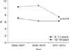
The total infection prevalence rate was 7.4% (187/2,530) with an overall prevalence of 7.6% and 7.2% in boys and girls, respectively; there was no difference in the prevalence in boys and girls over time. The prevalence rate in children was 8.0% in 2004-2007, 7.7% in 2008-2010, and 6.7% in 2011-2014, showing no significant difference among the time periods. The prevalence rate in the different age groups was 7.5% for those aged 4-5 years, 6.3% for those aged 6-11 years, and 9.2% for those aged 12-16 years, which showed a significant difference (p=0.04) (Table 1). No significant relation was observed between increasing age groups and prevalence. Nevertheless, the prevalence of H. pylori infection was 6.5% for those aged 4-11 years and 9.2% for those aged 12-16 years, demonstrating that the prevalence in children aged <12 years was significantly lower than that in children aged ≥12 years (p=0.018) (Table 2, Fig. 1).
Prevalence rate according to modified cut-off value
After endoscopy, 1 child <6 years old and 4 children aged 6-16 years old with a titer not more than 3‰ points over the cut-off value had negative findings for H. pylori, CLO test, and biopsy. Ten children <6 years old and 15 children ≥6 years were temporarily excluded for reanalysis by applying modified cut-off value. When cut-off values were modified, the prevalence rates were 7.0% for the period 2004-2007, 6.5% for the period 2008-2010, and 5.8% for the period 2011-2014, which showed no significant difference over time (p=0.27). However, a statistically significant decrease in the prevalence was observed in the younger group with prevalence values of 4.6% for those aged 4-5 years when compared to 5.3% for those aged 6-11 and 9.2% for 12-16 years (p<0.01; Table 3).
DISCUSSION
The socioeconomic status, numbers of infected family and the personal hygiene are thought to be important factors affecting the prevalence of H. pylori infection in childhood [9]. In developing countries, the infection begins in children <10 years old [10]. If it is not treated, since natural eradication is rare [11], childhood infections persist until adulthood, suggesting a relationship between a high prevalence in adults and previous infection in their childhood.
Korean prevalence rates of H. pylori in the late 1990s are between the rate values of developed and developing countries. The prevalence rate of older adults is similar to that of underdeveloped countries while children's prevalence close to that of developed countries [12]. In addition, an adult study reported that the prevalence of H. pylori was significantly decreased from 1998 to 2005 [13], probably due to the economic development of the country as well as the improved sanitary conditions.
H. pylori is transmitted from person to person, with infection within families being the main route of transmission [14]. According to a recent report, even in developing countries, the onset of infection occurs usually in childhood within the family, underestimating the importance of other infection routes, such as the fecal-oral transmission. Furthermore, according to Rocha et al. [15], H. pylori-infected mothers can act as a strong independent factor to their children. In this study, PCR analysis of the mother's saliva revealed that 78% of mothers of H. pylori-infected children were infected with H. pylori [16]. This suggests an oral-oral transmission, suggesting that contact between the mother and child is the major source of infection [17]. As a result, the analysis of the risk factors associated with H. pylori infection indicates that the socioeconomic status in adult age has only a limited effect, whereas is related in children as well as living standards, such as mother's education, drinking water hygiene, and family income. Differences in prevalence according to the socioeconomic status, and the housing conditions during their childhood period have been identified as important factors related to the infection rate in adults and children [18].
Here, we suggest that high prevalence of H. pylori in Koreans in the 1980s and 1990s was mainly owing to the underdeveloped state of the socioeconomic status, environment, and sanitation, as well as because of the mouth-to-mouth transmission between mother and child, mainly associated with the mastication culture in the weaning food period. According to a study conducted in 2005 with 15,916 healthy people >16 years old, the prevalence of H. pylori was 29.3% for those in their 20s, 49.1% for those in their 30s, 57.8% for those in their 40s, and 61.5% for those in their 50s, pointing to a significant increase in prevalence in those in their 40s and 50s [19].
In our study, there was a significant difference in prevalence in patients ≥12 years old and <12 years old. Thus, when the age of the mother with children ≥12 years old is estimated to be over 40s, she would have been over 30s in 2005. Therefore, 49.1% of mother's infection rate affects children; it was thought to reflect the current prevalence in children aged ≥12 years, relatively low prevalence in 20s in 2005 affects decrease of prevalence in children <12 years. Thus, infection rate in children can be expected to decrease further if the maternal infection rates are lowered along with a decrease in the rate of mouth-to-mouth transmission by disappearing mastication culture in weaning food period.
A reduction in H. pylori prevalence of domestic in the 2000s when compared to the 1990s has already been reported [20,21]. Nevertheless, these studies did not demonstrate a significant decrease in the prevalence in Korean children in the last 11 years.
First, these results can be linked to the country's economic slow growth. As reported by the Bank of Korea in 2014, the changes in the economic growth and development of our country increased sharply in the 80s and 90s. However, the economic growth has stunted after the late 90s. Thus, due to the rapid economic and environment growth, and improvements in hygiene in the 80s and 90s, the prevalence of H. pylori decreased rapidly. Nonetheless, there was no significant decrease in H. pylori infection in the last 11 years probably due to the economic depression in the late 90s. Secondly, public awareness is increased over the last 10 years, that mouth-to-mouth transmission by mastication culture during weaning period is one of the routes of H. pylori infection transmission. Moreover, the decrease in the H. pylori infection rate in children during the last decade was lower due to the increasing maternal age.
The use of endoscopic biopsy for CLO tests and histopathologic examination is the most accurate procedure in the diagnosis of H. pylori infection. However, unlike adults, endoscopy is not always recommended for children due to the possible complications during examination and the psychological burden of the patient and their parents, so that there would be an increased risk of selection bias for prevalence study. The use of H. pylori immunoglobulins is an alternative method with high sensitivity and specificity in adults; however, because of very low sensitivity in children, this method is not used for diagnosis of H. pylori infection in children [22]. On the other hand, the UBT is a non-invasive test that is commonly used easily and safely without the need of experienced personnel, in contrast to endoscopy. The UBT has been used in a relatively accurate way to determine treatment success, since it has a high sensitivity and specificity. Here, we tried to estimate the prevalence rate of H. pylori in patients with recurrent abdominal pain, based on the findings that recurrent abdominal pain in children has no direct association with H. pylori [23]. Our study conducted the UBT for screening patients with recurrent abdominal pain, followed by endoscopy in cases where screening results were positive. Thus, we were able to examine the change in the prevalence rate of H. pylori infection in children, while improving the test accuracy and minimizing the use of endoscopy.
Applying UBT for children has an interpreting week point because of the higher false positive rate in the younger children. In a previous study, children <6 years of age showed a 8.3% false positive rate after UBT, showing 10 times higher values than the rate of 0.85% in children ≥6 years old [24]. Yang et al. [8] suggested a 7.0‰ cut-off value for those <6 years old in order to reduce false positive rates.
In this study, the prevalence of H. pylori infection in children with recurrent abdominal pain was statistically similar when compared to that in normal children (p=0.74) [25]. An endoscopic biopsy study by Lee et al. [26] among children revealed a prevalence of 11.3% for the period of 2003-2005 and 10.8% for the period of 2006-2008. These results were comparable to our study results, with a prevalence of 8.01% for the period 2004-2007 (p=0.28).
During our study, no significant decrease in H. pylori prevalence infection was observed in children with recurrent abdominal pain. However, when comparing children >12 and ≤12 years old, the prevalence of H. pylori was significantly lower in children <12 years. Therefore, we predict a reduction of H. pylori prevalence in the future. According to Fig. 1, the age difference between the 2 groups is about 7 years. The prevalence of H. pylori in the group aged 4-12 years can be estimated to be 10% in the late 1990s; since H. pylori is not naturally eradicated, we assume that the prevalence in children aged 4-12 years was decreased from 10% in the late 1990s (7 years before 2004-2007) to 6% currently. Further, we raised the cut-off value in order to reduce the false positive rate based on early experience that children who present with values 3‰ points higher than the cut-off value tested negative for H. pylori after endoscopy. Hence, when applying the raised cut-off value, the prevalence increased significantly in the higher age groups.
This study has some limitations. Our study was performed in a single institution. Hence, the prevalence of H. pylori in Korean children may not be representative of the whole population. In addition, recent studies have reported a lack of association between H. pylori infection and abdominal pain [23,27]; however, our study included children with recurrent abdominal pain and cannot accurately reflect the prevalence in normal children.
Here, we performed a study to target children with functional gastrointestinal disorders, presenting as recurrent abdominal pain. During the last 11 years, we found that the rate of H. pylori infection in children <12 years was significantly lower than that in children ≥12 years old. The study results will help to predict the decrease in the prevalence rate of H. pylori in Korean children in the future. In addition, they may be used as the basis for a national multicenter study.


 XML Download