

 PDF
PDF ePub
ePub Citation
 Print
Print


INTRODUCTION
Neonatal late-onset hypocalcemia is defined as hypocalcemia developed after postnatal 3 days. The causes of neonatal late-onset hypocalcemia are hypoparathyroidism, hypomagensemia, high phosphate intakes, maternal hyperparathyroidisms, or vitamin D deficiency [1,2,3].
Recently, maternal vitamin D deficiency is a common issue around the world [4]. Newborn-serum vitamin D concentrations depend on the maternal vitamin D status [5]. Exclusive breast milk feeding without exposure to sunlight is another risk factor of vitamin D deficiency for the newborns [6]. We experienced the increments of neonatal late onset hypocalcemia over 1 year, especially in neonates who were born in winter and spring time. The purpose of the present study is to investigate the relation of late onset hypocalcemia in newborns and maternal vitamin D levels by measuring the level of 25-OH vitamin D (25OHD) and intact parathyroid hormone (iPTH) in neonate-mother pairs.
MATERIALS AND METHODS
The medical records of newborn with late-onset hypocalcemia (serum total Ca<7.5 mg/dL) admitted at Gyeongsang National University Hospital from January 2007 to July 2008 were reviewed after institutional review board-approved protocols (GNUHIRB-2012-07-014). They were admitted at our hospital because of several mild illnesses such as suspected sepsis, jaundice and tachypnea. They did not have any history of maternal diabetes mellitus, neonatal asphyxia, renal insufficiency, blood transfusions, or use of diuretics, anticonvulsants, Di George syndrome or ventilator care. Hypocalemia was detected by initial blood chemistry or follow up. No one presented hypomagnesemia, hypoalbuminemia or renal dysfunction.
The serum of newborn and mother were obtained after diagnosis of late-onset hypocalcemia and other laboratory findings were collected by chart reviews. The serum 25OHD levels of newborn and mother were measured using an enzyme immunoassay (Roche Modular E170; Roche, Basel, Switzerland) at the same time. The normal range for 25OHD using this kit was 10-80 ng/mL in the newborn and 20-200 ng/mL in the mother. The normal range for iPTH was defined as 15-65 pg/mL.
RESULTS
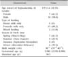
In total, 17 neonates from our hospital were enrolled in this study (Table 1). They consisted of 7 females (41.2%) and 10 males (58.8%) and the mean onset age was 5.9 days (range, 4-15 days). The mean age of mother was 30.5 years. Mean gestational age was 38+1 weeks (range, 35+5-40+5) and mean birth weight was 2,980 g (range, 2,350-3,900 g). Type of feeding was almost formula-fed (88.2%) and mixed-fed (11.8%). Nobody was exclusively breastfed. Most of them was born in the winter (35.3%) and the spring (41.2%).
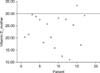
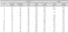
Table 2 showed the laboratory findings of 17 neonate-mother pairs. Their mean serum calcium level was 6.9±0.5 mg/dL, serum phosphorus was 8.6±0.9 mg/dL, serum alkaline phosphatase (ALP) was 196±65 U/L, and plasma iPTH level was 60±43 pg/mL. Their serum 25OHD was 24.8±8.4 ng/mL. Maternal serum 25OHD was 23.0±6.6 ng/mL and plasma iPTH level was 26±14 pg/mL. Vitamin D deficiency (25OHD<20 ng/mL) was observed in 29.4% of the subjects. Insufficient vitamin D status (20<25OHD<30 ng/mL) was 58.9% (Fig. 1). All 17 neonates were managed with oral calcium gluconate (calcium gluconate; Daihan Pharm Co., Ltd., Seoul, Korea) replacements for hypocalcemia and phosphate scavenger. Twelve neonates were followed up until 4 months and were regularly checked on body weight, height, and serum level of calcium, phosphorus, ALP and albumin levels were in normal range.
DISCUSSION
Maternal hypovitaminosis D is one of common causes of neonatal late-onset hypocalcemia [7,8]. Rickets is an uncommon problem in children and neonates as Korea is a developed country. Only patients with risk factors of cholestasis, mal-absorption syndromes, vitamin-D resistant diseases are considered for the risk of vitamin D deficiency.
This is the first study to evaluate the relationship between neonatal late onset hypocalcemia and maternal vitamin D status in Korea using paired neonatal-mother sera. In this study, neonatal late-onset hypocalcemia seems to be related with the maternal vitamin D deficiency and insufficiency, on the bases of their birth seasons and maternal vitamin D status. The fact is supported that most of the neonates born in spring and winter are short of sunlight exposures due to the late pregnancy periods of mothers [9]. No neonates born at autumn were diagnosed as late onset hypocalcemia, and it may be related with long and high sunlight exposures of mother during late pregnancy during summers [9]. In Iran, 100% of neonates with delayed hypocalcemia were born by mothers with vitamin D deficiency [10]. Previous studies showed same results that neonate who were born from mother of vitamin D deficiency or insufficiency have vitamin D deficiency and late onset hypocalcemia due to vitamin D deficiency [11,12]. In the present study, 76.5% of neonates who had late-onset hypocalcmia were born by mothers with vitamin D deficiency or insufficiency. Exclusive breast milk feeding without vitamin D supplementations is another risk factor of vitamin D deficiency for neonates and infants. Our report of neonatal vitamin-D deficiency indicates that the newborns intake the entire milk. Late onset hypocalcemia is usually caused by high phosphate intakes [13]. Late-onset hypocalcemia caused by excessive phosphate load is usually accompanied by raised PTH and ALP levels [12]. However, most of our patients with hypocalcemia and hyperphosphatemia represented normal or near normal ranges of iPTH and ALP. Only case 16 showed hypocalcemia, hyperphosphatemia and remarkably raised PTH level but serum ALP level was relatively low despite of hyperphosphatemia. Practically, vitamin D insufficiency or deficiency was identified in the mothers with hypocalcemic babies according to the preliminary data at our hospital. Therefore, we tentatively considered that maternal vitamin D insufficiency could be associated with the occurrence of late-onset hypocalcaemia even in the formula-fed neonates, although there was no statistically significant relationship (r=-0.131, Pearson correlation by PASW Statistics 18.0 [IBM Co., Armonk, NY, USA]).
Studies in adults have demonstrated that parathyroid hormone concentrations are at their ideal physiologic concentrations when 25OHD concentrations are above 32 ng/mL [14,15]. Similar data in children are unavailable [16]. According to Avery's disease of the newborn, iPTH levels are usually dependent on the serum calcium status. The normal ranges of iPTH are suggested as 10-60 pg/mL under normocalcemia, but hypocalcemia can stimulate iPTH maximally up to 100-150 pg/mL and hypercalcemia can supress iPTH to 2-5 pg/mL [17]. In Korea, there were several studies on the levels of PTH and hypocalcemia: One study was that serum PTH was elevated in the hypocalcemic infants with infectious diseases [16]. The other was that iPTH elevated in infants with subclinical rickets [18]. In the present study, most iPTH levels of neonates with late-onset hypocalcemia was not remarkably elevated except for only 1 case based on Avery's diseases of the newborn. The response of parathyroid hormone might be less sensitive in neonates as compared to the study in infants with subclinical rickets (range of iPTH: 74.7 to 462 pg/mL) [18]. In our study, no elevation of ALP or iPTH was observed in neonates with late-onset hypocalcemia despite maternal vitamin D insufficiency or deficiency. Given the results, we deduced that there might be an another cause for late-onset hypocalcaemia in the subject neonates, besides the maternal vitamin D deficiency and insuffiency. This might be caused by delayed responses of parathyroid hormones to hypocalcemia in neonates [18] and associated with transient hypoparathyroidism. However, it is difficult to make results clear due to the lack of studies regarding the responses of parathyroid gland in neonates. Therefore, more studies should be followed in the near future regarding the effects of vitamin D or hypocalcemia on the functions of parathyroid in neonates.
We could stop calcium supplementations to the hypocalcemic infants around the age of 3-4 months. Currently, 45 IU of vitamin D is contained in 100 mL of formula which is commercially available in Korea, indicating that about 1 L of milk should be ingested to meet the recommended vitamin D daily intakes (400 IU/day). In most cases, they could ingest around 1 L of milk when they are 3-4 months. Considering that calcium, phosphorus, and ALP values were observed to be normal in the examination around the fourth month after birth, the authors assumed that vitamin D insufficiency or deficiency would be recovered as the recommended vitamin D daily intakes was met due to an increase of milk intakes. On the basis of their clinical course, neonatal late onset hypocalcemia in this study might be affected by maternal vitamin D status rather than delayed response of parathyroid hormone.
There are several limitations in this study. First, the number of newborns with late-onset hypocalcemia was small. Second, we did not obtain information on each mother's amount of sunlight exposures, sunscreen uses and diets. Third, whether vitamin D concentration was normalized was not confirmed as vitamin D concentration was not examined around 3-4 months of age. Fourth, this study is not a case-control study and maternal vitamin D insufficiency or deficiency is difficult to determine a definitive cause of neonatal late-onset hypocalcemia in the present study. Regardless of these limitations, the present study is likely to be meaningful because it is the first study to evaluate the relationship between neonatal late onset hypocalcemia and maternal vitamin D status in Korea using paired neonatal-mother sera and late-onset hypocalcaemia could occur under the conditions of maternal hypovitaminosis D or transient hypoparathyroidism in formula-fed neonates besides phosphorus overload.
 XML Download