

 PDF
PDF ePub
ePub Citation
 Print
Print


INTRODUCTION
Vitamin K deficiency and impaired coagulation in cholestasis result from malabsorption of fat soluble vitamins caused by reduced bile drainage. It usually develops late in the course of the disease and can lead to severe bleeding [1].
Neonatal cholestasis can be life-threatening and presents usually with jaundice, dark urine, pale stools and hepatomegaly, resulting from decreased bile drainage caused by damage to hepatocytes or biliary obstruction [2].
Biliary atresia is the most important differential diagnosis of neonatal cholestasis [3]. The cause remains unknown. The clinical presentation of biliary atresia is jaundice, acholic stools and dark urine and hepatomegaly [4].
We report an atypical presentation of biliary atresia.
CASE REPORT
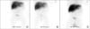
A 4-week-old girl was referred because of a blue, painful swelling in the right axilla (Fig. 1). There was no history of trauma and there were no other signs or symptoms.
She was term born by means of a cesarean section because of failure of delivery progress. Vitamin K was administered at birth (1 mg orally) and daily since the 7th day (150 µg orally) as recommended by the Belgian guideline for healthy, breastfed infants. The patient was exclusively breastfed.
On physical examination we saw a pale, only slightly jaundiced infant with a large, non-fluctuating swelling (10×7 cm) with a central hematoma in the right axilla. There were some petechiae on the palate. The liver was enlarged (4 cm under the costal margin), there was no splenomegaly. The remainder of the physical examination was unremarkable. Clay-coloured, fatty stools were reported since birth, jaundice had not been recognized earlier.
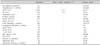
Bloodtests showed anemia, severe coagulation disorders, slightly elevated liver enzymes and a clear hyperbilirubinemia (Table 1).
Ultrasound of the axillary mass showed a lesion of 4 cm in diameter, without a flow pattern, with perilesional edema, suggesting a recent hematoma. Cerebral ultrasound showed no abnormalities. Abdominal ultrasound showed a diffusely enlarged liver (approximately 8.3 cm, normal value <7.2 cm). Neither the choledochal duct, nor the gallbladder could be visualized. Intrahepatic bile ducts seemed present.
The diagnosis was an axillary hematoma due to secondary vitamin K deficiency resulting from biliary atresia.
A hepatobiliary scintigraphy was performed and showed no filling of the intra- or extrahepatic bile ducts, nor excretion of the contrast fluid to the intestine up to 6 hours after injection (Fig. 2).
The clotting disorder was treated with 1 milligram vitamin K intravenously twice daily. Laboratory values showed normalisation of prothrombin time (PT) and activated partial thromboplastin time (aPTT) after administration of vitamin K. Fresh frozen plasma was not given at this point. The anemia was treated with an erythrocyte transfusion.
The patient was transferred to a university hospital where a liverbiopsy was performed showing extensive cholestasis, a ductulary reaction, giant cell transformation and moderate fibrosis, hence biliary atresia. The patient underwent a successful Kasai hepatoportoenterostomy at the age of 5 weeks.
DISCUSSION
Since vitamin K levels are low at birth and exclusively breastfed infants are at even greater risk of developing a vitamin K deficiency, the standard of care is to administer 1 milligram vitamin K orally to all infants at birth and 150 micrograms orally daily from the age of 1 week in case of breastfeeding [1,2].
Earlier reports of infants with a vitamin K deficiency hemorrhage secondary to cholestasis have been made. However, these infants had either not received vitamin K prophylaxis [4] or a lower dose [5].
The patient discussed in this article was exclusively breastfed and had therefore recieved an appropriate dose of vitamin K at birth and daily from the age of 1 week according to the Belgian guideline. However, she presented with a hematoma in the 4th week of life, which was shown to be caused by a vitamin K deficiency as a consequence of an extra-hepatic bile duct atresia. The hemorrhagic diathesis resulted from absent bile drainage and subsequent fat malabsorption that led to a vitamin K deficiency, since vitamin K is a fat-soluble vitamin. It acts as a cofactor for an enzyme that converts glutamyl residues in several coagulation factors (II, VII, IX, X). Therefore, hemorrhage results from a coagulopathy caused by a decrease in vitamin K-dependent coagulation factors [5]. Coagulation studies showed decreased activity in factors from the extrinsic pathway (factor VII), which resulted in a prolonged PT, as well as a prolonged aPTT due to deficient factor IX.
The patient showed clay-coloured stools as an early sign of diminished intestinal bile excretion, which had not been recognized before admission. There was only moderate clinical jaundice despite of the elevated bilirubin because of anemia. There was moderate hepatomegaly.
Neonatal cholestasis is defined as conjugated hyperbilirubinemia that persists beyond the first 2 to 3 weeks of life. The disorder commonly presents itself with jaundice, hepatomegaly and clay-coloured stools. The incidence of neonatal cholestasis is 1 : 2,500 live births. The most common causes are biliary atresia and neonatal hepatitis [2]. There are multiple causes of neonatal cholestasis, including infectious, genetic and metabolic diseases. However, biliary atresia must be differentiated from other causes because early intervention results in a better prognosis [4,6,7]. The incidence of biliary atresia varies from about 1 : 5,000 live births in Taiwan to about 1 : 18,000 live births in Western Europe [7]. Girls are slightly more effected [4]. The clinical triad of biliary atresia shortly after birth consists of jaundice, acholic stools and hepatomegaly. Thereafter, weight loss, splenomegaly and ascites (due to portal hypertension) and hemorrhage develop as later signs [4].
The first step in the acute management of a life threatening hemorrhage should consist of stabilizing the patient. It is important to acknowledge the underlying vitamin K deficiency and to swiftly administer vitamin K intravenously to enhance coagulation and stop the bleeding.
Secondly, meticulous history taking addressing jaundice, colour of stools and urine and signs of increased bleeding tendency, and a thorough physical examination are essential to detect signs of cholestasis. Swift diagnosis is not only important in the case of biliary atresia; early supportive measures can prevent complications and are beneficial for other causes of neonatal cholestasis [2].
The North American Society for Pediatric Gastroenterology, Hepatology and Nutrition (NASPGHAN) recommends that primary care providers screen for cholestasis in any infant that is jaundiced ≥ the age of 2 weeks [2].
Apart from laboratory studies that help assess hepatobiliary function (total and conjugated bilirubin, aspartate aminotransferase, alanine aminotransferase, alkaline phosphatase and gamma-glutamyl transpeptidase) abdominal ultrasound may be performed as an initial, easily available, non-invasive test. It is important to realize, however, that abdominal ultrasound alone cannot exclude biliary tract obstruction [3]. It may be difficult to differentiate between biliary atresia and neonatal hepatitis. Hepatobiliary scintigraphy may provide additional information to aide in differentiating biliary atresia from neonatal hepatitis and other causes of cholestasis. If after the abovementioned investigations biliary atresia is suspect, peroperative cholangiography, the gold standard in diagnosing biliary atresia, should be performed. There is no evidence that a diagnostic laparotomy is harmful to infants with idiopathic neonatal hepatitis [8].
According to the NASPGHAN no single pathway seems to be clearly superior for the diagnosis of conditions leading to cholestasis. Vigilance is crucial for detection of these patients as well as selecting appropriate and timely diagnostic testing and referral to a pediatric gastroenterologist [2].
Bleeding diathesis due to vitamin K deficiency is an unusual presenting symptom of neonatal cholestasis, that usually presents with jaundice and clay-coloured stools. Treatment of the bleeding consists of administration of vitamin K to enhance coagulation. It is of great importance to also acknowledge the underlying cholestasis along with causes that necessitate early treatment, since early intervention results in a better prognosis in certain cases, like with biliary atresia. Performing a methodical and comprehensive diagnostic investigation, preferably by a pediatric gastroenterologist, is essential. Furthermore, it is important to realise that abdominal ultrasound cannot exclude biliary atresia and that cholangiography should be carried out if in doubt.

 XML Download