

 PDF
PDF ePub
ePub Citation
 Print
Print


INTRODUCTION
Diarrhea is one of the most common symptoms of illness during childhood, and many children experience diarrheal episodes on average one to five times per year [1,2,3]. Infections from bacterial, viral, and parasitic pathogens are the most common causes of acute diarrheal disease, and viral pathogens are the most common cause of acute gastroenteritis (AGE) [1]. Traditionally, rotavirus was thought to be the most common identifiable viral pathogen around the world. However, since implementation of universal rotavirus vaccinations and the subsequent decrease in rotavirus infection, norovirus and rotavirus are now both common viral causes of AGE in children [4,5].
Viruses and enterotoxin-producing bacteria both affect predominantly the proximal small bowel and cause watery diarrhea via non-inflammatory mechanisms. Although these are bacterial infections, stool examinations reveal no fecal leukocytes. Therefore, it is difficult to clinically distinguish an enterotoxin-producing bacterial enteric infection from a viral enteric infection. Nevertheless, bacterial pathogens are often overlooked as a causative agent of AGE, especially in infants.
In this study, we investigated the enteropathogenic organisms, both viral and bacterial, that currently cause AGE in infants and documented their clinical and laboratory characteristics.
MATERIALS AND METHODS
Patients
From January 2013 to December 2013, we investigated infants younger than 1 year of age with acute-onset diarrhea who underwent stool examination at the Pusan National University Children's Hospital. Those with chronic diarrhea were excluded. Diarrhea is defined as an abnormally loose form of stool occurring in three or more bowel movements a day.
Clinical and laboratory data
Data were collected retrospectively by reviewing medical records of patients. Age, sex, previous rotavirus vaccination, need for hospitalization, underlying disease, and associated respiratory or neurological symptoms were recorded. Dates of symptom onset, admission, and stool collection were also collected.
Clinical data regarding vomiting, mucoid or bloody diarrhea, dehydration, irritability, and poor oral intake were investigated. Dehydration was defined as a decrease in normal body weight of 4% or more; poor oral intake was defined as less than half of the usual dietary amount.
Laboratory data regarding white blood cell count, percentage of segmented neutrophils, platelet count, albumin, and C-reactive protein were collected.
Methods for enteropathogenic evaluation
Stool specimens of at least 2 g were stored at 4℃ immediately after collection. All of the stool examinations except Clostridium difficile were conducted in the Division of Microbiology and Epidemiology, Gyeongnam Research Institute of Health & Environment, Korea. The specimens were delivered using a cooler bag to the laboratory, where tests were performed once a week.
The examinations were conducted by standard protocol according to The Practice Guidelines of Diagnosis of the Waterborne and Food Related Diseases (2013) [6]. The test techniques were as follows. In bacterial examinations, the collected feces were diluted in 1 mL of phosphate-buffered saline (pH 7.4). Each 100 µL of diluted specimen was incubated on 2-5 mL of selected enrichment media for enrichment culture. All cultured specimens underwent polymerase chain reaction (PCR) analysis. Positive results of both culture and PCR tests were considered as positives. Secondary biochemical PCR analysis was performed to identify the toxins isolated from the following enteropathogens: Salmonella, Shigella, Vibrio, enteropathogenic Escherichia coli, Bacillus, Listeria, Clostridium perfringens, Yersinia, Staphylococcus aureus, and Campylobacter. C. difficile was identified using the Seeplex Diarrhea ACE Detection kit (Seegene, Seoul, Korea) by a standard protocol in our institute that detects C. difficile toxin B. Sterile phosphate-buffered saline was used for the vial studies. Rotavirus and adenovirus were first identified by enzyme immunoassay antigen capture and then confirmed by PCR or reverse transcriptase PCR (RT-PCR). Norovirus was identified by real-time RT-PCR, and astrovirus and sapovirus were identified by duplex RT-PCR.
Statistical analysis
The confirmed stool pathogens were divided into isolated bacterial or isolated viral pathogen groups. Clinical symptoms and laboratory parameters were compared according to pathogen. Differences in clinical symptoms were analyzed by χ2 and Fisher's exact test; laboratory parameters were analyzed by t-test. All statistics were performed by using SPSS version 12.0.0 (SPSS Inc., Chicago, IL, USA). p-values less than 0.05 were considered statistically significant.
RESULTS
Demographic, clinical, and laboratory patient data
A total of 41 infants (24 males and 27 females) were included and 18 patients (45.0%) had previously completed rotavirus vaccination. Thirty-five patients (85.4%) were hospitalized. The mean age was 6.6±3.6 months (range: 0.6-11.9 months). Eleven patients (26.8%) had underlying diseases, six had a history of bowel surgery, two had neurological disorders, two had chronic lung disease, and one had a history of Kawasaki disease. Thirteen patients (31.7%) had respiratory symptoms, such as rhinorrhea or cough. No patients showed neurological symptoms, such as seizures. All patients showed diarrhea, and fever was the second most common symptom (25 patients, 61.0%), followed by vomiting (19 patients, 46.3%), poor oral intake (18 patients, 43.9%), and irritability (10 patients, 24.4%).
Laboratory data showed mild leukocytosis, normal percentage of segmented neutrophils, and normal platelet counts. Albumin was normal (4.1±0.5 g/dL), and C-reactive protein was mildly increased at 2.4±3.9 mg/dL in all patients.
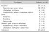
Enteropathogens Twenty-one (51.2%) of 41 cases were positive for one or more enteropathogens. Seventeen cases (41.5%) were positive for bacterial pathogens, whereas seven (17.1%) were positive for viral pathogens. Three cases (7.3%) were positive for both bacterial and viral pathogens (Table 1).
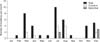
S. aureus was the most common pathogen (13 cases, 31.7%). Eight (61.5%) cases developed AGE in the summer (Fig. 1). All 13 of the cases were positive for enterotoxin. Two cases had Staphylococcus enterotoxin (SE) G; one had SEG and SEI; one had SEG, SEI, and SEN; five had enterotoxin SEG, SEI, SEM, and SEN; one had SEG, SEI, SEN, and SEO; and three had SEG, SEI, SEM, SEN, and SEO. C. perfringens (four cases, 9.8%) was the second most common bacterial pathogen, and all cases were positive for alpha toxin and beta-2 toxin. The case with C. difficile (one patient, 2.4%) was positive for Clostridium toxin B. Norovirus (five cases, 12.2%) was the most common viral pathogen. Three cases showed both bacteria and virus in their stools.
Correlations between clinical data, laboratory data, and enteropathogens
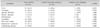
Fever and respiratory symptoms were more common in the isolated viral infection group (p=0.023 and 0.044, respectively). However, the frequencies of vomiting, dehydration, irritability, and poor oral intake were not different between the isolated bacterial and viral infection groups. In cases of isolated viral infection, mucoid stool was observed in only a single patient, and bloody stool was not observed. However, mucoid or bloody diarrhea was not statistically different between the isolated bacterial and viral infection (Table 2). The symptomatic data were also not significantly different between patients with isolated S. aureus (n=13) and C. perfringens (n=4).
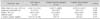
White blood cell count, percentage of segmented neutrophils, and platelet count were slightly higher in the isolated viral pathogen group; C-reactive protein was slightly higher in the isolated bacterial pathogen group. However, this was not statistically significant (Table 3).
DISCUSSION
The present study revealed 31.7% of enterotoxin-positive S. aureus-related gastroenteritis in infants. There were no cases of inflammatory diarrhea, such as Salmonella, Shigella, Vibrio, enteropathogenic E. coli, Bacillus, Listeria, and Yersinia in our study. According to the 2013 Korea Centers for Disease Control and Prevention reports of acute diarrheal disease surveillance study with whole age group, S. aureus, E. coli, and Salmonella were 4.77%, 4.23%, and 2.38%, respectively (http://www.cdc.go.kr).
S. aureus-related enteropathy is generally thought to be food poisoning and is frequently reported in outbreaks [7]. It is also suspected of causing antibiotic-associated diarrhea or nosocomial diarrhea [8]. Staphylococcus is commonly found among gut microbiota, especially in breast-fed infants, and is acquired from parental skin [9]. The colonization rate of S. aureus was reported to be from 0% to 54.8% in infants in various studies [9,10,11]. In a study of sporadic diarrhea in patients aged 5 years or younger, the enterotoxigenic strains of S. aureus were found in 3.5% [12]. In our study, the rate of S. aureus and SE was 31.7%, which is relatively high compared with other studies. It is unclear whether the source of Staphylococcus was pre-existing gut microbiota or ingested pathogens. In the 2013 Korea Centers for Disease Control and Prevention reports of acute diarrheal disease surveillance study with whole age group, S. aureus gastroenteritis did not show specific seasonal differences. However, our study revealed seasonal variation, with higher rates in summer, which indicates a high possibility of infection from contaminated food or water. Most infants in this study were fed breast milk or whole milk combined with weaning food. Therefore, control of food safety seems to be especially important during the summer season.
About 95% of food-borne S. aureus outbreaks are associated with changes in expression from SEA to SEE. However, we did not observe this in our study. SEG and SEI, which are newly discovered superantigens belonging to the enterotoxin gene cluster operon, have been reported as having a minor role in food poisoning [13,14,15] and some association with toxic shock syndrome [16]. SEJ to SEQ are related to the staphylococcal enterotoxin-like gene, but their effect on the host is not yet clear. There is a report of severe neonatal enteropathy associated with SEG and SEI [17]. In our study, all patients who presented with S. aureus had SEG, and all except two patients showed two or more enterotoxins; however, no patient showed severe symptoms. SEM, SEN, and SEO have not yet been associated with specific pathogenic functions, but are frequently found in combination with SEG and SEI [16]. In our opinion, it is unclear whether SEM, SEN, or SEO has pathogenic potential; further data are required.
In the present study, C. perfringens was the second most prevalent bacterial pathogen of AGE in infants. Although C. perfringens and C. difficile are considered normal floras [11], they are associated with gastrointestinal infection and nosocomial diarrhea, and detecting the toxin is important. C. perfringens gastroenteritis is usually more prevalent in the elderly in the community [18], and is not unfrequently found in children [19].
In previous Korean epidemiological studies, rotavirus was found to be the most common viral pathogen in children with AGE [4,20]. However, according to the surveillance data of acute diarrhea in children younger than age 5 years from the Korea Centers for Disease Control and Prevention, norovirus (12.9%) was comparable to rotavirus (13.5%) in the past 5 years, and norovirus showed a higher prevalence than rotavirus in 2010 and 2012 (http://www.cdc.go.kr). A 2011 study in the Iksan, Korea, area demonstrated that norovirus was more prevalent in infants than in any other age group, whereas rotavirus was more common in children between 1 and 2 years old [4]. A recent community-based study performed in Nicaragua, where the area of rotavirus vaccine coverage is more than 90% of the country, demonstrated a markedly higher prevalence of norovirus (24%) compared with rotavirus (8%) in children younger than age 5 years with AGE [5], and norovirus was more commonly detected than rotavirus among children younger than age 2 years [3]. Our study revealed a rotavirus vaccination rate of 45%. There are many factors influencing the increasing incidence of norovirus, with the introduction of rotavirus vaccinations being one of the possibilities affecting the overall changing of AGE viral etiology. Further nationwide studies of AGE etiology correlated with rotavirus vaccination would be helpful.
In our study, most patients underwent stool collection on the first day of admission. However, four patients underwent stool collection 3 days after admission during hospitalization for other symptoms. Two patients were diagnosed with S. aureus, which suggests the possibility of nosocomial infection, and one patient was diagnosed with concomitant norovirus infection. Another patient, who was diagnosed with intussusceptions, initially had diarrhea for 3 days after hospitalization and had adenovirus in his stool. However, he also had respiratory symptoms, and PCR analysis of his sputum was positive for adenovirus. The other patient's stool was not positive for enteropathogens by our examination. One patient who presented with both S. aureus and C. difficile had a history of previous antibiotic uses, which suggested the possibility of antibiotic-related disease.
In this study, fever was observed in six of seven cases with viral pathogens. Fever and respiratory symptoms were more commonly observed in isolated viral infections (Table 1). However, the other clinical and laboratory parameters were not different between the isolated bacterial infection and isolated viral infection groups. It is therefore difficult to clinically distinguish between viral infection and bacterial infection in infantile AGE.
This study has several limitations. First, in our study, 51.2% of cases revealed at least one enteropathogen. We only conducted stool examinations once a week and stored stool specimens in a 4℃ refrigerator. For maintaining high sensitivity and specificity in the viral pathogens, specimens should be stored at -70℃ after 72 hours. We acknowledge that the diagnostic yield especially in the viral pathogens was perhaps underestimated. Second, this study did not distinguish between community-acquired gastroenteritis and nosocomial infections. Two patients were suspected of having nosocomial infection but their prior history of hospitalization was not investigated. Third, we did not test for protozoan infections. Finally, we tried to collect samples from all infants who had visited with acute diarrhea; however, there were instances of unexpected loss of samples. In addition, this study was conducted retrospectively limited to 1 year in a single center and thus might have some selection bias.
In conclusion, we observed a relatively high incidence of S. aureus and norovirus gastroenteritis in infants. Without stool examination, clinical and laboratory data were insufficient to distinguish bacterial pathogens from viral pathogens.
 XML Download