

 PDF
PDF ePub
ePub Citation
 Print
Print


INTRODUCTION
We often see children with abnormal liver function in pediatric outpatient clinics [1]. The children have variable complaints, such as incidental finding of abnormal liver function test on routine health check-up in their school, and jaundice or hepatomegaly suspected dysfunction of the hepatobiliary system. Occasionally, we have to identify the possibility of liver dysfunction in some children, to whom long-term medication had been undertaken, for treatment of some intractable disease. During the growth of children who have chronic infection of hepatitis B virus (HBV) caused by vertical infection, liver injury may occur without hepatobiliary symptoms. Recently, as childhood obesity has increased, liver dysfunction may accompany obesity.
More frequently, hospitalized children develop an abnormal liver function. We have to find which liver functions are abnormal in children, who are admitted to be diagnosed or treated for their major hepatobiliary symptoms. Variable febrile illness caused by viral or bacterial infection in children is often accompanied by some liver dysfunction.
BASIC UNDERSTANDING ABOUT THE LIVER
In a brief review of the anatomy of the liver, it was traditionally divided into four lobes, of right, left, caudate and quadrate lobe. According to Couinaud nomenclature, the division of the liver into 8 segments is frequently used. The liver is divided into right and left lobe by the line between the gallbladder and inferior vena cava. Each lobe is partitioned into 2 sub-lobes, and each sub-lobe into 2 segments. This divides the liver into 8 segments clockwise from the caudate lobe [2]. Blood and oxygen supply to the liver is attributed to the portal vein from the superior vena cava, and hepatic artery from the heart. While the portal vein supplies 70% blood and 40% oxygen, the hepatic artery is responsible for 30% blood and 40% oxygen [2]. The pathway of bile excretion from the liver to the duodenum is the common hepatic duct. The portal vein, hepatic artery and common hepatic duct are triple structures of the porta hepatis. In the internal structure of the liver, the porta hepatis is connected to the portal tract, one of three components of the hepatic lobule [2,3].
The core structure of liver histology is a hepatic lobule with hexagonal shape [2,3]. The central vein and portal tract are located at the center, and three angular points of the hepatic lobule, respectively. Liver cells compose three groups [3,4]. The first is the parenchymal cells, consisting of hepatocytes and bile duct epithelia. The second is sinusoidal cells, including the hepatic sinusoidal endothelial cells and Kupper cells (hepatic macrophages). The third is perisinusoidal cells, consisting of hepatic stellate cells and pit cells.
MAJOR TESTS FOR LIVER FUNCTION
To evaluate the degree of liver injury or liver disease, the most common 'liver function tests' are aspartate aminotransferase (AST), and alanine aminotransferase (ALT). But, they represent 'liver biochemical tests', rather than tests for the known functions of the liver.
The most useful biochemical test to discover liver disease is the standard battery test. The test consists of total bilirubin, albumin, prothrombin time, and serum enzymes. Serum enzymes, which include AST, ALT and alkaline phosphatase (ALP), are usually measured. Gamma glutamyl transpeptidase (GGTP) and 5'-Nucleotidase (5'NT) are occasionally measured [6].
Bilirubin
Total bilirubin ranges 1.0 to 1.5 mg/dL normally, and decreases to the level 0.2 to 0.9 mg/dL in 95% of the population. The normal value of indirect bilirubin is 0.8 to 1.2 mg/dL. The normal upper limit of direct bilirubin is 0.3 mg/dL. Even a small increase of direct bilirubin means the possibility of liver injury. In patients with jaundice, the ratio of direct bilirubin to total bilirubin does not differentiate obstructive jaundice from liver parenchymal jaundice. A ratio over 20% traditionally means cholestasis in children. The degree and duration of hyperbilirubinemia is not a prognostic factor for liver disease. But, the higher the serum bilirubin is, the deeper the severity of liver injury is.
Aminotransferase
Serum aminotransferases was called transaminases in the past. It is the most sensitive marker for acute liver injury. AST and ALT catalyze the α-amino group of L-aspartic acid and alanine, respectively, to move to the α-keto group. AST, which was previously called serum glutamic oxaloacetic transaminase, are in the cytosol and mitochondria of cells. It most commonly distributes to cardiac muscle, followed by the skeletal muscle, kidney, brain, pancreas, lung, leukocyte and erythrocytes. ALT, which was previously called serum glutamic pyruvic transaminase, is cytoplasmic enzyme, and exists most commonly in hepatocyte. So, it is a more specific marker for the evaluation of liver injury, than AST. The normal value of ALT is generally less than 30 U/L in men, and less than 19 U/L in women. But, the value is dependent on the laboratories.
Alkaline phosphatase
Most serum ALP is made in liver and bones. The normal value of ALP depends on the age. Adolescents have two times higher level than adults. The difference between adolescents and adults seems to be due to bone growth. A high level of ALP, when the increase of GGTP and 5'NT is identified, must originate from liver, rather than the bone.
Gamma glutamyl transpeptidase
GGTP is on the cell membrane of the liver (hepatocyte and bile duct cell), kidney, pancreas, spleen, heart and brain, etc. A high concentration of serum GGTP has a limitation for clinical use; because although the sensitivity is high, the specificity is low for hepatobiliary diseases. The increase of GGTP can be detected in patients taking phenytoin and barbiturates.
5'-Nucleotidase
5'-NT is associated with canalicular among hepatocytes, and sinusoidal plasma membrane neighboring hepatocytes. The function of 5'NT is not well known. 5'NT exists in the small bowel, brain, heart, blood vessel and pancreas. The normal value of serum 5'NT increases with aging. 5'NT, as well as GGTP, is used for differential diagnosis of high serum ALP level alone.
Albumin
Albumin is the most important plasma protein, in terms of quantity. It is responsible for 75% of plasma colloid osmotic pressure, and is synthesized only in hepatocytes. When albumin loss occurs rapidly, the liver can make 2 times the usual production. The half life of albumin is 14 to 20 days. The final site of break down is not known. Albumin synthesis is regulated by nutritional status, osmotic pressure, systemic inflammation, and hormone concentration in the blood. Therefore, when hypoalbuminemia is detected, differential diagnosis should include live cell dysfunction, protein-losing enteropathy, nephrotic syndrome, chronic systemic inflammation, and imbalance of hormone.
A long half-life of albumin is the cause of low usability for liver synthetic function, when acute liver injury has developed. In chronic liver disease or liver cirrhosis; however, albumin is an excellent marker for the synthetic function of the liver.
Prothrombin time
All coagulation factors, except factor VIII, are synthesized in the liver. Prothrombin time measures the extrinsic pathway of hemosatasis. Factors II, V, VII and X are clotting factors involved in prothrombin production. Prolongation of prothrombin time may occur from other liver diseases, beside of liver synthetic dysfunction. Vitamin K deficiency and disseminated intravascular coagulation are representative causes of prolonged prothrombin time. The measuring of prothrombin time is most useful in patients with acute liver disease. In contrast to serum albumin, prothrombin time can evaluate the actual liver synthetic function. Prothrombin time is also a valuable prognostic factor of liver failure.
DISEASES CAUSING LIVER DYSFUNTION
When liver dysfunction occurs, the most common laboratory finding is an increase of AST and ALT, representative of serum enzyme associated with liver injury [1,7]. Abnormal AST and ALT levels are occasionally accompanied by cholestatic jaundice in variable liver diseases. Without the increase of AST and ALT; however, jaundice alone may appear [8]. Diseases of abnormal bilirubin metabolism are classified to two types such as the conjugated hyperbilirubinemia and unconjugated hyperbilirubinemia without hepatitis. The former include Gilbert syndrome, and Crigller Najjar types I and II. The latter contains Rotor syndrome and Dubin-Johnson syndrome.
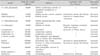
The review will focus on diseases with increased AST and ALT (Table 2) [1]. The representative disease is hepatitis, caused by viral infection, such as hepatotrophic viruses and other viruses inducing systemic febrile infection [1]. Besides of hepatitis caused by virus infection, there is hepatitis caused by bacterial sepsis or parasitic infection. Other diseases are autoimmune hepatitis, metabolic liver disease, such as Wilson disease, toxic hepatitis caused by drugs, cholestatic hepatitis from anatomic problems of the hepatobiliary system, and idiopathic hepatitis caused by non-alcoholic fatty liver disease (NAFLD) of obese children.
Hepatitis A virus
In the domestic area, the prevalence of hepatitis A virus (HAV) infection has shown several outbreaks since the second half of the 1990s, and tremendous increase since year 2000 [9-11]. The possibility of meeting the patients is rising in outpatient clinics. The earlier child may be a little bit sick, without jaundice. In contrast, the older child and adults prominently complain of hepatobiliary symptoms [9,11]. Symptoms include fever, anorexia, nausea, vomiting, fatigue and jaundice. The typical symptoms may continue for 1 to 2 weeks. AST, ALT, bilirubin, ALP, 5'NT and GTTP become over the normal limits. The disease can be easily confirmed by positive anti-HAV antibody (immunogrobulin M [IgM]). ALT rapidly increases to the top, before symptom development. From this point, symptoms such as jaundice begin. After this, ALT gradually decreases, and normalizes, when jaundice disappears.
Hepatitis B virus
The hepatitis B surface antigen (HBsAg) positive rate of school ages, born before the induction of HBV vaccine, was 3.2% in 1988. But, the rate decreased to 0.9% in a survey (Seoul area, 1995) of infant and toddlers born after the vaccine induction [12,13]. At present, domestic children of preschool ages have 70% to 80% of positive hepatitis B surface antibody (HBsAb). The positive rates of HBsAg are 0.4% in 20s, and 0.2% in adolescents [14]. But, we can occasionally meet children with HBV infection in outpatient clinics.
The most common pathway of HBV transmission in childhood is vertical infection from an HBsAg pospositive mother [15]. Over 90% of vertically infected children develop chronic HBV infection. In their natural history, the change from the immune tolerance phase to the immune clearance phase occurs in 15% of patients before 20 years of age [16]. The immune tolerance phase is a period of normal AST, ALT, positive HBeAg and high concentration of HBV DNA in the serum. The immune clearance phase means the period of elevated AST, ALT, positive HBeAg, and decreasing concentration of HBV DNA. In the immune clearance phase, the mean of AST and ALT can increase 3 to 4 times over that of the immune tolerance phase [15,17].
Hepatitis caused by other viral systemic infection
We can often see the disease in children with viral respiratory infection and viral gastroenteritis [1]. The disease is usually accompanied by fever. Most patients don't have other liver dysfunction, except for elevated AST and ALT. Rarely, AST and ALT of 10 to 20 times higher than normal value can be seen. In that case, it may take 6 to 12 months, until the enzymes normalize. Viruses can be identified, using polymerase chain reaction for respiratory infection or gastroenteritis. Hepatitis caused by cytomegalovirus or Epstein bar virus etc. may occur, and the tests to identify these viruses are necessary [1].
Wilson disease
Wilson disease is an autosomal recessive genetic disorder, which is caused by difficulty of copper excretion to bile duct from the liver cell, and is accompanied by liver and neurologic disease [18]. The domestic prevalence of children is approximately 1 per 37,000 persons. In East Asia, including Korea, the most common mutation is R778L (Arg778Leu) of the ATP7B gene [19]. The disease usually does not show abnormal liver function until 5 years old. So, most patients visit outpatient clinics with abnormal liver function in the health check-up of elementary or middle school. In particular, when siblings with abnormal liver function visit the clinic together, we can easily suspect Wilson disease. In most patients, other liver dysfunctions, other than elevated AST and ALT, are not detected. The screening test is the measure of serum ceruloplasmin. If the value is under 20 mg/dL, the confirmative test should be done. The patients should take drugs for Wilson disease throughout life. Fortunately, if the disease is diagnosed before it is accompanied by neurologic complication, most patients can maintain health through life. Earlier diagnosis for Wilson disease can prevent severe neurologic complication. Therefore, suspicion and diagnosis is crucial for Wilson disease.
Non-alcoholic fatty liver disease
Most NAFLDs are discovered in obese children. Yang et al. [20] reported that 33 of 111 children with NAFLD had elevated hepatic enzymes and nonalcoholic steatohepatitis. In this way, obese children visiting clinics tend to have the possibility of abnormal liver function. Other abnormal liver functions are rare, except for elevated hepatic enzymes. If habits of diet, exercise, life and mind are appropriately managed, the obesity will be improved, and followed by normalization of the liver function.
Diseases with abnormal bilirubin metabolism
Diseases that have only jaundice, without elevated hepatic enzymes, are divided into two groups [8]. One is the disease with increased indirect bilirubin. These include Gilbert syndrome, and Crigller Najjar type I and II. Another is the disease with increased direct bilirubin. That includes Rotor syndrome and Dubin-Johnson syndrome.
DISEASE MIMICKING LIVER DYSFUNCTION
Muscular dystrophy
In the earlier child with Duchenne muscular dystrophy, elevated levels of AST and ALT can always be seen [7]. Muscular dystrophy can be easily discovered in the child with marked delay of motor development, and musculoskeletal symptoms. With only elevated AST and ALT levels, and without perception of sign of motor dysfunction; however, some children can be referred to the gastroenterologist. The elevation of AST and ALT level in these children originates in excessive excretion from the musculoskeletal muscle. Serum creatine kinase (CK) excreted from muscle usually ranges 15,000 to 35,000 IU/L (normal<160 IU/L) [21]. For this reason, serum CK should be included in the screening test for the earlier child with abnormal AST and ALT levels.
CONCLUSION
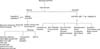
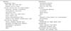
The screening tests for children with abnormal liver function usually consist of anti-HAV IgM, HBsAg/Ab, anti-hepatitis C virus, cytomegalovirus IgM/culture, Epstein bar virus viral capsid antigen IgM, Rubella IgM, herpes simplex virus IgM, CK/lactate dehydronase, and ceruloplasmin and liver sonography, etc (Fig. 1).
The etiologies of abnormal liver function are variable. We can first think hepatitis is caused by viral infection, followed by non-viral infection, autoimmune, metabolic, toxic and anatomic liver diseases. Finally, NAFLDs can be considered. Once abnormal liver function is detected, screening tests should immediately be done for differential diagnosis.
 XML Download