

 PDF
PDF ePub
ePub Citation
 Print
Print


INTRODUCTION
Infantile hepatitis is classified as infectious, metabolic, genetic, idiopathic, and miscellaneous hepatitis. However, in a majority of cases with an unknown etiology, viral infection was the main cause. More than 25 viruses (including Adenovirus, Coxsackie viruses, Cytomegalovirus [CMV], Echovirus type 20, Enterovirus, Epstein-Barr virus, hepatitis A-E virus, Herpes simplex virus, Human herpesvirus 6, Human herpesvirus 7, Influenza virus, Parvovirus B19, Rubella virus, TT virus, and Varicella-Zoster virus) are associated with hepatitis in infants [1].
CMV infection is the main cause of non A-E viral infantile hepatitis. A Japanese study [2] demonstrated that CMV DNA was detected in whole-blood samples of twenty-five (58.1%) of 43 infants (mean age 4.6 months) with hepatitis. Another Japanese study [3] showed that the DNA of CMV was detected in the plasma of six (23%) of 26 infants (median, 7 months old; range, 1 to 24 months). Among 45 Korean children with acute non-A,B,C viral hepatitis, the etiology was unknown for 26 infants (58%) and CMV in 14 (31%) infants [4].
The prevalence of infection increases with age, and is higher for those with a lower socioeconomic status, and people living in developing countries. Congenital CMV infection occurs in approximately 0.2 to 2.4% of all live births [5-7]. Perinatal infection occurs more frequently than congenital infection and thus many more are infected within the first year. In a study conducted in France [8], excretion of CMV in urine was detected in forty-eight of 246 infants (19.5%) aged 6-12 months attending 29 daycare centers.
CMV is the most common virus that causes congenital infections. Approximately 90% of newborns with congenital infection have no clinical evidence of disease at birth; 5% have disease involving the neurologic, hematopoietic, respiratory, gastrointestinal and other organ systems causing high mortality and long-term sequelae [5]. In the remaining 5% of newborns, mild focal organ involvements such as ventriculomegaly, optic atrophy, cytopenia, hepatitis, and others are observed. The most common clinical manifestation of symptomatic congenital CMV infection is liver function abnormalities in small for gestational age premature infants [9]. In perinatal CMV infection, the majority of infants will be asymptomatic, and diseases such as hepatitis, pneumonitis, and mononucleosis occur in some infants.
Congenital CMV infection is the most common viral cause of hearing loss in children [10], and hearing loss will be a long-term sequelae for 50-60% of patients with symptomatic congenital CMV infection [11]. 10-15% of patients with asymptomatic congenital CMV infection develop long-term sequelae, the most common being hearing loss [12]. On the other hand, perinatal CMV infection in infants can result in neurologic sequelae and psychomotor retardation, but the risk of hearing loss does not increase [5,13].
The prognosis of CMV hepatitis has been known to be good in immunocompetent infants, but there are few studies that describe the clinical features of CMV hepatitis. Guidelines for the use of antiviral therapy such as ganciclovir have not been established. Also, little is known about the differences between CMV hepatitis and viral hepatitis of uncertain etiology, where tests for TORCH, EBV and Hepatitis A, B, C viruses, commonly checked in infantile hepatitis, are negative.
The objective of this study was to compare and evaluate the clinical characteristics, laboratory data, and prognosis for infants under age 1 year with CMV hepatitis and those with viral hepatitis of uncertain etiology.
MATERIALS AND METHODS
A retrospective study of infants under 1 year of age who were admitted with acute hepatitis to the Department of Pediatrics, Dongguk University Ilsan Hospital, from April 2004 to February 2012 was conducted. The exclusion criteria consisted of: autoimmune, genetic, metabolic, toxic hepatitis, Exanthem subitum, extrahepatic bilary atresia, Kawasaki disease, muscular diseases, sepsis, and systemic disorders such as urinary tract infections. Of these, 30 patients, where markers for CMV (IgM anti-CMV, CMV PCR in urine, CMV culture in urine) were obtained and negative IgM anti-HAV, HBsAg, IgM anti-HBc, anti-HCV, IgM anti-toxoplasma, IgM anti-rubella virus, IgM anti-HSV, HSV PCR, IgG anti-EBV, IgM anti-EBV, EBV PCR tests, were analyzed. For toxic hepatitis, medication histories for two months were evaluated.
CMV serostatus was determined by enzyme immunoassay (VIDAS-bioMérieux, Marcy-l'Etoile, France). Qualitative CMV PCR assay was performed. The shell vial culture procedure was used for the isolation of CMV from urine specimens.
None of the infants suspected of infectious hepatitis, excluding CMV, had positive tests for IgM anti-toxoplasma and the viruses mentioned above.
In this study, the reasons for limiting the age of onset to 1 year are as follows: First, the author attempted to clarify the association between CMV infection and hepatitis by excluding those who were in latent states with prolonged CMV excretion regardless of the current hepatitis. Second, the author attempted to evaluate the correlation between CMV infection and major contributory factors in transmission (such as breast milk feeding or spontaneous delivery) by minimizing confounding factors (like childcare centers).
The 30 patients included were divided into two groups based on markers for CMV. CMV hepatitis was identified in any of the following results: a positive IgM anti-CMV, detection of CMV DNA in urine specimens by PCR assay, or isolation of CMV in urine specimens. The following were reviewed in all the patients from both groups: age at onset, alanine aminotransferase (ALT), aspartate aminotransferase (AST), breast milk intake, educational background of the parents, gestational age, mode of delivery, number of siblings, physical examination, sex of the patient, symptoms and, total bilirubin.
Statistical analysis
Statistical analyses were performed using SPSS program version 17.0. A univariate analysis for discrete variables was performed using Fisher's exact test. A nonparametric Mann-Whitney test was employed to compare continuous variables between the groups. Significance was established as p<0.05 in all tests.
RESULTS
Patient group
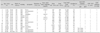
Fifteen patients showing 3 positive markers for CMV were enrolled and analyzed for this study (Table 1). The patient median age was 2.8 months (range 1.3 to 12 months) and nine (60%) of fifteen patients were younger than 3 months of age. There was a ratio of seven males to eight females. One patient was born at 28 weeks and one day of gestation; all other patients were born at term. Birth weights were within normal range. Ten (67%) patients were born by vaginal delivery. Overall the rate of breast-feeding was 67%. No patients received blood transfusions or blood products.
Clinical symptoms at presentation included fever (60%), followed by vomiting, and jaundice. Nine patients had a fever of one to two days duration before admission, of whom three were given antipyretics but there were no records of the medication given. All symptomatic patients had one symptom; four patients were asymptomatic at presentation. None had coagulopathy, hepatosplenomegaly or thrombocytopenia. No other organ involvement was detected in any patient. After admission, fever duration ranged from 1 to 5 days (median 3 days).
Four patients had hyperbilirubinemia, all of them under 3 months of age. Peak serum total bilirubin levels ranged from 2.6 to 6.7 mg/dL (median 5.3 mg/dL). Peak serum direct bilirubin levels ranged from 2.3 to 4.5 mg/dL (median 4.0 mg/dL). The duration of hyperbilirubinemia ranged from 3 to 9 weeks (median 6 weeks) as shown in Table 2. Peak serum AST levels ranged from 59 to 1,092 IU/L (median 320 IU/L). Peak serum ALT levels ranged from 51 to 1,581 IU/L (median 310 IU/L). The duration of AST elevation ranged from 1.5 to 34 weeks (median 12 weeks). The duration of ALT elevation ranged from 1.5 to 26 weeks (median 9 weeks). All had recovered in full without ganciclovir.
Control group
A total of 15 patients were enrolled as controls. The patient median age was 2.5 months (range 0.8 to 12 months), nine (60%) of 15 patients were younger than 3 months of age. All were born at term and the majority of patients were born by vaginal delivery. Overall the rate of breast-feeding was 73.3%.
At presentation, clinical symptoms included fever (40%), followed by cough, rhinorrhea, and vomiting. The patient that presented with a cough or rhinorrhea was diagnosed with upper respiratory infection, bronchiolitis, or pneumonia. No patients had coagulopathy, hepatosplenomegaly, and thrombocytopenia. The duration of the fever ranged from 1 to 5 days (median 1 day).
Four patients of varied ages had hyperbilirubinemia. Peak serum total bilirubin levels ranged from 1.6 to 9.1 mg/dL (median 3.4 mg/dL). Peak serum direct bilirubin levels ranged from 0.4 to 6.3 mg/dL (median 1.1 mg/dL). The duration of hyperbilirubinemia ranged from 0.7 to 6 weeks (median 2.8 weeks) as shown in Table 2.
Peak serum AST levels ranged from 64 to 801 IU/L (median 429 IU/L). Peak serum ALT levels ranged from 26 to 1,794 IU/L (median 425 IU/L). The duration of AST elevation ranged from 0.5 to 44 weeks (median 5 weeks). The duration of ALT elevation ranged from 0.3 to 44 weeks (median 5 weeks). All had recovered in full without complications.
Comparison of the clinical characteristics between the patient group and the control
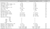
No significant differences were observed between both groups regarding the educational background of parents, number of siblings, rate of breast-feeding, and ratio of spontaneous delivery (Table 2).
Also, there were no significant differences between the groups with respect to age at presentation, fever duration, distribution of patients upon peak serum ALT levels, peak serum ALT levels, peak serum AST levels, peak serum direct bilirubin levels, and peak serum total bilirubin levels.
The patient group had higher values for duration of ALT elevation, duration of AST elevation, and duration of hyperbilirubinemia compared with the control group; however the differences were not statistically significant.
DISCUSSION
The confirmative diagnostic test for CMV infection is virus isolation or demonstration of CMV genetic material by a polymerase chain reaction (PCR) from urine or saliva [5,14]. In this study, CMV infection was confirmed in all patient groups by CMV DNA detection in urine specimens by PCR assay or virus isolation in urine specimens.
Although CMV infection was documented in patients with hepatitis, confirmation of the association of CMV infection with hepatitis must be interpreted with caution. This is because CMV infection leads to a lifelong latent phase in normal hosts [5] and infected hosts excrete the virus for considerable periods in their saliva and urine [14]. Therefore, viral excretion in the urine alone does not necessarily indicate an etiological role for the virus in hepatitis. A longitudinal cohort study [15] showed the median duration of viral urinary excretion in children born with asymptomatic and in those with symptomatic congenital CMV infection were 4.55 years and 2.97 years, respectively. However, in general, high to medium levels of IgM antibody (peak titers) can be detected during the first 1 to 3 months after the onset of infection, after which the titer starts declining but remains positive for more than 9 months after the onset of primary infection [16]. Therefore, a positive IgM anti-CMV result alone is insufficient to confirm an association with hepatitis. Although a finding of typical cytomegalic inclusions is diagnostic for CMV hepatitis, they are rarely detected in a liver biopsy of immunocompetent individuals. Also numerous other findings on the liver biopsy have been documented [17,18]. So, the direct detection of CMV DNA or antigen in liver tissue by means of a virus culture, immunohistochemistry, in situ hybridization or PCR is recommended to confirm the diagnosis [19]. However, liver biopsy was not performed in this study because all patients with hyperbilirubinemia had peak serum total bilirubin levels on the first hospital day, which had continued to decline during follow-up. More specifically, in patients with relatively high levels of total bilirubin, serum total bilirubin levels fell to within 4 to 5 mg/dL in the first week and within 3 to 4 mg/dL in the second week. Then they had normalized by the seventh week and the ninth week, respectively. In addition, 3 patients also presented with a fever suggestive of infection, and the fever abated within the 3rd hospital day. Likewise, a liver biopsy in typical acute viral hepatitis is not usually necessary. The remainder of the patients, namely the patients without hyperbilirubinemia, liver biopsy was not indicated because serum ALT reached their peak within the third hospital day, and had continued to decline during follow-up.
There were some investigations of the causal relationship between CMV infection and disease in patients with acute hepatitis in whom liver biopsy was not performed. In one study, the CMV DNA was detected in the plasma specimens of four (15.4%) of twenty-six children (median age, 7 months old) with hepatitis, compared with none of the 48 normal control infant (median, 10.5 months) [3]. In another study, the detection rate of CMV DNA in whole-blood samples of 43 infants (mean age 4.6 months) with hepatitis was 58.1% by conventional PCR assay and 67.4% by RT-PCR assay, respectively [2]. In contrast, no CMV DNA was detected in any of the 97 healthy subjects (mean age 6.3 months) which was statistically significant. In addition, 12 (27.9%) patients showed a significant elevation in the IgG anti-CMV antibody titre, compared to none in the controls. Five (13.9%) patients showed significant positive results on IgM Anti-CMV antibody, compared to none in the controls. In this study, the urine CMV PCR or urine CMV virus culture was positive for everyone in the patient group, but IgM anti-CMV was negative for some of the patients. The IgM anti-CMV negative patients were mainly those under 3 months of age; young infants have immature immune systems which may fail to produce antibodies in acute infections and IgM anti-CMV can come back negative [20].
The incubation period of CMV ranges from 4 to 12 weeks (mean 8 weeks). Congenital CMV infection is diagnosed when the virus is isolated in infants within the first 2 weeks of life [5,21]. Therefore, it is difficult to differentiate perinatal infection from congenital infection in infants more than 2 weeks of age, unless clinical features of the former such as chorioretinitis, hearing loss, intracranial calcification, and microcephaly are present [21,22]. Since negative IgG anti-CMV indicates the absence of congenital infection, and all IgG anti-CMV tests performed in this study were positive, we were unable to distinguish perinatal infection from congenital infection. A 5-week-old patient, the youngest in the patient group, in whom a marker for CMV were examined in the second week of life. Therefore, it is impossible to differentiate congenital infection from perinatal infection. There were no hepatosplenomegaly, intrauterine growth retardation, microcephaly, prematurity (excluding patient 15) or thrombocytopenia, in the patient group. Examination of the fundus and a hearing test also revealed no abnormalities in the patient group.
In order to date the onset of infection, a review of the literature on the excretion of CMV in urine after primary infection was done. In a prospective study [23], the rate of early-postnatal CMV viruria in infants with median 31 weeks gestation (n=95) born to CMV-seropositive mothers without congenital CMV infection was 22%. Among these infected infants, the time of the first detection of CMV viruria ranged from 32 to 140 days (median 75 days). Another prospective study [24] on babies born to mothers whose cervical cultures were positive at late pregnancy, indicated that the babies were virus negative at birth and started to excrete virus in urine between 3 and 12 months after birth. A study of 45 seronegative adolescents with asymptomatic primary CMV infection found that the time of first detection of CMV viruria ranged from 2 to 80 weeks after infection [25]. As mentioned previously, the diverse range of first detection of CMV viruria was not applicable to our patients in differentiating congenital infection from perinatal infection.
Transmission of CMV occurs 1) in utero by transplacental passage from hematogenous spread to the fetus during maternal viremia, 2) at birth by passage through an infected maternal genital tract, or 3) postnatally by ingestion of CMV-positive human milk, saliva, or by transfusion. The most frequent perinatal sources of virus are genital tract secretions at delivery and breast milk. Around 2-28% of seropositive mothers shed CMV in cervical and vaginal secretions during delivery, and approximately 50% of exposed infants were infected [26]. Around 9-88% of seropositive mothers shed CMV into their milk, and approximately 50-60% of infants fed breast milk that contains the virus became infected [26,27]. A study on risk factors of early post-natal CMV infection revealed that receiving breast milk within the first 30 days (OR=4.5, p=0.02) or for greater than 30 days (OR=7.9, p<0.01) were independently associated with CMV infection [23]. Birth weight, blood transfusion and vaginal delivery were not associated factors [23]. In this study, no significant differences were observed between both groups regarding the educational background of parents, the rate of breast-feeding, the ratio of spontaneous delivery, and the number of siblings. No patients in patient group attended childcare centers or received blood transfusions or blood products. However, due to inherent limitations of a retrospective study, the CMV status of siblings and mothers and the economic status of the parents are unknown and limit the results of the analysis.
In this study, the incidence of CMV hepatitis was 50% (15 of 30 patients), for none of the tests for hepatitis A, B, C virus, toxoplasma, rubella virus, herpes viruses, EBV were positive in infants with suspected infectious hepatitis. There are few studies reporting the frequency of CMV hepatitis in patients with acute viral hepatitis. There is only one such study in Korea, and it reported that CMV infection was confirmed in 31% of acute non-A, B, C viral hepatitis patients (14 out of 45 patients) [4].
Clinical manifestations of CMV hepatitis in immunocompetent hosts as overt jaundice are rare; aminotransferase elevation is moderate, it peaks at 2-3 weeks and mostly returns to normal at 5 weeks [28]. In infants with CMV hepatitis, fever was the most common symptom at presentation, hepatosplenomegaly was not found, and hyperbilirubinemia was not common and only found in infants under 3 months of age. The median peak total bilirubin was 5.25 mg/dL, peak ALT ranged from 51 to 1,581 IU/L. Both reached the maximum within 3 days of onset of symptoms and it is different from what was previously known. The median duration of hyperbilirubinemia was 6 weeks. The median duration of ALT elevation was 9 weeks.
In this study, treatment with ganciclovir was determined to be unnecessary, for all patients with jaundice and ALT elevation showed early improvement and there were no neurologic or multi-organ involvement. All patients fully recovered. Patients with jaundice were given ursodeoxycholic acid. In general, CMV hepatitis in immunocompetent patients does not require antiviral therapy. It has been reported that, rarely, acute CMV infection in patients with normal immune systems can cause massive hepatocellular necrosis and coagulopathy [28]. There are reports of effective ganciclovir treatment of acute CMV hepatitis in immunocompetent children [29,30]. Also, biochemical, serological, virological improvements have been reported in cases of neonatal cytomegalovirus cholestatic hepatitis treated with ganciclovir [31]. Thus, administration of ganciclovir can be considered in CMV hepatitis patients with severe jaundice, coagulopathy or severe ALT elevation; however, specific levels of bilirubin, ALT, PT (INR) have not been established. Also, it needs to be taken into consideration that neutropenia, thrombocytopenia, and other toxicities of ganciclovir are frequent and often severe, and in Korea, insurance applies only to immunocompromised patients or patients with CMV retinitis.
On the other hand, the need for antiviral therapy in symptomatic congenital CMV infection in infants has not been established due to insufficient data [13,15]. Other references state that administration of ganciclovir could be considered in cases of symptomatic congenital CMV infection involving the central nervous system [26], and ganciclovir may be helpful in perinatal infection of critically ill infants [14].
No significant differences were observed between both groups with respect to the duration of ALT elevation, distribution of patients upon peak serum ALT levels, duration of hyperbilirubinemia, peak serum ALT levels, and peak serum bilirubin levels. First, this could be due to the small number of patients in the study. Second, other causes of viral hepatitis not tested for in this study could have been included in the patient group. Since very little of the incidence of viral hepatitis of unknown etiology in children is known, estimating the number of patients included is difficult. However, there were no patients with respiratory symptoms in the patient group; the rotavirus stool antigen, adenovirus stool antigen, stool culture of patient number 12 presenting with vomiting for one day was negative, vomiting subsided on the day of admission, and there was no fever or diarrhea during the hospital stay.
There was no chorioretinitis or hearing loss in our patient group. Sensorineural hearing loss is the most common sequelae of congenital CMV infection, and occurs in symptomatic congenital CMV infection as well as in patients with asymptomatic congenital CMV infection. In particular, delayed onset of sensorineural hearing loss in childhood appears in 5-18% of patients with asymptomatic congenital CMV infection [13,32,33]. In addition, in 50% of patients with delayed hearing loss in asymptomatic congenital CMV infection, hearing loss worsened during childhood (median age at first progression at 18 months [range, 2 to 70 months]) [33]. Therefore, regular hearing test is recommended for patients with a history of congenital CMV infection until elementary school [10].
In conclusion, although congenital CMV infection and perinatal CMV infection were not discernible in this study, prognosis was good without ganciclovir treatment. There were no significant differences in the clinical features between infants with CMV hepatitis and those with viral hepatitis of unknown etiology. However, this study has a small number of patients, and future studies with more patients are needed. Congenital CMV infections cause severe symptoms at birth that lead to death in some cases, and long-term sequelae such as hearing loss in more. Determination of CMV infection in infants with viral hepatitis is important for administration of antiviral medications can improve the course of the illness and reduce complications.
 XML Download