

 PDF
PDF ePub
ePub Citation
 Print
Print


INTRODUCTION
It had been reported that the terminal ileal-only Crohn's disease (CD) (L1 according to the Montreal classification [1]) was identified in 6-25% of patients with CD diagnosed in childhood [2]. In Korea, L1 was identified in 13% of patients with pediatric CD [3]. The terminal ileal-only CD (L1) diagnosed in childhood was decreased from 14% to 9% after the 10-year follow-up [4]. Also, The terminal ileal-only CD (L1) was observed in 36% of adult patients with CD [5]. Thus, though the difference between two prevalences of adults and childhood CD exist, the whole small-bowel examination is certainly needed for the definite diagnosis of the terminal ileal-only CD (L1). Moreover, because whether newly diagnosed small-bowel disease be existent in colonic CD (colon only or ileocolonic; L2 or L3 according to the Montreal classification) may change its treatment [6] and the newly diagnosed small-bowel disease may lead to reclassification inflammatory bowel disease (IBD) from ulcerative colitis (UC) or indeterminate colitis (IC) to definitive CD [7], it is very important for diagnosis of CD to evaluate the whole small-bowel.
It is difficult to access to the small intestine by endoscope owing to its anatomical location and length. There are many methods for evaluation of small intestine; These are small bowel barium radiography, enteroclysis, CT enterography, MR enteroclysis, Sonde enteroscopy, push enteroscopy, and double balloon enteroscopy, but these methods are insufficient to satisfy requirements that include non-invasive procedure, high sensitive detection, and the exposure to low-dose radiation [8].
Since the US Food and Drug Administration approved the capsule endoscopy (CE) in adults in 2001 and pediatric patients aged 10-18 years in January 2004, CE has been recognized as a relatively better tool for the evaluation of small bowel bacause of the capability to observe the intestinal wall non-invasively with high-resolution. Also, In Korea, CE has come into wide use in adult patients. The guidelines for CE were suggested by the Gut Image Study Group under the Korean Society of Gastrointestinal Endoscopy in 2008 [9].
The purpose of this article is to introduce the role of capsule endoscopy in the diagnosis of pediatric CD with focuses on the application, safety, and limitation of capsule endoscopy by the review of published studies.
TYPES OF CAPSULE ENDOSCOPE
Two types of capsule endoscope including PillCam SB (Given Imaging, Israel) and MiroCam (IntroMedic, Korea) are available in Korea, but EndoCapsule (Olympus, Japan) is not in Korea. The capsule endoscope measures 1.1×2.4-2.6 cm, weighs 3.7 g, and takes images forward in the range of 140-165 angle degree. After swallowing the capsule endoscope, it is propelled by peristalsis through the gastrointestinal (GI) tract. The video images (60,000-100,000 pixels per one image) are taken at 2-3 frames per second and transmitted to the data recorder on patient's belt, so about 50,000 images are stored for 8-11 hours of battery life, transferred to computer, and interpreted. There is no difference of the diagnostic yield between two capsule endoscopes available in Korea [10].
THE PROCEDURE OF CE
The informed consent about the purpose of CE, preparation, the course of procedure, the instructions after ingestion, and possible complications must be optained from patients and guardians. Contraindications of CE are bowel obstruction, dysphagia, the possibility of pregnancy, and an appointed MRI exam during CE. History taking include the medication history of NSAID and iron suppliments, past surgical histories, the history of radiation therapy, the diagnostic history of CD, the volume of blood transfusion, and color of the stool is achieved before CE, and helps to the preparation and the interpretation of CE [9].
A 12-hour fast is recommended before the capsule ingestion. Patients should stop eating iron suppliments, beans, berries, corns, and popcorns 3 days before the ingestion. Red-colored foods could be avoided because of mimicking GI bleeding. Whether active bowel preparations using polyethylene glycol or simethicone help to improve the diagnostic yields is controversial [11,12].
Water or clear fluids can be taken after 2 hours and food and medication can be taken 4 hours after ingestion of the capsule. After 8-9 hours, patients are asked to return to exam rooms. The sensor arrays with belts and data recorder are removed and the recorded images are downloaded and processed on the computer. The capsule is excreted with bowel movements within 24-48 hours. If the capsule is not passed in 3 days, the abdominal plain film should be taken for the identification of the capsule.
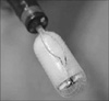
In a study of children aged under 8 years, 24% of children aged 4.0-7.9 years were able to swallow the capsule endoscope. The youngest child to swallow the capsule was a boy of 4 years of age. Of Children not to swallow the capsule in whom the capsule was introduced endoscopically into the stomach or the duodenum with net retrieval catheter (Roth net, US Endoscopy, USA) (Fig. 1), Advance introducer (US endoscopy, USA), and custom-made device. The youngest child to be achieved an endoscopic placement of the capsule was 1.5 years old [13-15]. It was reported that the endoscopic replacement into the stomach with polyp snare was performed in a 42-month-old girl in Korea [16].
THE APPLICATION OF CE TO EVALUATE SMALL-BOWEL CD
It is important to diagnose small-bowel CD because the evaluations of the activity and the localization of CD influence the policy of treatment. In a study to evaluate the diagnostic yield of CE compared with other modalities in patients with small-bowel CD using a meta-analysis, CE was superior to small-bowel barium radiography by 37% in the diagnostic yield, colonoscopy with ileoscopy by 15%, push enteroscopy by 42%, CT enterography by 39%, and MR enterography by 7%. The difference of the diagnostic yield between CE and MR enterography was not statistically significant. Therefore, CE had a significantly higher yield of diagnosis compared with most of other modalities for small-bowel CD [17].
Indications for performing CE in pediatric patients are known and suspected IBD, occult GI bleeding, survey of known polyposis syndrome, persistent vomiting and abdominal pain, and post-transplantation lymphoproliferative disease. Among these indications, rates of suspected and known IBD were 60.7% and 15.4% [18]. Also, indications for CE in CD patients were abdominal pain (45%), diarrhea (15%), GI bleeding and iron deficiency anemia (13%), abdominal pain plus GI bleeding (10%), abdominal pain plus diarrhea (9%), weight loss and/or abdominal pain (3%), and combination (4%) [19].
13 (62%) of 21 pediatric patients with known CD were found at the time of CE to have more extensive small bowel disease and newly-diagnosed jejunal disease were found in 12 (92%) of 13 patients. 4 of 5 patients with UC and 1 of 2 patients with IC had their disease reclassified to CD based upon CE mucosal lesions. All of them had therapeutic changes made [7].
THE SAFETY AND LIMITATIONS OF CE
The most side effect of CE is capsule retention due to intestinal strictures. The capsule retention is defined as failure of the natural excretion of the capsule within 2 weeks. The capsule may be delayed, but not impacted actually on the physiological narrowing of bowel, gastroesophageal junction, pyloric opening, and ileocecal valve. Patients who have the following clinical conditions are at higher risk of capsule retention: suspected CD, history of intestinal surgery, and abdominal radiation therapy [9]. In adults, 14 (1.4%) of a total of 1000 patients underwent CE were confirmed capsule retention. 3 of 22 patients with suspected CD were found capsule retention but none of these 22 patients was small-bowel CD [20]. In padiatric patients, capsule retention occurred in 3 (1.4%) of 207 patients. All of these 3 patients had known CD and 41 of 207 had known CD before CE. Thus, the capsule retention rate in pediatric CD was 7.3% (3 of 41 patients with known CD). This study had revealed that patients with previous small bowel follow-through demonstration small bowel CD (37.5%, retention risk) and BMI <5th percentile with known IBD (43%) had the potential for the capsule retention [21]. 22,840 procedures used PillCam SB of 22,753 patients collected from original articles relevant to small-bowel CE from 2000 to 2008 were evaluated. Capsule retention rates were 1.4% for overall, 1.2% for obscure GI bleeding, 2.1% for neoplastic lesions, and 2.6% for CD. The CD was the most common reason for capsule retention (35.3%) [22]. Therefore, pediatric capsule retention rate was similar to that in adults (1.4% vs. 1.4%) but the capsule retention rate in pediatric CD was assumed higher than that in adults (7.3% vs. 0%).
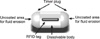
The possibility of capsule retention in patients with suspected small bowel stricture is evaluated with an identically-sized patency capsule (PC). If PC become impacted within strictures of the small bowel, PC is disintegrated by intestinal fluids within 40 hours. Thus, If the PC should disintegrate or cause pain during its passage despite intact expulsion, strictures of the small bowel is indicated. The study with Agile PC (Given Imaging, Israel) (Fig. 2) on a total of 18 pediatric patients including 5 with known CD, 3 with IC, 1 with UC, and 9 with suspected CD was reported. In this study, 15 patients excreted intact PC (mean 34.7 hours) and two of these patients who passed the PC at 52 and 57.5 hours were considered failures. Partial implosion of a PC was seen in one patient of the remaining except 15 patients at 38 hours and full disintegration was seen in one of the remaining at 49.5 hours. The latter patient was considered failure. 12 patients of a total of 18 patients excreted within 40 hours and the others over 40 hours. Thereafter, all of 18 patients underwent CE and consequently, small-bowel CD was diagnosed in 9 (75%) of 12 patients excreted within 40 hours and all of 6 patients excreted over 40 hours [23].
Limitations of CE are the cost for its use and the inability to take biopsies or perform any therapeutic procedures. The latter problem is complemented by double-balloon enteroscopy with the advantage of taking biopsies and performing therapeutic procedures after the diagnosis with CE.
It is reported that the so-called "back to back" interpretation, that is, a second reading by an experienced viewer, improve the diagostic accuracy for CE. The considerable experience (having more than 20 readings to verify the concurrence in the comparision with the interpreting of an expert) is recommended [9].
CONCLUSION
In pediatric CD, CE is the non-invasive test with a higher diagnostic yield than any other modalities in patients with small-bowel disease, and useful in the differential diagnosis and the decision of treatment. Retention of the capsule endoscope appears to be infrequent but feasible. Therefore, we pay careful attention to evaluate the stricture of bowel in suspected small-bowel CD.
 XML Download