

 PDF
PDF ePub
ePub Citation
 Print
Print


INTRODUCTION
Epstein-Barr virus (EBV) asymptomatically infects humans primarily during childhood. A subsequent immunopathological reaction can evolve into infectious mononucleosis, and about 50% of such patients have clinical symptoms including fever, pharyngitis, lymphadenopathy, splenomegaly, and hepatocellular dysfunction [1]. Most patients experience slightly elevated serum levels of aminotransferase with consistent parenchymal injury of the liver, with decreased bile flow. However, severe cholestasis has been rarely reported [2]. EBV-induced hepatitis can lead to cholestasis, which might be a cause of the acute cholecystitis in all the recently reported cases [3]. To date, no case reports have described cholelithiasis and choledocholithiasis associated with EBV infection in pediatric population. We report a case of EBV infection with a GB stone and a CBD stone.
CASE REPORT
A 14-year-old female was admitted to the National Medical Center (NMC), Korea due to a 5-day history of right upper quadrant (RUQ) abdominal pain. Laboratory studies by the NMC, emergency room revealed elevated liver enzymes. The patient had been admitted to the same hospital 15 days prior to this admission, with the same symptoms. The NMC had discharged her after her symptoms eased and her liver enzymes began to decrease, with the aspartate/alanine aminotransferase (AST/ALT) dropped from 550/953 IU/L to 109/338 IU/L (normal range: AST 5 to 45 IU/L , ALT 5 to 45 IU/L) over 5 days. Other laboratory data at her first admission showed her serum gamma-glutamyl transpeptidase (GGT) at 308 IU/L (nomal range: 5 to 24 IU/L), a direct bilirubin of 4.5 mg/dl (nomal range: 0 to 0.3 mg/dl), a total bilirubin of 7.8 mg/dl (nomal range: 0.2 to 1.2 mg/dl), and ammonia at 130 µL/dl (normal range: 19 to 71 µL/dl), with negative hepatitis-B surface antigen (HBsAg), positive hepatitis B core antibody (HBcAb), and negative hepatitis C antibody (HCVAb). At the same time, the result of her abdominal ultrasonography (US) was unremarkable.
Five days prior to her second admission, the patient's fever and RUQ abdominal pain became aggravated, with nausea and vomiting. Upon admission, her vital signs were unremarkable. Her body temperature was 36.7℃, heart rate was 80 beats/min, blood pressure was 133/80 mmHg, and respiratory rate was 20 breaths/min.
Her physical examination upon this second admission revealed icteric conjunctivae and skin color. Neither her liver nor spleen was palpable. We noted a tenderness on the RUQ area, but the patient's rebound tenderness was unremarkable. She showed a positive Murphy's sign. Her breathing sounds were clear without crackles, and no cardiac murmur was audible. Her chest radiograph was also unremarkable, with a normal-appearing mediastinum and no infiltrates.
The laboratory findings on this patient upon this admission day revealed hemoglobin (Hb) at 13.6 mg/dl, white blood cells (WBC) at 6,400/mm3 (67% neutrophils, 17.8% lymphocytes, and 10.6% monocytes), platelets at 288,000/mm3, and erythrocyte sedimentation rate (ESR) at 34 mm/hr. Her blood chemistry showed an AST/ALT of 302/768 IU/L, a total bilirubin of 4.7 mg/dl, albumin at 3.9 g/dL, alkaline phosphatase at 250 IU/L, GGT at 444 IU/L, and C-reactive protein (CRP) at 5.9 mg/L.
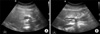
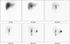
On her second day of hospitalization, the patient received empiric parenteral antibiotics: ampicillin/sulbactam (100 mg ampicillin/kg/day) and gentamicin (7.5 mg/kg/day). Her laboratory blood chemistry results this day showed AST/ALT was at 213/602 IU/L, hepatitis-A virus (HAV) IgG and IgM were both negative, and EBV viral capsid antigen (VCA) was positive for both IgG and IgM. The peripheral blood smear didn't show atypical lymphocytosis. Abdominal US revealed as follows: an arcuate echogenic density (about 1 cm), with posterior shadowing, in the GB neck; proximal dilation of the CBD (maximum about 1 cm), with relatively smooth tapering; and no definite wall thickening or severe dilatation of the GB (Fig. 1).
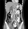
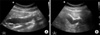
On day 4 of her hospitalization, we performed an abdominal CT scan to confirm other abdominal lesions. This CT scan revealed dilation of the intrahepatic duct (IHD) and CBD but no delineating stone anywhere (Fig. 2). For a final confirmation, we performed a follow-up abdominal US, which revealed a stone that had descended further in the distal CBD, just proximal to the ampulla of Vater, greater dilation (2 cm) of both the IHD and CBD, and a persistent stone in the GB neck (Fig. 3). The laboratory findings were as follows: AST/ALT was 118/357 IU/L; total bilirubin, 3.3 mg/dl; and direct bilirubin, 2.4 mg/dl.
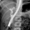
On day 7, the patient's laboratory findings showed improvement, as follows: AST/ALT, 58/184 IU/L; total bilirubin, 1.2 mg/dl; and CRP, 0.3 ml/L. The patient still complained abdominal pain. For removal of stones we performed the ERCP, which revealed moderate CBD dilatation but a normalized IHD size as compared to previous CT scans. The balloon cholangiography did not reveal any stones in the region, possibly because the stone spontaneously traveled down. In addition, the GB was invisible on this examination, despite the fact that we initially assumed the stone obstructed the GB neck (Fig. 4).
On day 9, the patient underwent a diisopropyl iminodiacetic acid (DISIDA) scan, which showed that her liver, CBD, and small bowel uptake were normal, but her GB was invisible (Fig. 5).
On day 14, the patient underwent a laparoscopic cholecystectomy without complications. During this operation, we observed no adhesion between the GB and other organs (such as the duodenum, colon, and omentum). The surgical sample showed a severely distended and slightly thickened GB. In addition, the cystic duct was dilated (0.8 cm). There was no pus collection, and we found no stones. The patient was started on a low fat diet from the first day post-operation. On post-operation day 5, her general condition had improved, with relief of symptoms, and we discharged her from the hospital.
DISCUSSION
To date, researches have not clearly explained the epidemiology of cholelithiasis in pediatric population. Although cholelithiasis remains an uncommon condition in children, recent publications reveal clinicians detecting this disorder with increasing frequency. Previous attempts (about 20 years ago) to estimate childhood gallstone frequency found a prevalence of 0.13% [4]. However, a study reported in 2000 revealed a prevalence reaching as high as 1.9%; the increased use of routine abdominal US may have produced this result [5]. The etiologies of cholelithiasis are hemolytic (20-30%); other known etiology (40-50%), such as total parenteral nutrition; ileal disease; the congenital biliary diseases; and idiopathic (30-40%) [6].
In our case, the patient had a gall bladder stone as well as a common bile duct stone, accompanied by colicky pain and jaundice. The laboratory results revealed an EBV infection. Finally, her symptoms and abdominal US supported a diagnosis of cholecystitis. In EBV infections, obstruction in biliary ducts and cholestatic hepatitis may result from the inflammation or lymphadenomegaly caused by viral replication. Furthermore, such replication may also affect the mesenteric lymph nodes, leading to severe abdominal pain [7]. EBV has infected more than 90% of adults worldwide, who carry the virus as a life-long persistent infection. In most cases, a primary infection occurs and resolves subclinically, during childhood [1]. Researchers have postulated high concentrations of enzyme-inhibiting autoantibodies against the antioxidative enzyme, manganese-superoxide dismutase, play a role in cholestasis, but limited support exists for this idea [2].
Some cases, reported in the international literature, show an acute acalculous cholecystitis with EBV infection [8-10]. Clinicians have recently recognized EBV-induced hepatitis as an important cause of cholestasis [11]. However, the exact mechanism that induces cholestasis remains to be determined.
In our case, the EBV infection coexisted with GB and CBD stones, accompanied by acute cholecystitis. We assumed the patient may have had GB and CBD stones prior to her EBV infection rather than EBV infection induced stones because it takes much time to form stones.
In conclusion, we treated the first case of acute cholecystitis coexisting with a GB stone, a CBD stone and an EBV infection in Korea. Further studies need to evaluate whether these stones make such a patient more susceptible to EBV infection or synergically promote acute cholecystitis. Furthermore, clinicians should consider evaluating the possibilities of EBV infection when pediatric patients present with cholecystitis or stone with unknown cause.
 XML Download