

 PDF
PDF ePub
ePub Citation
 Print
Print


INTRODUCTION
Peptic ulcer generally refers to gastric and duodenal ulcers. According to etiology, primary peptic ulcers, which are chronic and more often duodenal, are most often associated with Helicobacter pylori infection. In contrast, secondary peptic ulcers, which are more acute and resulted from underlying disease, mainly invade gastric mucosa and affect young children such as neonates and infants [1].
The prevalence of H. pylori infection in children is <10% in developed countries but up to 80% in developing countries [2]. The prevalence of H. pylori infection in children affected by duodenal ulcer is 92% globally [3]. The prevalence of H. pylori infection in Korean school-aged children is about 16.5% [4,5]. Approximately 27% of children with recurrent abdominal pain and 65% of children with duodenal ulcers in Korea suffer from H. pylori infection [4,5]. Therefore, duodenal ulcers are highly associated with H. pylori infection regardless of prevalence. In contrast, nonsteroidal anti-inflammatory drugs (NSAIDs), hypergastrinemia, and Crohn's disease are rare causes of peptic ulcer [6].
Peptic ulcer is caused by an imbalance between mucosal attack factors such as gastric acid and mucosal defense factors such as the epithelial layer, intracellular tight junctions, the mucosal layer, secretion of bicarbonate, and epithelial growth factor and prostaglandin production. Among them, gastric acid is highly related to duodenal ulcers [7].
H. pylori can only survive on gastric mucosa. But, excessive gastric acid secretion caused by increased gastrin secretion by H. pylori induces gastric metaplasia. As a result, colonization of H. pylori and duodenal ulceration may occur. Therefore, ulcerative lesions are often located in the duodenal bulb adjacent to the stomach, thus, an ulcer associated with H. pylori is rare below the ampulla of Vater, except for cases of gastric acid hypersecretion such as Zollinger-Ellison syndrome, as H. pylori growth is inhibited in proportion to bile juice [8].
Ulcers are rare in children, and symptoms are mainly abdominal pain, vomiting, and bleeding. In this article, we report a case of improved symptoms of a small bowel ulcer associated with H. pylori infection in an 11-year-old girl who had undergone a gastrotomy to remove a bezoar and, a few years later, received antisecretory and H. pylori eradication therapy.
CASE REPORT
Present illness
An 11-year-old girl presented with a history of stabbing epigastric pain every 10 minutes and nonbilous vomiting more than 10 times a day. She did not have fever or diarrhea.
Past medical history
She had undergone a gastrotomy and removal of an 8 cm trichobezoar 6 years previously. She ate hair on the floor at that time habitually. She had not been administered medication during the past 3 months.
Physical examination
She was 140 cm (25-50th percentile) in height and 34 kg (25-50th percentile) in weight, and her vital signs were: blood pressure, 100/60 mmHg; pulse, 88 beats per minute; respiratory rate, 20 breaths per minute; and temperature, 36.2℃. On admission, she had a clear mental status but seemed acutely ill. Neither conjunctiva were pale, and both sclera were icteric. Respiratory and heart sounds were normal. Abdominal distention was not observed, but bowel sounds were mildly activated. She complained of severe tenderness in the epigastric area with no rebound tenderness. No hepatosplenomegaly or other abdominal mass was detected on abdominal palpation. There was no patchy baldness or abnormalities of the skin or mucosa.
Laboratory findings
On admission, initial laboratory tests showed hemoglobin, 13.2 g/dL; hematocrit, 38.3%; white blood cell count, 13,260/mm3 (neutrophils 88.5%); platelet count, 272,000/mm3; erythrocyte sedimentation rate, 9 mm/hr; and C-reactive protein, 1.6 mg/L. The biochemical profile consisted of serum protein, 6.7 g/dL; albumin, 4.7 g/dL; total bilirubin, 0.5 mg/dL; direct bilirubin, 0.2 mg/dL; aspartate aminotransferase, 17 IU/L; alanine aminotransferase, 11 IU/L; blood urea nitrogen, 6.8 mg/dL; creatinine, 0.5 mg/dL; and amylase, 55 U/mL. A blood coagulation test showed a prothrombin time-internationalized normal ratio, 1.17; and activated partial thromboplastin time, 27.0 sec. Urine analysis showed specific gravity, 1.025; pH, 6.0; negative for protein, glucose, blood, urobilinogen, bilirubin, and nitrite, but positive for ketone bodies. No white blood cells or red blood cells were observed in the urine.
Radiologic findings
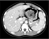
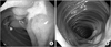
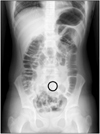
A simple abdominal X-ray was unremarkable at admission. Abdominal computed tomography (CT) revealed a 4 cm sized low attenuating mass-like lesion in the stomach (Fig. 1). We performed esophagogastroduodenoscopy (EGD) for identifying a recurrence of trichobezoar (Fig. 2). Mucosal erythema was observed in the gastric antrum, but no ulcerative lesions were found in the stomach. A scar, which was considered a gastrotomy suture line, and a duodenal bulb with structural deformity were noted. We suspected transition of the mass below the stomach, so we inserted a longer endoscopic fiber using a CF-Q260AL (Olympus, Tokyo, Japan). We inserted the fiber 60 cm from the incisor without kinking and approached the proximal jejunum. We found multiple longitudinal ulcerative lesions from the duodenal bulb to the proximal jejunum, although there was no trichobezoar or mass causing a mechanical bowel obstruction. We performed a small bowel series after marking the endoscopic approach level using clips (Fig. 3) and found normal contrast transition without obstruction or abnormal findings near the mass. Partial hypertrophy of the bowel mucosal fold was noted, but no abnormality was found in the ileocecal portion. The rapid urease test in the duodenal bulb was negative.
Pathological findings
Chronic inflammation with lymphoid follicles was noted on pathological examination of the ulcerative lesion (Fig. 4).
Hospital course
Serum gastrin level and the autoimmune antibody test were performed to determine the cause for the small bowel ulcer. The laboratory findings showed serum gastrin, 57.0 pg/mL; negative anti-neutrophil cytoplasm antibodies and anti-Saccaromyces cerevisiae antibodies; and negative Tb-polymerase chain reaction of the bowel mucosa tissue. The patient received lansoprazole (15 mg/dose, twice a day). On the day of discharge, 4 days after admission, the epigastric pain improved, and she returned to a normal diet. After discharge, she received H. pylori eradication therapy, including lansoprazole (15 mg/dose, twice per day), amoxicillin (50 mg/kg/day in two divided doses), and clarithromycin (30 mg/kg/day in two divided doses) for 2 weeks. She took additional lansoprazole (15 mg/dose, once per day) for 4 weeks, subsequently. Eight weeks after stopping the medication, the urea breath test was negative. The patient has not had any specific symptoms for 18 months.
DISCUSSION
Before it was determined that peptic ulcers were related to H. pylori, there was only the hypothesis that peptic ulcers resulted from an imbalance between gastric acid secretion and the mucosal defense mechanism. Thus, peptic ulcers are known as primary ulcers. After Warren and Marshall found H. pylori in the gastric mucosa of a patient with gastritis and a duodenal ulcer in 1982 [9], the etiology of peptic ulcer in children and adults was identified as H. pylori infection [10,11]. H. pylori mainly chronically affects children >10 years old and often causes duodenal ulcers without systemic symptoms. The exact role of H. pylori in peptic ulcer has not been clearly elucidated. And it is curious that H. pylori infection precedes symptoms or is related to recurrence after eradication therapy. This is because peptic ulcers have a multifactorial correlation with virulence, quantitative colonization of the bacteria itself, genetic vulnerability of the host, a drug attenuating mucosal defense mechanism, psychological stress, and smoking [12].
In our case, because the CT image showed a mass-like lesion in the stomach, we suspected a mechanical obstruction by a mass and performed EGD. The operative record indicated gastrotomy of the anterior wall of the stomach and evacuation of a trichobezoar. We assumed that the gastrotomy incision included the duodenal bulb, as we detected a longitudinal suture scar in the duodenal bulb during endoscopy. However, no mass was found, so predicting distal transition of the mass, we inserted the endoscopic fiber more forward than usual into the small bowel. Although no mass was found in the small bowel, we found multiple longitudinal ulcerative lesions from the duodenal bulb to the proximal jejunum. We marked the endoscopic approach level using clips and performed a small bowel series immediately to identify transition of the mass and the range of the ulcer. No problem occurred by passing contrast, and mass-like bezoar or structural anomaly of the ileocecal portion were not found. But, mild mucosal fold hypertrophy was suspected in the partial proximal jejunum. A biopsy performed at the margin of the ulcer and a histological examination showed nonspecific chronic inflammation with lymphoid follicles but no gastric metaplasia.
A jejunal ulcer is rare, but some restrictive reports are available about this type of ulcer. Among them, a report showed that jejunal ulcer occurred in an adult with Zollinger-Ellison syndrome, and another report presented an experimental animal model with duodenal and jejunal ulcers caused by hypersecretion of gastric acid as in Zollinger-Ellison syndrome. Aspirin has been associated with jejunal ulcers in some reports [13-15].
In this case, the patient had not taken any medications including NSAIDs for 3 months from the onset of abdominal pain and vomiting. We checked serum gastrin level as a cause for the small bowel ulcer, but it was normal. No evidence of Crohn's disease was found. Gastric acid loading in the duodenum caused by gastric acid hypersecretion should precede colonization of H. pylori in the duodenum. The duodenal bulb is the main site for peptic ulcer, as bile juice secreted into the ampulla of the duodenum is a mucosal defense factor against H. pylori growth. In contrast, increased gastric acid loading in the duodenal bulb can induce weakness of the mucosal defense mechanism, because gastric acid-precipitated bile acid combines with glycine in the form of bile salts. As a result, highly polarized bipolar phospholipids make it easy for gastric acid to infiltrate the mucosa [7].
In our case, the patient may have had a weak mucosal defense mechanism against H. pylori, because a structural deformity of the duodenal bulb caused by gastrotomy can lead to gastric acid overexposure of the mucosa below the duodenal bulb, although bile juice is secreted into the ampulla of Vater.
As the endoscopic fiber is usually introduced up to the second portion of the duodenum during EGD, a diagnosis of peptic ulcer below that level can be restricted. We observed ulcerative lesions below the duodenal bulb, as we performed the endoscopic approach more forward than a conventional procedure due to suspicion of a false mass shown on the CT image.
The patient was administered a proton pump inhibitor (lansoprazole), and the abdominal pain subsided gradually after day 4 of hospitalization. The proton pump inhibitor was maintained for more 4 weeks after eradication of H. pylori. After termination of therapy, symptoms disappeared completely and her parents did not want an endoscopic follow-up, so we confirmed eradication of H. pylori by a negative result on the urease breath test.
Although this patient developed abrupt symptoms, had no systemic symptoms and showed normal ileocecal findings in a small bowel series, we thought there was a possibility for early stage Crohn's disease localized in the small bowel, so we followed her closely. The patient has had no symptoms, and laboratory findings such as inflammatory markers, hemoglobin, and albumin have been maintained in the normal range for 18 months after eradication therapy.
There is a limitation for revealing that a peptic ulcer is associated with H. pylori infection in this article such as other reports. Nevertheless, as we experienced that the patient who did not have any systemic disease before and had a structural deformity of the duodenal bulb caused by a gastrotomy, was improved by eradication and antisecretory therapy. Therefore, we report a case of a distal duodenal and proximal jejunal ulcer associated with H. pylori infection.

 XML Download