

 PDF
PDF ePub
ePub Citation
 Print
Print


INTRODUCTION
Intestinal obstruction in the infants and older children may be caused by a variety of disorders, which include atresia and stenosis, annular pancreas, malrotation, duplication cyst, meconium ileus, meconium plug syndrome and neonatal small left colon syndrome, Hirschsprung's disease, neoplasia, trauma [1]. Small bowel obstruction (SBO) caused by an anomalous congenital band is very rare in children [2]. Because obscure symptoms such as nonbilious vomiting and abdominal pain are common sufferings, diagnosis may be delayed and thus patients can be in danger of a life-threatening state. To prevent this aggravation, rapid diagnosis should be made by the upper gastrointestinal series or abdominal sonography. Abdominal computed tomography (CT) with intravenous contrast is frequently used as a method of additional radiologic studies for the identification of undiagnosed strangulation [3,4]. We report a 4-year-old male with the terminal ileal obstruction by an anomalous congenital band, who had no history of previous surgery.
CASE REPORT
A 4-year-old male with colicky abdominal pain and non-projectile vomiting for a day presented to our emergency department. Having experienced no similar episode of abdominal pain before, he presented with infrequent bowel movement. He underwent no abdominal surgery and had no history or evidence of abdominal trauma or intra-abdominal inflammation. He was born vaginally at 41 weeks gestation weighing 3,250 g. At birth, he had a history of admission owing to aspiration pneumonia. Thereafter, he experienced severe gastroesophageal reflux in infancy, but showed normal growth and development. He was 109 cm (50-75 percentile) in height and 19 kg (50-75 percentile) in weight and vital signs were: heart rate of 120 beats per minute, respiratory rate of 24 breaths per minute, body temperature of 37.1℃, and oxygen saturation 100% on room air. Mental state was alert. Mild dehydration with dried lip and tongue was observed. Abdominal mass was not palpated and bowel sound increased. Tenderness but no rebound tenderness or muscle guarding over the whole abdomen were revealed. He did not have bowel movement since the onset of symptoms.
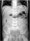
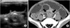
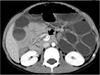
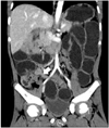
The initial laboratory findings were as follows: hemoglobin 12.8 mg/dL, white blood count 10,970/mm3 (neutrophils 85.4%, lymphocytes 10.6%, monocytes 2.4%), platelets 326,000/mm3, C-reactive protein 2.86 mg/dL (normal range, 0-0.5 mg/dL), amylase level of 8 U/L (normal range, 30-100 U/L), and a lipase level of 39 U/L (normal range, 145-216 U/L). The abdominal radiography demonstrated dilated small bowel loops with air-fluid level in the left upper abdomen and lack of large bowel gas shadows (Fig. 1). The patient was initially observed under treatment with intravenous fluids and electrolyte supplementation but his condition did not improve. The next day, the abdomen was progressively distended and bowel sound was absent on physical examination. Abdominal ultrasonography showed whirlpool-like appearance in the lower abdomen. Axial contrast enhanced abdominal CT image revealed similar whirlpool-like appearance at the same level of ultrasonography (Fig. 2A and Fig. 2B). At the superior mesenteric artery (SMA) level, axial contrast enhanced abdominal CT image showed that superior mesenteric vein (SMV) branch moves counterclockwise around the SMA as it goes away from the SMV (Fig. 3). The coronal multiplanar reformat CT image revealed collapsed proximal ascending colon with short segmental thickened terminal ileum, and dilatation of fluid filled small bowel loops in the whole abdomen (Fig. 4).
Based on the impression of acute SBO due to malrotation, an emergency explorative laparotomy was performed. During operation, hemorrhagic changes and severe distention of the small intestine was observed, and some part of proximal ileum was found to be twisted. The zigzag membranous band developed between the antimesenteric side of terminal ileum and sigmoid colon, extending to proximally 30 cm from ileocecal valve. An acute angle of terminal ileum due to adhesion of the band induced dilatation of the proximal ileum. The band was separated, and the proximal ileum dilatation was not compromised. When the bowel lumen was suctioned and decompressed by enterotomy, neither intrinsic obstruction nor Meckel's diverticulum was identified. Pathologic analysis showed a fibrotic band without blood vessels inside. The patient has remained asymptomatic since the surgery. He was discharged 10 days after the operation. No recurrence was observed during the 8-month follow-up period.
DISCUSSION
SBO is a common disorder, especially for those patients with previous abdominal surgery that can result in postoperative adhesions. Occurrence of intestinal obstruction due to congenital band may be rare in the patients who have not undergone surgery. In our case, the absence of abdominal or pelvic surgery and evidence of intraperitoneal inflammation explains that the adhesive band was an anomalous congenital band. Before the operation, the patient was suspected of having SBO associated with malrotation because of whirlpool-like appearance on ultrasonography (USG) and CT, but malrotation was not observed. The membranous band existed between sigmoid colon and terminal ileum. The kinked and twisted proximal ileum made the angle more acute, and that's the reason why the patient's symptoms began abruptly.
The cause of a congenital band cannot be precisely determined. Embryonic remnants, such as vitelline vessels or omphalomesenteric ducts, can be differentiated according to their localization and pathologic findings [2,5]. On the other hand, a congenital band may be explained as remnants of ventral mesenterium that failed to reabsorb completely past the fourth week of gestation [2]. A congenital band can be single vascular band or multiple dense or adhesive bands like our case [6-9]. According to previous related reports in the literatures, terminal ileum is the most common at the level of obstruction [4,6,7, 10-12]. SBO is mostly caused by compression of the bowel by the band, and may develop in case that intestinal loop is trapped band and the mesentery [2,12]. In the present case, SBO was associated with dilatation of the proximal ileum induced by an acute angle of the terminal ileum due to a membranous band.
At the initial stage, signs and symptoms are indicative of intestinal obstruction, but chronic abdominal pain or constipation history may be observed in patients over two years of age. If patients admitted with these chronic symptoms are treated only with conservative therapy without thorough evaluation, they may lapse into a life-threatening condition [4]. Thus, clinicians should consider the potential occurrence of SBO with congenital band in patients with common symptoms such as nonbilious vomiting and abdominal cramping. The accurate diagnosis was not made until the abdominal USG and CT scan were performed respectively. Abdominal USG is not so a satisfactory method for the evaluation of gas-containing structures. Thus, abdominal CT with intravenous contrast is frequently employed. CT scan can detect the etiology of obstruction in over three-fourths of cases and serve to exclude a strangulated bowel [4]. Although radiologic examination such as abdominal radiography or abdominal CT scans is likely to help to differentiate various diseases, it is not worthy of making a correct preoperative diagnosis of SBO resulting from a congenital band. Once a congenital band was suspected, it should be confirmed whether malrotation was accompanied or not. Radiologic estimation of suspected malrotation is usually accomplished by use of an upper gastrointestinal series. Radiography using water-soluble contrast has some disadvantages such as inability to ingest large amounts of contrast and difficulty in assessing of small bowel abnormality. Recently, sonographic display of the third portion of the duodenum between the SMA and aorta has been recommended as a possible method for locating malrotation [13]. Besides, position of SMV relative to SMA and a clockwise mesenteric vascular swirl-like appearance on CT findings are useful for diagnosis of malrotation [14,15]. Among the whirlpool signs observed in our case, one proved to be a normal finding (Fig. 3) and the other was confirmed as temporally twisted proximal ileum, which aided in diagnosis of SBO (Fig. 2). Thus, it must be remembered that swirl-like appearance on USG or CT can be a normal and nonspecific finding due to a counterclockwise rotation of the SMV on the SMA [16,17].
Laparoscopic surgery in children with SBO is difficult to perform because of limited intraperitoneal space and high risk of injuring the bowel. Recently, the laparoscopic approach has been gradually accepted as a safe and viable option for selected cases of SBO. But, laparotomy should be conducted in patients with bowel gangrene, dense or massive adhesion, the obstruction of unknown cause or location, or bowel perforation during the procedure [18].
A congenital band is a very rare cause of SBO, but it should be recognized as a potential etiology. Although differential symptoms are not observed, the diagnosis should be based on the prompt detection of obstructive manifestations by the clinician and the accurate evaluation of radiographic findings such as USG and CT scan.
 XML Download