

 PDF
PDF ePub
ePub Citation
 Print
Print


INTRODUCTION
Esophageal hiatal hernia is the hernia of a part of or the whole of stomach to posterior mediastinum through esophageal hiatus. It can be classified according to the result of radio test and operation. It is broadly classified as sliding hiatal hernia (type I) and paraesophageal (type II) [1]. When sliding hiatal hernia and paraesophageal hernia are found at the same time, it is called combined sliding and paraesophageal hernia (type III) and the case with other organic hiatal hernia is called complex paraesophageal hernia (type IV). Type III and IV are clinically classified as paraesophageal hernia (PEH) [2]. Esophageal hiatal hernia in infants is mostly congenital resulted from various abnomalities during the development of diaphragm. Most of cases are Bochdalek type which is the result of the posteroexternal defect of diaphragm and esophageal hiatal hernia is very rare. Only several cases have been reported in Korea up to now [3-7]. The treatment of hernia is different according to the type of hernia, damaged part, and the kind, location and size of the organism that is in hernia. In case of type I, conservative therapy is tried in most cases but in case of PEH of type II, III and IV, operational treatment is performed considering the possibilities of complications such as acute occlusion, volvulus, strangulation, hemorrhage, and perforation even where there is no visible symptom [8].
The authors by chance found a case of complex PEH (type IV) and corrected it with operation during the treatment of mycoplasma pneumonia of 10 month-old patient. Thus, it is to report this case with literature review.
CASE REPORT
Patient: Chang ○○, 10 month-old-girl patient
Cardinal symptoms: Fever persisted for 4 days, Cough and Sputum
Present illness: While the patient was hospitalized because of scald in the left wrist (profundus level 2, 1%) at plastic surgery department in this hospital, she was transferred to pediatric department because of fever, cough and sputum.
Birth history: The gestational period was 41 weeks and 2 days. The weight at birth was 3.06 kg (25-50 percentile), height 46 cm (5-10 percentile), head circumference 32 cm (5-10 percentile). She was delivered through normal vaginal delivery and there were no problems in perinatal period.
Past medical history: There were no abnormalities such as vomiting or backflow in feeding and bowel disorder. She had 7-8 times bottle feeding a day at 100-120 cc per bottle.
Symptoms: Her development was good as 8 kg in weight (25-50 percentile), 65 cm in height (25-50 percentile) and in good nutrient conditions. At stethoscope examination, rale was heard from right lung field. Although bowel sound had not been heard from the chest at the time of transfer, it became heard intermittently from right chest with the improvement of pneumonia.
Examination opinions: In the blood test performed at the time of transfer, hemoglobin was 10.7 g/dL, the number of leukocytes 6,730/mm3, and platelet 443,000/mm3, and electolyte test, liver function test, and renal function test returned normal values. CRP was elevated to 25 mg/dL (Normal range 5-0 mg/dL) and Anti-Mycoplasma antibody IgM was positive.
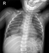
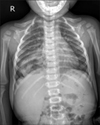
Radioactive examination: In the first simple chest X-ray examination, lobar pneumonia was detected in the lower lobe of the right lung and in the second simple chest X-ray taken 8 days after, a cystic mass filled with air was observed (Fig. 1 and 2).
Accordingly chest tomography was performed for a differential diagnosis. The gastric fundus was located at the right intrathoracic and posterior mediastinum and body of stomach was partly narrowed (Fig. 3). Through the above tests, it was diagnosed as PEH.
Treatment and process: The patient was admitted because of scald and pneumonia and discharged after 11 days' treatment with antibiotics and conservative therapy. In the follow-up outpatient visits, a cystic mass filled with air was observed with the traces of pneumonia on chest X-ray although respiratory symptoms were improved. Therefore, chest tomography was performed and diagnosed as PEH. For the operation, she was transferred to Children's Hospital in Seoul National University Hospital. For the operation, she was accessed via upper midline. It was found that the stomach and a part of colon were moved to thoracic cavity through hiatus and the hernial sac existed. It was tried to return the stomach to the peritoneal cavity and to remove the hernial sac. In the removal trial, however, pleura damage was made and it was opening repaired with hernia sac. It was finally diagnosed as congenital PEH type IV - stomach and colon in the throracic cavity after operation. The patient was recovered without complications and is under observation as outpatient.
DISCUSSION
Congenital diaphragmatic hernia is classified as Bochdalek hernia, Esophageal hiatal hernia, Morgagni hernia and Central part damage according to the location of the hernia. Among them, esophageal hiatal hernia is classified as two subtypes such as sliding hiatal hernia and PEH according to the anatomic location of stomach and gastroesophageal junction.
Type I is the sliding hiatal hernia in which the phrenoesophageal membrane around the esophagus becomes weak and a part of stomach is herniated. Here, the gastroesophageal junction moves up and is located at the upper part of diaphragm. It is observed in around 95% of esophageal hiatal hernia [9]. Type II is the PEH in which a part of the stomach is herniated into intrathoracic area but gastroesophageal junction and stomach cardia are located under the diaphragm. Type III is a complex hiatal hernia in which type I and type II is mixed, and type IV is a multiple hiatal hernia accompanying the herniation of peritoneal organs such as colon, intestine, spleen and pancreas with type II or type III esophageal hiatal hernia [9]. The case in this study was diagnosed as type IV PEH, as the gastric fundus was herniated to the right thorax on the chest computed tomography and the herniation of transverse colon was detected during the operation.
Type I mostly does not show clinical symptoms but sometimes it can accompany vomiting, respiratory symptoms because of regurgitation and delayed growth. Type II can bring vomiting, abdominal discomport, dyspnea and dysphagia after meals, and when the herniation progresses, it can accompany the complications such as abdominal pain, ileus, incarceration, hemorrhage, ulcer and perforation. Type III and IV can accompany the symptoms of type I and II [5]. In infants, vomiting and drooling arise hours after the birth or from the first feeding or cyanosis by asphyxia may occur after the first feeding, which requires esophageal atresia and the differential diagnosis [7,10]. In Korea, Jeon et al. [3] reported a sliding hiatal hernia of which the chief complaint of which was cough in three-month-old female infant, and Hong et al. [4] reported a type III PEH with vomiting and anemia, Rhou et al. [5] reported a type III PEH whose chief complaints were dehydration and malnutrition. In case of type IV PEH, Hong et al. [6] reported one in 12 month old girl whose chief complaint was cough. In the case of this study, the patient showed fair development although the feeding amount was 100-120 cc per bottle, which was less than the average at the same month and was fed more frequently such as 7 to 8 times a day. She did not show any abnormalities until she got a mycoplasma pneumonia and a cystic mass filled with air was observed by chance during the treatment of pnemonia.
There are many different reports regarding the mechanism and pathophysiology. Some propose that it is acquired because it is found mostly among adults [11] and others address that it may occur with congenital defect around the esophagus [9].
Although it is very rare for infants to have esophageal hiatal hernia, it can be divided into acquired and congenital. The acquired ones occur as complications of the operation owing to gastroesophageal reflux [10], while the congenital ones may result from an embryonic defect during the development of the lumbar part of diaphragm in the mesoderm around the aorta that gastroesophageal junction is attached to [12].
As esophageal hiatal hernia symptoms are non-specific, it can be diagnosed by radioactive examinations [2,9]. In chest radioactive examination, the air-fluid level in the thoracic cavity is helpful for the diagnosis, and it can be definitely diagnosed with the increasing contrast agent in the stomach located in thoracic cavity in contrast barium study [2,9]. A tomography can be performed to identify the accurate location of the anatomically damaged part and the herniated organisms, and to evaluate whether to accompany complications, and a colon study can be performed to find out if colon herniation is accompanied [2]. In this case, air-fluid level in the thoracic cavity was not detected in upright position. However, there was a cystic mass filled with air. In the chest computed tomography performed for the differential diagnosis, gastric fundus was located in the right thorax and the stomach body was partly narrowed. Although the herniation of transverse colon was not detected, it was confirmed to be present in this case from the operation. Therefore, it was finally diagnosed as congenital PEH type IV.
For the treatment, supportive treatments are recommended for the sliding hiatal hernia when it shows symptoms such as gastroesophageal reflux and esophagitis [1,2,10], while for PEH type II, III, and IV operational treatment is highly recommended right after diagnosis as they may accompany complications [1,2,11].
Skinner and Belsey [1] reported that 6 out of 21 (28%) who were diagnosed as PEH but did not have operation and received physician's treatment only were dead because of acute gastric expansion, strangulation, perforation or hemorrhage. The mortality rate of elective operation was 0.3%, while that of emergency operation was 14%. As the mortality rate is very high in emergency operation, it is better to have an elective operation when it is first diagnosed [2,11].
The operation can be done by transthoracic access, transabdominal access, or cervicothoracic access [8,13], and with the recent development in laparoscopic surgery, 90 to 100% treatment effects can be achieved with less invasive method [14]. The method of operation returning the herniated organism to the original position and repairing hiatus defects with non-absorbent mesh or suture is determined according to the preference of the operator and the status of the patient [1,8,15]. In this case, transabdominal access operational method was used.
Conclusively, the authors experienced PEH accompanying transverse colon herniation during the treatment of mycoplasma pneumonia and as it is very rare in infants, the authors report this case with literature review.

 XML Download