

 PDF
PDF ePub
ePub Citation
 Print
Print


Abstract
Strabismus is defined as an ocular misalignment. Since it can cause not only impaired visual function but also social handicap and tremendous emotional stress, the care of patients with strabismus should include psychological and social aspects. Although strabismus is one of the major fields in pediatric ophthalmology and neuro-ophthalmology, its precise mechanism and etiology are still unknown. It can be inherited from strabismic parents, or be derived from the anomalous structure, neurologic deficits, and refractive errors. The diagnosis of strabismus can be made by covering one eye, and the degree of strabismus can be quantified by the alternate prism cover test. Recently MRI is used widely for the diagnosis of various anomalous orbital and muscular structures, especially to investigate heterotopia of extraocular muscle pulley. The treatment modalities for strabismus are either surgical or nonsurgical. Surgical treatments can be made by recession or resection of the involved extraocular muscle. The adjustable suture technique was introduced in 1970s, which has been the gold standard among surgical treatment modalities. Nonsurgical treatments include prism, glasses, bifocal lenses, and drugs. A young strabismic patient may have amblyopia and decreased stereoacuity due to abnormal interaction between the sound eye and the deviating eye. Once amblyopia is detected, immediate treatment is needed to correct the visual dysfunction. Recent efforts to elucidate the mechanisms of strabismus are believed to unravel the mysterious pathophysiology in the near future.
Figures and Tables
Figure 1
A) The drawing of rectus muscle recession. The effect of the recession is greatest when the eye rotates toward the recessed muscle.(KW Wright, PH Spiegel. Pediatric ophthalmology and strabismus. 2nd ed. Springer-Verlag New York, Inc., 2003: 279)
B) Recessed rectus muscle sutured to the sclera with three scleral attachments (arrows). (Rosenbaum, Santiago. Clinical strabismus management; Principles and surgical techniques. 1st ed. W.B. Saunders company, 1999: 444)
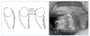
Figure 2
A) The effect of a rectus muscle resection. The resection has its greatest effect on gaze away from the resection.(KW Wright, PH Spiegel. Pediatric ophthalmology and strabismus. 2nd ed. Springer-Verlag New York, Inc., 2003: 281)
B) Resected muscle attached to insertional stump. (Rosenbaum, Santiago. Clinical strabismus management; Principles and surgical techniques. 1st ed. W.B. Saunders company, 1999: 445)
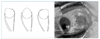
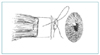
Figure 3
Bow tie adjustable suture technique. After the sutures have been passed through the scleral insertion, they are tied together in a single-loop bow tie. This bow tie can be untied postoperatively to adjust the muscle. (KW Wright, PH Spiegel. Pediatric ophthalmology and strabismus. 2nd ed. New York: Springer-Verlag, Inc., 2003: 281)
References
1. Hippocrates . Airs, waters and places. The Genuine Works of Hippocrates. 1886. New York: William Wood & Co;171.
3. Schlossmann A, Priestley BS. Role of heredity in etiology and treatment of strabismus. Arch Ophthalmol. 1952. 47:1–20.

4. Ziakas NG, Woodruff G, Smith LK, et al. A study of heredity as a risk factor in strabismus. Eye. 2002. 16:519–521.
5. Paul TO. The heritability of strabismus. Presented at the annual meeting of the International Society for Genetic Eye Disease. June 1~3, 1992; Siena, Italy:
6. Miller JM, Demer JL, Rosenbaum AL. Effect of transposition surgery on rectus muscle paths by magnetic resonance imaging. Ophthalmology. 1993. 100:475–487.
7. Demer JL, Oh SY, Poukens V. Evidence for active control of rectus extraocular pulleys. Invest Ophthalmol Vis Sci. 2000. 41:1280–1290.
8. Oh SY, Poukens V, Demer JL. Quantitative analysis of rectus extraocular muscle layers in monkey and humans. Invest Ophthalmol Vis Sci. 2001. 42:17–22.
9. Krzizok TH, Kaufmann H, Traupe H. Elucidation of restrictive motility in high myopia by magnetic resonance imaging. Arch Ophthalmol. 1997. 115:1019–1027.
10. Jampolsky A. Strabismus re-operation technique. Trans Sect Ophthalmol Am Acad Ophthalmol Otolaryngol. 1975. 79:704–717.
11. Spierer A, Barequet I, Rosner M. Reattachment of extraocular muscles using fibrin glue in a rabbit model. Invest Ophthalmol Vis Sci. 1997. 38:543–546.
12. Pediatric Eye Disease Investigator group. A Randomized trial of prescribed patching regimens for treatment of severe amblyopia in children. Ophthalmology. 2003. 110:2075–2087.
13. Hasse W, Wenzel F. The natural course of untreated functional amblyopia; Does it progress between childhood and adulthood. Binocular Vis Strabismus Q. 1997. 12:17–12.
14. Leguire LE, Walson PD, Rogers GL. Levodopa/carbidopa treatment for amblyopia in older children. J Pediatric Ophthalmol Strabismus. 1995. 32:143–151.
15. Campos EC, Schiavi C, Beneditti P. Effect of citicholine on visual acuity in amblyopia; Preliminary results. Graefe's Arch Clin Exp Ophthalmol. 1995. 233:307–312.
16. Scott AB, Rosenbaum AL, Collins CC. Pharmacologic weakening of extraocular muscles. Invest Ophthalmol. 1973. 12:924.
17. Magoon EH. Jankovic J, editor. Clinical use of botulinum toxin; clinical trials for strabismus. Therapy with botulinum toxin. 1994. 1st ed. New York: Marcel Denker Inc;371–376.
18. BJ Kushner. Pediatric ophthalmology in the new millennium. Arch Ophthalmol. 2000. 118:1277–1280.
 XML Download