

 PDF
PDF ePub
ePub Citation
 Print
Print


Abstract
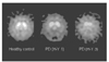
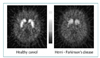
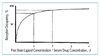
Functional brain imaging with single photon emission computed tomography (SPECT) and positron emission tomography (PET) enables the in vivo study of specific neurochemical processes in the context of normal regulatory mechanisms and pathophysiological alterations of the brain. Receptor studies with SPECT and PET have focused on the occupancy during drug treatment, quantification in neuropsychiatric diseases and visualization of specific pathology. SPECT and PET imaging of dopamine transporters can play an important role in detecting neuronal degeneration in parkinsonian syndromes and in drug-induced neurotoxicity. This article summarizes the SPECT and PET studies to evaluate the dopaminergic system in the human brain and their clinical applications.

References
1. Wagner HN Jr, Burns HD, Dannals RF, Wong DF, Langstrom B, Kuhar MJ, et al. Imaging dopamine receptors in the human brain by positron tomography. Science. 1983. 221:1264–1266.

2. Garnett ES, Firnau G, Nahmias C. Dopamine visualized on the basal ganglia of living man. Nature. 1983. 305:137–138.
3. Brooks DJ. Functional imaging in relation to parkinsonian syndromes. J Neurol Sci. 1993. 115:1–17.
7. Ring HA. The value of positron emission tomography in psychopharmacology. Hum Psychopharmacol. 1995. 10:79–87.
8. Kim SE, Conley RR, Tamminga CA, Chan B, Ravert HT, Wong DF, et al. Serotonin-2 receptor occupancy in schizophrenic patients treated with clozapine as measured by positron emission tomography using C-11 NMSP. J Nucl Med. 1994. 35:74.
9. Farde LO, Wiesel F-A, Halldin C, Sedvall G. Central D2-dopamine receptor occupancy in schizophrenic patients treated with antipsychotic drugs. Arch Gen Psychiatry. 1988. 45:71–76.
 XML Download