

 PDF
PDF ePub
ePub Citation
 Print
Print


INTRODUCTION
More than 50% of epithelial ovarian cancer (EOC) cases are diagnosed at advanced stages, and advanced EOC is currently associated with poor patient prognosis. Approximately 75% to 80% of patients with advanced EOC relapse within 5 years after initial aggressive treatment [1], and recurrent EOC is generally incurable. Therefore, to improve the prognosis of patients with advanced EOC, it is important to eradicate cancer cells completely during the initial intensive treatment.
Primary debulking surgery (PDS) followed by adjuvant chemotherapy (ACT) with platinum and taxane (PDS-ACT therapy) is regarded as standard treatment for advanced EOC [2], whereas neoadjuvant chemotherapy (NACT) followed by interval debulking surgery (IDS) and ACT (NACT-IDS therapy) is conventionally regarded as salvage treatment for advanced EOC cases deemed unresectable due to the presence of widespread invasive disease or a poor performance status. It is difficult to directly compare NACT-IDS with PDS-ACT with regard to benefit and patient outcomes because the patient population that receives NACT-IDS has a worse prognosis; however, several retrospective studies have shown that PDS-ACT and NACT-IDS therapy result in similar outcomes. Moreover, two international prospective randomized trials recently demonstrated that NACT-IDS therapy is also an acceptable treatment strategy for EOC [34].
The significant value of complete resection with no gross residual disease (NGRD) during PDS has been reported previously [567]. Recently, several reports have also shown that achievement of NGRD is as important during IDS as it is during PDS [38]. Nevertheless, the therapeutic benefit of systematic lymphadenectomy for advanced EOC remains controversial [910111213]. Compared to the resection of only bulky nodes, Panici et al. [14] reported that systematic retroperitoneal lymph-adenectomy during PDS improved progression-free survival (PFS) but not overall survival (OS) in patients with optimally debulked advanced ovarian cancer; however, there have been few reports of lymphadenectomy during IDS.
Therefore, we retrospectively analyzed the characteristics and prognoses of advanced EOC patients who received NACT-IDS therapy to investigate the clinical significance of systematic retroperitoneal lymphadenectomy during IDS.
MATERIALS AND METHODS
After obtaining Institutional Review Board approval, we reviewed the medical records of 146 patients with advanced EOC who received NACT-IDS therapy at the Cancer Institute Hospital (Tokyo, Japan) between 2000 and 2008. In this study, we originally applied the following exclusion criteria: synchronous or metachronous (within 5 years) malignancies other than carcinoma in situ, missing data because patients were referred to a different institution for initial treatment, received only palliative therapy after exploratory laparotomy, stage III disease without macroscopic peritoneal dissemination (e.g., pT1N1, pT2N1, pT3aN0, and pT3aN1), and received PDS-ACT therapy as initial treatment. Finally, excluding 22 patients who were not able to undergo IDS because of disease progression during NACT, we retrospectively included 124 patients and analyzed treatment regimens, the number of cycles of NACT, details of IDS (e.g., surgical procedure, operation time, blood loss, size of residual disease, and the number of resected nodes), postoperative treatment, and prognoses.
Our strategy for NACT-IDS therapy consisted of intensive chemotherapy (six or more cycles) aimed at complete resection during IDS and pathological complete response followed by maximum debulking surgery included systematic retroperitoneal lymphadenectomy in principle. The first-line NACT regimen before 2005 included ifosfamide, epirubicin, and cisplatin (IEP), and the regimen after 2005 included paclitaxel and carboplatin (TC). In cases of allergy to paclitaxel, docetaxel was administered instead of paclitaxel with carboplatin (DC). After about six cycles of NACT, we then performed IDS unless the disease had progressed. Surgical procedures for IDS included total abdominal hysterectomy (TAH), bilateral salpingo-oophorectomy (BSO), and partial/subtotal omentectomy (OM). Furthermore, as far as possible, we attempted retroperitoneal lymphadenectomies, such as pelvic lymphadenectomy and/or para-aortic lymphadenectomy, and resection of other organs (e.g., sigmoid colon, rectum, liver, and small intestine) to achieve complete resection. However, although it was planned for all cases in principle, lymphadenectomy was left to the discretion of the surgeon and was not performed in some cases such as highly invasive surgery due to resection of multiple organs, massive hemorrhage, elderly patients, and serious complications. After IDS, ACT was generally administered for about three cycles using the same regimen. However, some patients did not receive three cycles of ACT due to having undergone intensive chemotherapy before surgery or having undergone highly invasive surgery. Conversely, more than three cycles of ACT were necessary in the case of some patients for whom complete resection was not achieved.
Between-group differences were analyzed using the chi-square test. Survival curves and rates were calculated using the Kaplan-Meier method, and differences in survival were evaluated using the log-rank test. A two-sided p<0.05 was considered statistically significant. The Cox proportional hazards model was used to assess various prognostic factors for PFS and OS. Statistical analysis was carried out using R version 3.0.0 (http://www.r-project.org/).
RESULTS
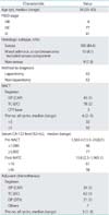
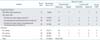
The clinical characteristics of 124 patients are summarized in Table 1. The median age was 58 years (range, 29 to 83 years), and the median follow-up period was 39.5 months (range, 5 to 142 months). International Federation of Gynecology and Obstetrics (FIGO) stages were as follows: six cases (4.8%), stage IIIB; 77 cases (62.1%), stage IIIC; and 41 cases (33.1%), stage IV. Regarding histologic subtypes, serous adenocarcinoma accounted for 85.0% (105/124) of cases; furthermore, 92.7% (115/124) of cases if 10 cases with mixed adenocarcinoma and carcinosarcoma with serous components were included. We diagnosed advanced disease using exploratory laparotomy in 62 cases, whereas 62 cases were diagnosed by non-laparotomy methods, such as cytology of ascites or pleural effusion with diagnostic imaging. Regimens for NACT were IEP (including cyclophosphamide, adriamycin, and cisplatin [CAP]) in 44 cases, TC (including DC) in 80 cases, and irinotecan-based chemotherapy in three cases. The median number of NACT cycles was 6 (range, 2 to 9). The median serum cancer antigen 125 (CA-125) level was 1,569.4 U/mL (range, 13.5 to 24,821 U/mL) before NACT and 15.8 U/mL (range, 2.3 to 1,965.1 U/mL) after NACT. Regimens for ACT were IEP (including CAP) in 25 cases, TC (including DC) in 65 cases, docetaxel and cisplatin or docetaxel alone in 22, and other regimens in seven cases. Five patients refused adjuvant treatment after IDS. The median number of ACT cycles was 3 (range, 1 to 8).
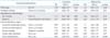
The IDS surgical procedures and results are summarized in Table 2. Exploratory laparotomy was performed in 11 patients, TAH+BSO+OM (minimum) in 10 patients, TAH+BSO+OM+excision of other organs (radical-1) such as the sigmoid colon, rectum, liver, and small intestine in 16 patients, TAH+BSO+OM+retroperitoneal lymphadenectomy (radical-2) in 38 patients, and TAH+BSO+OM+excision of other organs and retroperitoneal lymphadenectomy (radical-3) in 48 patients. As a result, 86 patients underwent systematic retroperitoneal lymphadenectomy, and the mean number of dissected pelvic and/or para-aortic nodes was 46 (range, 19 to 96). Positive lymph nodes (LNs) were detected in 49 patients, including two patients for whom we only sampled bulky LNs. While, negative LNs were detected 41 patients, also including two patients for whom we only sampled bulky LNs. The each background data (e.g., stage, histology, ascites cytology during IDS, the rate of NGRD, and resected nodes) were almost similar between these two groups. Regarding the presence of residual disease following IDS, 98 patients (79%) had NGRD, 15 had residual disease sized <1 cm (optimal), and 11 had residual disease sized ≥1 cm (suboptimal). Only four patients were diagnosed with pathological complete remission. Among the radical surgery groups (radical-1, radical-2, and radical-3; 102 patients), the mean operative time was 419 minutes (range, 185 to 611 minutes), the mean estimated blood loss was 1,291 mL (range, 220 to 5,640 mL), and 72 patients (70.6%) required non-autologous blood transfusions.
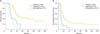
OS and PFS according to the maximum size of the residual tumor are shown in Fig. 1. The 2-year OS, 5-year OS, and 2-year PFS rates were 88.8%, 43.4%, and 39.8% in the NGRD group; 40.0%, 0%, and 13.3% in the optimal group; and 36.3%, 0%, and 0% in the suboptimal group, respectively. Both OS and PFS rates in the NGRD group were significantly higher than those in the optimal group (p<0.001 for both).
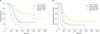
OS and PFS according to presence of LN metastasis are shown in Fig. 2. The 5-year OS and 2-year PFS rates were 62% and 56.1% in the LN-negative (LN-) group, 26.2% and 24.5% in the LN-positive (LN+) group, and 19.1% and 26.1% in the unknown LN status group (U-LN) group, respectively. Both 5-year OS and 2-year PFS rates in the LN+ group were significantly lower than those in the LN- group (p<0.001 for both). Furthermore, there were no differences in both 5-year OS and 2-year PFS between the LN+ and U-LN groups (p=0.616 and p=0.895, respectively).
The sites of first recurrence according to IDS surgical procedure (except for the exploratory laparotomy group) are shown in Table 3. Recurrence occurred in 91 of 113 patients, and irrespective of surgical procedure, recurrence rates were high, ranging from 75% to 94%. Peritoneal dissemination was detected in about 62% of cases. LN recurrence was detected in 8/27 patients (29.6%) in the non-lymphadenectomy group (minimum and radical-1) and 17/86 patients (19.8%) in the lymphadenectomy group (radical-2 and radical-3); this difference was not statistically significant (p=0.534).
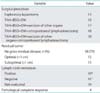
We subsequently evaluated the effect of multiple prognostic factors on OS using univariate and multivariate analyses (Table 4). In the univariate analysis, FIGO stage, histologic subtype, the number of NACT cycles, and treatment regimens were not found to be associated with survival. With regard to IDS, no systematic lymphadenectomy, positive ascites cytology, and positive LN metastasis were also not found to be associated with survival; however, gross residual lesions resulted in a significantly elevated risk for poor OS (hazard ratio [HR], 4.03; 95% confidence interval [CI], 2.46 to 6.61 compared with NGRD). Moreover, the multivariate analysis identified only one independent predictor of OS: gross residual tumor during IDS (HR, 4.14; 95% CI, 2.39 to 7.16 compared with NGRD).
DISCUSSION
Our data demonstrated that lymphadenectomy during IDS does not improve patient prognosis. However, the prognosis of patients with negative LNs, as determined by systematic lymphadenectomy, was relatively good. If there is any therapeutic value of lymphadenectomy, the survival of patients in the LN+ group must be improved because their positive LNs were removed during surgery. Nevertheless, our results showed that survival in the LN+ group was the same as that in the U-LN group but not the LN- group. The better prognosis of the LN- group may be attributed to negative LNs before treatment or a cure of positive LNs by means of remarkably effective chemotherapy. Furthermore, regarding sites of first recurrence, about 80% of patients had peritoneal dissemination, regardless of lymphadenectomy performed during IDS. The number of patients whose site of first recurrence was localized to the pelvic nodes and/or para-aortic nodes was only 4/49 in LN+ group, 2/41 in LN- group, and 2/23 in U-LN group. The remaining patients with LN metastasis at the first recurrence also had peritoneal dissemination. Thus, many patients recurred with peritoneal dissemination and the rate of recurrence in the LNs was not different regardless of lymphadenectomy.
With regard to the number of resected nodes, Panici et al. [14] defined pelvic lymphadenectomy as when at least 25 nodes were removed and aortic lymphadenectomy as when at least 15 nodes were removed in their randomized clinical trial: systematic aortic and pelvic lymphadenectomy versus resection of bulky nodes only in optimally debulked advanced ovarian cancer. As aresult of strict classification, they concluded that systematic lymphadenectomy improved PFS but not OS in women with optimally debulked advanced ovarian carcinoma. The number of resected nodes in our study was 19 to 76 (median, 46) in the LN+ group and 24 to 96 (median, 46) in the no-lymphadenectomy group. However, although only 62 of 86 patients (72%) met Panici's criteria, we removed the sufficient number of LNs in the remaining 24 patients, and the number of patients who underwent "biopsy" was only 4, with 1 to 5 resected LNs. Therefore, we cannot simply compare the benefit of patients who undergone lymphadenectomy and those whose bulky nodes were removed only. On the other hand, 1,876 nodes were resected in the LN- group, which we confirmed as "negative," while 2,161 nodes were resected in the LN+ group, and we confirmed that 274 nodes were "positive." Moreover, 31 of 49 patients in the LN+ group had only 1 to 5 positive nodes as a result of lymphadenectomy meaning that these nodes were resected by means of bulky node biopsy instead of systematic lymphadenectomy, and we believe that the therapeutic value will be the same between them. Therefore, if lymphadenectomy in IDS has therapeutic value, only patients with many positive nodes may benefit from lymphadenectomy, because resecting many positive nodes means maximum debulking. However, these situations are rare and not applicable to all patients with IDS.
The therapeutic benefit of systematic lymphadenectomy for advanced EOC remains controversial [910111213]. Panici et al. [14] reported that systematic retroperitoneal lymphadenectomy during PDS, compared to the resection of only bulky nodes, improved PFS but not OS in patients with optimally debulked advanced ovarian cancer during PDS; however, there have been few reports of lymphadenectomy during IDS. Recently, Fagotti et al. [15] reported the prognostic role of systematic lymphadenectomy in advanced EOC patients at the time of IDS in a double-institution case-control study. They reported that systematic pelvic lymphadenectomy and para-aortic lymphadenectomy during IDS had no value in terms of improvement in PFS and OS and resulted in higher rates of complications, such as longer operative time, blood transfusion, and lymphocele occurrence. Thus, successful complete resection with NGRD and continued sensitivity to anticancer agents may be important for the improvement of prognosis, and the removal of bulky LNs provides therapeutic benefit during both IDS and PDS.
There is no doubt that NACT plays an integral role in the treatment of advanced ovarian cancer; however, there were no correlations between treatment regimens or the number of cycles and prognosis in our study. Furthermore, systematic lymphadenectomy and excision of other organs were also found to be associated with survival. According to the multivariate analysis, only achievement of NGRD during IDS contributed strongly to prolonging OS. In particular, the achievement of NGRD during IDS was independently associated with improved survival (p<0.001). Consistently with our data, a prospective randomized trial reported by Vergote et al. [3] recently demonstrated that complete resection of all macroscopic disease during primary or IDS was the strongest independent variable in predicting OS. In other words, debulking surgery with complete resection, by either PDS or IDS, is the most important determinant of outcome in patients with advanced EOC. Resection of the bulky LNs must be done at least from the standpoint of debulking aimed at achieving NGRD. Mikami [16] recently reported that patients with optimal reduction who underwent complete systematic retroperitoneal lymphadenectomy showed better OS than those who underwent only pelvic node dissection or those who did not undergo LNs resection (p<0.001). He recommended that systematic retroperitoneal lymphadenectomy should be performed in all patients who are likely to achieve optimal cytoreduction.
There were critical limitations to this study. In particular, this was a retrospective study with a small sample size at a single institution, and there is a great deal of selection bias due to the heterogeneity of advanced ovarian cancer disease and patient characteristics, in addition to heterogeneous management methods, which included several chemotherapeutic regimens and indications for lymphadenectomy. Indeed, it is difficult to establish an optimal generalized treatment strategy of NACT-IDS therapy. Two prospectively randomized trials investigating the treatment strategy in patients with advanced ovarian cancer are currently ongoing. One is the LION trial (Arbeitsgemeinschaft Gynäkologische Onkologie group), which is comparing systematic lymphadenectomy and no lymphadenectomy during debulking surgery in patients without both macroscopic residual intra-abdominal tumor and bulky LNs. The other is a Japanese Clinical Oncology Group trial, which is comparing PDS-ACT versus NACT-IDS in patients with advanced ovarian cancer, peritoneal cavity cancer, or fallopian tube cancer [17]. The results of these trials are expected to provide more meaningful information about the therapeutic value of lymphadenectomy or optimal IDS procedure in patients with advanced EOC.
In conclusion, our data suggest that systematic retroperitoneal lymphadenectomy during IDS predicts patient outcome but provides no therapeutic benefit. However, it may be critical to remove bulky LNs in order to achieve NGRD.
 XML Download