

 PDF
PDF ePub
ePub Citation
 Print
Print


INTRODUCTION
The incidence of female genital cancers varies by ethnicity. In more developed regions, endometrial cancer (EC) is the most common female genital tract malignancy with an estimated incidence of 12.9 per 100,000 women. In less-developed regions where cervical cancer is more prevalent, the incidence of EC is less common with an estimated incidence of 5.9 per 100,000 [1].
EC has two main pathophysiologic pathways [2]. Type I EC involves exposure to high estrogen levels, and is represented by endometrioid histology. Several risk factors are reported with type I EC, including diabetes mellitus, obesity, or metabolic syndrome [345]. Type II EC, which is hormone-independent, has high grade histology features of more aggressive behavior; e.g., serous or clear cell carcinomas. Another less common EC is related to genetic predisposition [67]. Among genetic inherited disorders, familial colorectal cancer or hereditary non-polyposis colorectal cancer (HNPCC) appear to carry the highest degree of association with EC. A lifetime cumulative risk of EC among women with HNPCC ranges from 40% to 60%, which may exceed their risk of colorectal cancer [7]. There are conflicting data regarding other genetic risks of EC in women without HNPCC. For instance, some authors reported that 5% of EC are associated with family history of EC alone and 2% occur with HNPCC [8], while others found no genetic risk of postmenopausal EC without history of HNPCC or other cancers [9].
Patients with EC develop other cancers from many reasons, as features that are risk factors for EC development are also risk factors for other cancers. For example, hormonal imbalance, obesity, or metabolic syndrome found in EC patients are also risk factors for breast cancer [4], while previous pelvic radiation therapy can also cause EC [10]. Regarding genetic risk, individuals with HNPCC have a 75% risk of other cancers aside from colon and EC; e.g., ovarian, gastric, biliary tract, uro-epithelial, and kidney cancers, or central nervous system tumors [111213].
The majority of EC patients present in early stages at diagnosis, so the overall prognosis is generally good. Other cancers may affect the survival of EC patients particularly when they are more aggressive or are in advanced stages and especially when they develop after EC cure.
Most data of other cancers among EC patients come from Western countries and there are limited reports from Asia. There may be variations in the incidence of other cancers in EC patients in other regions with different ethnic or environmental backgrounds. As there is a current lack of information, it is important that data from Asian countries are collected. Herein, we aimed to evaluate the prevalence of other cancers, the possibility of a familial association, and their impact on patient survival.
MATERIALS AND METHODS
We obtained approval from the Ethics Committee for Research involving human subjects of Vajira Hospital prior to commencing our study. The Archives of the Gynecologic Oncology Unit, Department of Obstetrics and Gynecology were searched to identify EC patients treated between January 1995 and December 2012. We included patients with EC or carcinosarcoma who had treatment and follow-up visits at our institution. Exclusion criteria were patients who had uterine sarcoma or no available medical records.
Data collected were age, medical morbidities (particularly components of metabolic syndrome such as diabetes mellitus, dyslipidemia, hypertension, and obesity), International Federation of Gynecology and Obstetrics (FIGO) stage of disease according to the FIGO 2009 criteria, fundus/body or lower uterine segment (LUS) site of tumor, histopathology, tumor grade, adjuvant therapy, other cancers, follow-up period, and living status. Data of medical morbidities were partly taken from our data set of a previous report that focused on medical morbidities of EC patients [14]. Stages of patients who were treated prior to 2009 were reassigned according to FIGO 2009 staging criteria. LUS tumors in this study referred to tumors that developed in the LUS (which might expand to the lower uterine corpus or upper cervix), but not tumors that extended from the fundus/body downward. Other cancers in this study did not include preinvasive cervical, vaginal, or vulvar lesions. Data of other cancers were recorded for their site of origin, histopathology, timing of occurrence in relation to EC (prior to, during, or after EC diagnosis), and their treatment. Pertinent family history of cancers was also recorded.
As a general practice in our institution, when ovarian cancers are found at the same time as EC, operative and pathological findings as well as pathological reports/slides are reviewed and discussed in the multi-disciplinary clinic to verify the primary site(s) and stage(s) of cancer(s) for appropriate postoperative adjuvant treatment. The criteria to differentiate second primary or metastatic cancers followed the description by Young and Clement [15].
Progression-free survival (PFS) was calculated from date of diagnosis to the date of progression, death, or last evaluation in the patients who were lost to follow-up. For the patients who died, cause of death was explored as EC or non-EC-related. Overall survival (OS) or EC-specific survival was obtained from date of diagnosis to date of death from any causes or from EC, or last evaluation in patients who were alive at the end of study.
Data were analyzed using IBM SPSS ver. 22 (IBM Co., Armonk, NY, USA). Descriptive statistics were used to analyze demographic data and are summarized as numbers with percentages, mean with standard deviation (SD), or median with range. OS and PFS were analyzed by the Kaplan-Meier method and were compared between groups by log rank test. A p<0.05 was considered significant.
RESULTS
We identified 446 EC patients during the study period. One-hundred and two patients were excluded as they had other types of uterine cancers or had no medical records. Mean age of the 344 patients included in the study was 56.8±10.8 years. Twenty-two patients (6.4%) were aged ≤40 years and 87 patients (25.3%) were aged ≤50 years.
With respect to EC, eight patients (2.3%) had either radiation or chemotherapy prior to surgery because of poor performance status or far advanced disease. Among 336 patients who had primary surgical treatment, early stage (stage I-II) and high grade (grade II-III) tumors were more commonly found in 265 patients (77.0%) and 257 patients (74.7%), respectively. The most common histopathology was endometrioid carcinoma with or without other minor components in 298 patients (86.6%). A total of 157/341 patients with available data (46.0%) had adjuvant treatment after primary surgery.
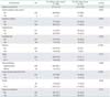
Of 344 EC patients, nine had preinvasive cervical (n=7) or vaginal lesions (n=2). A total of 50 patients (14.5%) had other non-EC invasive cancers. Mean age of these 50 patients with other cancers was not significantly different from those without other cancers (55.7±10.04 and 57.1±11.0 years, respectively; p=0.358). Although the prevalence of other cancers was higher in EC patients aged <50 or <40 years compared with corresponding older age groups (17.2% vs. 13.6% or 18.2% vs. 14.3%, respectively), the differences were not statistically significant. History of any cancer among family members was more commonly found among patients with other cancers (6.0% [3 patients] vs. 1.7% [5 patients]; p=0.095). Cancers reported among family members were endometrial, colon, breast, thyroid, and prostate cancer. Except for hypertension, which was significantly more common in patients without other cancers, there was no difference between the two groups regarding diabetes mellitus, dyslipidemia, obesity, stage of disease, tumor grade, or the use of adjuvant therapy. Of note, LUS tumors were found significantly more frequent in the patients with other cancers than those without (12.0% vs. 1.0%, p<0.001). Characteristic features of EC patients who had or did not have other cancers are shown in Table 1.
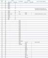
Of the 50 patients who had other cancers, 44 patients had one other cancer, while six had ≥2 other cancers in different settings. A summary of other cancers by setting in relation to EC in 50 patients is shown in Table 2. Five patients (patients no. 8, 9, and 36-38) had two other cancers aside from EC, while one patient (patient no. 10) had three other cancers; these resulted in 58 total events of other cancers. These other cancers were synchronous with EC in 25 cases (43.1%) and metachronous prior to or after EC in the other 15 cases (25.9%) and 18 cases (31.0%), respectively. Ovarian, breast, and colon were the three most common sites of other cancers found in 19 (38.0%), 11 (22.0%), and 8 patients (16.0%), respectively. Other less common cancers found were cancers of the lung (n=5), thyroid (n=2), skin (n=2), cervix (n=2), vagina (n=2), vulva (n=1), gall bladder (n=1), brain (n=1), appendix (n=1), and pancreas (n=1), as well as non-Hodgkin lymphoma (n=1).
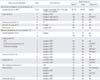
The mean age of the 25 EC patients who had synchronous cancer was 51.2±10.4 years, and 47.9±10.6 years for the 19 patients with ovarian cancers. Synchronous ovarian cancer, which was the most common other cancer, was found slightly more frequently than metastatic ovarian cancer (19 patients compared with 16 patients; a ratio of 1.2). Three of them also had breast, colon, or thyroid cancers in either pre- or post-EC diagnosis. Histopathology of EC and ovarian cancer were endometrioid carcinoma at both sites (15 patients), endometrioid EC with serous carcinoma, Sertoli cell tumor, or struma carcinoid of ovary (one each), and mixed endometrioid/serous EC with mixed serous/clear cell carcinoma of ovary (one case). Sixteen patients had EC limited to the uterus (stage I), while three had EC spreading to the serosal surface or outside the uterus (stage III). Their epithelial ovarian cancer stages were stage I (IA, seven patients; IC, six patients), stage II (one patient) or stage III-IV (five patients). In comparison, ovarian cancer stage was more advanced than EC in seven (36.8%), less advanced in five (26.4%), and similar in the other seven (36.8%) patients. Details of the 19 ovarian and 6 other synchronous cancers in the 25 EC patients are shown in Table 3.
Among the 30 patients with 33 metachronous cancers, mean age when they had other preceding cancer(s) was 48.3±12.2 years (aged 59.1±11.3 years when they had EC). Mean interval before they developed EC was 9.5 years (range, 3 to 21 years). Three of them had pelvic radiation for colon cancer (patients no. 11 and 12) or cervical cancer (patient no. 13). Conversely, the mean age of the patients when they had other subsequent cancer(s) was 62.1±8.5 years (aged 57.0±7.3 years when they had EC). The mean interval was 5.1 years (range, 1 to 9 years) after EC diagnosis. Table 4 shows details of metachronous cancers encountered in our EC patients.
Because breast and colon were the two most common metachronous cancers, we explored their data in detail. The mean age of 8/10 patients who had preceding invasive ductal breast carcinoma was 50.8±9.44 years. All ten preceding breast cancer patients had taken tamoxifen for 2 to 10 years with a 3 to 10-year interval from breast cancer to EC diagnosis (patients no. 1-10). Another patient developed breast cancer 4 years after EC (Table 2). Four of them were nulliparous (single status) while six had one or more components of metabolic syndrome. Two of them (patients #8 and 9) also had squamous cell carcinoma of the buttocks or colon cancer aside from breast cancer and EC. Of interest, one patient (patient #10), who had breast and colon cancers at 33 years old or 9 years prior to EC, had co-incidental ovarian cancer at 42 years of age and subsequently had contralateral breast cancer at age 44 or 2 years after EC. This patient also had eight family members (in their 40s or 50s) from three successive generations who had various cancers, including EC, ovarian, breast, and colon cancer. This patient met the Amsterdam II criteria for HNPCC [12]. Data of EC in 11 patients who had breast cancer were: four had stage III-IV EC and all except one patient with grade 1 tumor had grade 2-3 EC (being carcinosarcoma, clear cell, or serous carcinoma in three) (Table 4).
Among eight EC patients who had colon cancers, three occurred 7-9 years prior to EC (when they were aged 33, 40, and 57 years), three developed it 2-6 years after EC (when they were aged 52, 55, 66 years), and one was coincidentally found with EC at 54 years of age (Table 2). Among five patients with available data, colon cancers were rectosigmoid (two patients), ascending (two patients), or transverse (one patient). Three of them also had other cancers (patients no. 9, 10, and 38). Two patients had breast cancer with or without ovarian cancer as described earlier, while another had a carcinoid tumor of the appendix at the time of EC. These patients had EC with the following features: endometrioid histology (all eight), grade 2-3 (six patients), and stage III-IV (two patients).
After a median follow-up of 57.1 months (range, 0.07 to 236.8 months), 18 (5.2%) had progressive diseases while 27 (7.8%) had recurrences. The 5-year disease-free survival was 87.4% (95% confidence interval [CI], 83.8 to 91.0). A total of 63 patients (18.3%) had died: 48 EC patients with no other cancers (16.3%) and 15 patients with other cancers (30.0%). Causes of death among patients with no other cancers were from EC in 33 patients (11.2%) and from medical illnesses in 15 (5.1%). However, deaths among patients with other cancers were from EC in seven patients (14.0%), from other cancers in the other seven (14.0%), and from medical illness in another one (2.0%). The 5-year OS and EC-specific survival of all patients were 84.8% (95% CI, 80.8 to 88.9) and 88.4% (95% CI, 84.8 to 92.0), respectively. The 5-year OS of EC patients who had other cancers was significantly lower than that of those without: 79.3% (95% CI, 68.3 to 90.3) compared with 86.0% (95% CI, 81.7 to 90.3), respectively (p=0.023). The corresponding EC-specific survivals were 85.1% (95% CI, 75.5 to 94.7) compared with 89.0% (95% CI, 85.1 to 92.9), respectively (p=0.514).
DISCUSSION
Most reports of EC are from the more-developed regions where its incidence is high, while there are fewer reports of EC in less-developed areas where cervical cancer is more prevalent [1]. Along with differences in overall incidence, studies involving other co-morbidities, particularly of other cancers with EC, are more common in more-developed regions.
Our study demonstrated approximately 15% of Thai EC patients had other cancers, approximately half of which were discovered at the time of EC diagnosis (synchronous cancers), while the other half were found either before or after EC (metachronous cancers). Although we found a higher prevalence of other cancers in younger age groups (<50 or <40 years) of EC patients, the differences were not statistically significant. The prevalence of other cancers in our study was higher than one report from Korea that found 7% of their EC patients had secondary cancers [16]. This may be because our study included all other cancers identified, while their study focused on "hereditary predisposition" cancer [16]. Nevertheless, their study had similar findings as our study in that ovarian cancer was the most common other cancer followed by breast and colon [16].
The occurrence of other cancers in EC patients may be a result of various causes, as mentioned earlier. The first reason is a high estrogen milieu, which is a common risk factor of both EC and breast cancer [34517]. In the circumstances when EC occurred first, salpingo-oophorectomy done at surgical staging for EC may reduce the risk of breast cancer by 37% to 100% by removing the source of endogenous estrogen [18]. However, two of our EC patients still developed breast cancer after EC. This indirectly reflects the fact that there are other risk factors for breast cancer development aside from hormonal factors. The second reason is the effect of treatment for other preceding cancers. One example is tamoxifen, which is commonly used as adjuvant therapy for breast cancer, is a well-recognized risk factor for EC due to its estrogenic effect on endometrial tissue [19]. All ten EC patients in our study who previously had breast cancers had received tamoxifen. Another example is radiation therapy, which is notorious for inducing genetic instability and cancer [1020]. We found three of our patients had pelvic radiation therapy for colon or cervical cancers prior to EC. The third reason is genetic risk. One well recognized genetic abnormality closely related with EC is Lynch syndrome or HNPCC. The incidence of cancer in individuals with lynch syndrome is approximately 80% for colorectal cancer, 60% for EC, and 12% for ovarian cancer [11]. One study found that the risk of familial EC is 3-fold higher in EC patients aged 20-54 years and 1.5-fold higher with a family history of cancer in a first-degree relative [21]. Non-HNPCC family members rarely have synchronous or metachronous EC or colon cancer. Age at diagnosis of HNPCC-EC patients was approximately 10-15 years younger than sporadic cases with 57% occurring at <50 years [22]. Approximately 60% of these EC women with HNPCC developed a second primary cancer and 15% had more than two primary cancers [22]. Our study showed some findings suggestive of a genetic or familial association aside from other risk factors. Although not to significant levels, our patients with other cancers tended to be younger and had higher percentages of cancers in the family. LUS tumors frequently associated with Lynch syndrome [23] were also found significantly more frequently in patients with other cancers. One patient in this study who was 42 years of age with several family members with cancers seemed to meet the Amsterdam II criteria; however, she did not have a LUS tumor [12].
We found very few studies that directly addressed the prevalence and features of other preceding cancers prior to EC, so we searched for studies that reported subsequent cancers (including EC) after breast cancer. One study found that women with early-onset breast cancer (aged <40 years) were at a 3.9-fold increased risk of developing a new cancer later in life [24]. The risk was 7.4-fold for a second breast cancer and 6.1-fold for ovarian cancer. However, the risks for EC or colorectal cancer were not significantly elevated: 2.7-fold for endometrial and 2.4-fold for colon [24]. The authors raised the hypothesis of genetic susceptibility in these young breast cancer patients. Our patients had a mean age of approximately 51 years when they had breast cancer prior to EC and only two were in their forties. The tamoxifen stimulating effect on endometrium played a more obvious role than genetic predisposition in our patients because all ten EC patients who had preceding breast cancer had received this hormonal treatment.
Focusing on synchronous ovarian cancer, other studies have also reported a higher incidence among younger vs. older women (7%-30% vs. 2%-3%) [2526272829]. Most synchronous ovarian cancers in previous studies were of low grade and early stage. Although we also found that our EC patients with synchronous ovarian cancer had mean age of 48 years, which was younger than the 57 years of all EC patients, our study also found more advanced ovarian cancer than EC in nearly 40% of patients.
One previous study from the US using SEER data reported subsequent cancers after EC [30]. Significantly elevated risks for cancer of the colon, breast, and urinary bladder, as well as leukemia were found. The cumulative incidence of a second cancer after EC was 17.5% at 25 years. The two most common were breast and colon cancers, but the risks varied by age at EC diagnosis. Moreover, elevated risks were limited to younger women (47% increase for women diagnosed at younger than 40 years of age, and 25% for those diagnosed at younger than 50 years) [30]. A tendency of early onset of multiple primary cancers may suggest a genetic association. We found one study from Asia using population-based data from the Taiwan Cancer Registry that reported 4.8% prevalence of second primary malignancy after uterine cancer [31]. The two most common cancers were vagina/vulva and small intestine. In contrary to the study from the US, the report from Taiwan found significantly higher risk of secondary cancer in older patients ≥50 years than in younger patients [31]. Our patients had lung cancer as the most common cancer after EC (n=5) followed by colon (n=4), thyroid (n=2), breast (n=2), and others. The mean age when they had EC was 57 years (or 51.6 years particular to those with colon cancer). Our data were similar to the Taiwan study rather than data from the US. Ethnic influence may contribute to the difference. Of note, older age incidence of EC with other cancers does not support a genetic association in our patients.
Regarding the survival of EC patients with other cancers, a study from Korea found a 5-year OS (defined as EC-specific survival in their study) of 95% in both groups of patients who had other cancers (defined as hereditary predisposition cases) or had not (sporadic cases) [16]. Survival of the former group with stage I EC was slightly but significantly lower than the latter group of the same stage (94% vs. 98%, p=0.027). Our study could not demonstrate a significant difference in 5-year EC-specific survival of patients with or without other cancer (85% and 89%, respectively). This may be due to a few possibilities, including a small number of events in each group that did not allow a detection of survival difference statistically, or the patients actually had similar prognoses of EC as reflected by their similar prognostic features (Table 1). Nevertheless, we found that EC patients who had other cancers had worse overall prognosis with a lower 5-year OS than those without (79% compared to 87%, respectively). Because nearly half of the deaths in this "other cancer" group were from other cancers (14% of the 30% of deaths in this group), we conclude that the prognosis of other cancers has an impact on EC patient survival.
Some limitations of our study need recognition. First, the number of patients in our study was limited compare with studies from Western countries where EC is more common. Second, data of other cancers could not be detailed in all patients; e.g., details of colorectal or breast cancers or history of genetic aberration among these patients. Because current data suggest that universal screening by microsatellite instability testing and immunohistochemistry is important to identify Lynch syndrome patients [32], our future work may explore the possibility of a genetic association by these tests in our patients. Third, a complete pedigree chart was not possible when data were obtained from medical charts that only had pieces of available information noted over a period of time and by different physicians. The doctors in our country were probably not aware of familial cancer risk as in Western countries, while the patients might be uncertain about their relatives' detailed health conditions or history. We were aware of a few studies from Asia that described HNPCC-related EC in Koreans [163334]. Findings from their studies and our study might raise awareness of this topic in Thai and other Asian populations.
In conclusion, other cancers in Thai EC are not uncommon. When present, they may complicate treatment planning and affect EC patient prognosis. A thorough personal and family history taking as well as careful surveillance after EC treatment are important, as this will help to identify other cancers in EC patients and their family members. EC patients with risks of familial association should undergo genetic counseling and testing as appropriate.

 XML Download