

 PDF
PDF ePub
ePub Citation
 Print
Print


To the editor: The macroscopic rendering of a surgical gynecologic specimen can sometimes be difficult, particularly if the medical practitioner is faced with a rare and complex condition. In the last years the radiological imaging has made great strides in this direction. However, these sophisticated techniques are not without risks for the patients (e.g., ionizing radiation, contrast medium) and they cannot always be easily accessible. Under this perspective, a new generation of three-dimensional (3D) imaging softwares for the anatomical rendering of the human body, and related surgical pathologies, are in the development phase [1]. We have recently tested one of these platforms with encouraging results in a 39-year-old female, affected by a high-grade endometrial stromal sarcoma, arisen in the context of an extrapelvic endometriotic focus at the level of the right inguinal ligament. Inguinal endometriosis is a rare condition occurring in only 0.6% of women and it is usually associated with previous pelvic surgery [2]. Our patient in fact was submitted to the surgical removal of an endometriotic scar of the right parametrium 9 years ago. The occurrence of an endometrial stromal sarcoma within an endometriotic nodule is a rare, but possible, event, previously described at the level of vulva, vagina, ovary, round ligament, rectosigmoid colon, terminal ileum, abdominal wall, and sciatic nerve [3,4,5,6,7]. In our case, the sarcoma had grown from the right inguinal ligament until reaching 23.5 cm of maximal diameter. It was bordered by a discontinuous pseudo-capsule and the presurgical computed tomography (CT)-scan showed a diffuse infiltrative front of growth in the surrounding deep tissues. For this reason, the patient was promptly submitted to a software-assisted surgery with abdominal wall reconstruction. Starting from the CT images, the software has been developed to upload them and to assign a predefined color in relation to the signal intensity. Then, it has worked using different subtraction filters and a 3D-space vision. More in detail, it has been possible to tip and rotate the neoplasia through 360°, removing anatomical layers, in order to view any pathological structure, and making easier to explain complex surgical correlations. The subtraction filters available were: dermal subtraction filter, superficial muscular subtraction filter, deep muscular subtraction filter, and visceral subtraction filter. The software has been able to detect the neoplasia origin from the right inguinal ligament and to better define the infiltration front, favoring a radical surgery (Fig. 1). It has been proved a useful tool in the surgical and pathological approach to this unique case, never reported in literature. Its major advantages appear to be the running on portable electronic devices, such as notebook or tablet, an easy-touse interface with intuitive functionality, and the opportunity to purchase for each unit. For the future, an implementation with standardization of software-assisted procedures could be useful in the gynecologic management of the patients affected by uncommon malignancies.
Figures and Tables
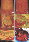 | Fig. 1(A) A longitudinal digitalized image of the abdomen, centered on the umbilicus, is seen without application of subtraction filters. (B) The dermal subtraction filter allows to remove the skin surface, which does not show pathological alterations. (C) The superficial muscle subtraction filter is able to subtract the superficial muscle layer and the sarcoma appears as a right-sided abdominal bulky mass (asterisk). The neoplasia shows a pseudo-capsule (yellow arrows), an area of colliquation (white arrow) and take its origin from the right inguinal ligament (white circle). The green pointer of spatial orientation is also reported. (D) The deep muscle subtraction filter points out the infiltration of the surrounding deep tissues, which appears as a color distortion (asterisk). (E) On a transversal section, the visceral subtraction filter displays the presence of a massive front of neoplastic infiltration towards the intestine (asterisk). (F) The gross appearance confirms what has been previously observed by the digitalized images.
|
References
1. Zeller JL. New 3D imaging software opens new vistas. JAMA. 2006; 296:2908–2913.
2. Bergamini A, Almirante G, Taccagni G, Mangili G, Vigano P, Candiani M. Endometriosis-associated tumor at the inguinal site: report of a case diagnosed during pregnancy and literature review. J Obstet Gynaecol Res. 2014; 40:1132–1136.
3. Kim JY, Hong SY, Sung HJ, Oh HK, Koh SB. A case of multiple metastatic low-grade endometrial stromal sarcoma arising from an ovarian endometriotic lesion. J Gynecol Oncol. 2009; 20:122–125.
4. Alcazar JL, Guerriero S, Ajossa S, Parodo G, Piras B, Peiretti M, et al. Extragenital endometrial stromal sarcoma arising in endometriosis. Gynecol Obstet Invest. 2012; 73:265–271.
5. Sato K, Ueda Y, Sugaya J, Ozaki M, Hisaoka M, Katsuda S. Extrauterine endometrial stromal sarcoma with JAZF1/JJAZ1 fusion confirmed by RT-PCR and interphase FISH presenting as an inguinal tumor. Virchows Arch. 2007; 450:349–353.
6. Sinha R, Sundaram M. Endometrial stromal sarcoma from endometriosis. J Minim Invasive Gynecol. 2010; 17:541–542.
7. Usta TA, Sonmez SE, Oztarhan A, Karacan T. Endometrial stromal sarcoma in the abdominal wall arising from scar endometriosis. J Obstet Gynaecol. 2014; 34:541–542.
 XML Download