

 PDF
PDF ePub
ePub Citation
 Print
Print


INTRODUCTION
Ovarian cancer has the highest mortality rate among gynecologic cancers, and the incidence of ovarian cancer has increased in the last decade. Combination therapy with platinum and taxane is used as postoperative standard chemotherapy for epithelial ovarian cancer [1]. In the 1980s, the 5-year survival rate for stage III ovarian cancer was 30%. After the introduction of the platinum-taxane combination therapy, the survival rate increased to 43% [2]. However, the survival rate of 43% is still too low in terms of longevity. The survival of patients with ovarian cancer can be improved by adding bevacizumab to the paclitaxel-carboplatin (TC) therapy and then using bevacizumab alone as the subsequent maintenance therapy [3]. Moreover, it was reported that the disease-free survival and overall survival (OS) rate of patients who received dose-dense weekly TC therapy were significantly better than those of patients who received TC therapy [4]. On the other hand, the efficacy of intraperitoneal chemotherapy was demonstrated in a randomized comparative study [5]. With the use of molecular-targeted drugs and modification of therapeutic regimen, the outcome of primary chemotherapy for epithelial ovarian cancer has improved.
Clinical complete response (CR) is achieved by primary chemotherapy in approximately 75% patients with epithelial ovarian cancer. On the other hand, approximately 25% patients with epithelial ovarian cancer fail to achieve CR. Moreover, approximately 60% patients who have achieved clinical CR relapse, and many of these patients do not respond to second-line and third-line chemotherapy, leading to a clinical course similar to that of chronic disease and eventual death.
BAF250a, a protein encoded by AT-rich interactive domain 1A (SWI-like) gene (ARID1A), is a chromatin remodeling factor that belongs to the SWI/SNF family [6,7]. BAF250a is involved in DNA repair. ARID1A is believed to be involved in DNA repair through ATP-dependent induction of chromatin migration and dissociation [8]. ARID1A mutations are frequently found in ovarian clear cell adenocarcinoma and endometrioid adenocarcinoma of the ovary; however, no ARID1A mutation has been found in serous adenocarcinoma [7]. Moreover, loss of BAF250a protein has been strongly correlated with ovarian clear cell adenocarcinoma, endometrioid adenocarcinoma, and the presence of ARID1A mutations [9]. On the other hand, ARID1A mutations and loss of BAF250a protein are clearly noticeable in these tumors and adjacent atypical endometriosis; however, ARID1A mutations and loss of BAF250a protein have not been found in the distal portion of the endometriosis lesion [7]. On the basis of some studies, it has almost been confirmed that loss of ARID1A is an early molecular phenomenon in the oncogenic transformation of endometriosis [6,7,10-14]. Moreover, loss of ARID1A is reportedly involved not only in these tissue types but also in mucinous ovarian tumors and endometrial carcinoma [15-17].
A recent study indicated that loss of ARID1A is related to short disease-free survival and chemoresistance in ovarian clear cell adenocarcinoma [18]. Thus, our present study was conducted in epithelial ovarian cancer of all tissue types to investigate whether an increased or decreased expression level of ARID1A can be a prognostic factor for ovarian cancer or can influence the sensitivity to anticancer drugs.
MATERIALS AND METHODS
1. Study population and tissues
Immunohistochemical examination was performed retrospectively on 111 epithelial ovarian carcinomas obtained from women who were surgically treated at the Hirosaki University Hospital between 2006 and 2011 after informed consent had been obtained. One slide from each case was reviewed by the study gynecologic pathologist (MF) to confirm the diagnosis of epithelial ovarian carcinoma. The tissue specimens included 44 serous adenocarcinomas, 18 endometrioid adenocarcinomas, 30 clear cell adenocarcinomas, 14 mucinous adenocarcinomas, and 5 other typed carcinomas. All patients were primarily treated with cytoreductive surgery and adjuvant TC chemotherapy (paclitaxel 175 mg/m2 and carboplatin area under the curve [AUC] 6). They all received 6 to 9 cycles of this combination regimen. Sixty nine of the 111 patients were surgically staged in accordance with the 1988 International Federation of Gynecology and Obstetrics (FIGO) criteria. The breakdown for stages of ovarian carcinomas consisted of 57 patients with stage I, 8 with stage II, 42 with stage III, 4 with stage IV. The duration of follow-up ranged from 21 to 92 months (median, 64 months). The mean age of patients with ovarian carcinoma at surgery was 54.1 years (range, 24 to 79 years). Here, chemosensitivity was defined as achieving CR by primary chemotherapy and chemoresistance was defined as failing to achieve CR by primary chemotherapy. The acquisition of the tissue material was approved by the Instituional Review Board of Hirosaki University Graduate School of Medicine.
2. Immunohistochemistry
Six-µm sections of formalin-fixed and paraffin-embedded tissue specimens were stained by established method as described previously [19]. Sections were incubated with antibody specific for ARID1A (Santa Cruz Biotechnology, Santa Cruz, CA, USA) overnight 4℃ after antigen retrieval in a sodium citrate buffer. Slides were incubated with biotinylated species-specific secondary antibodies for 30 minutes and then exposed to avidin-biotin-peroxidase complex. Sections were treated with 0.02% diaminobenzidine as a chromogen, and counterstained with hematoxylin. The expression level of ARID1A was graded using staining scores, which were calculated by multiplying the staining intensity of the nuclei by the stain-positive area within the tumor. As shown in Fig. 1, the staining intensity of nuclei was classified as negative (score 0), weakly positive (score 1), moderately positive (score 2), or strongly positive (score 3). Stain-positive areas were graded as follows: score 0 was given to a specimen with 0% stain-positive area, score 1 to a specimen with ≥1% to <25% stain-positive area, score 2 to a specimen with ≥25% to <50% stain-positive area, and score 3 to a specimen with ≥50% stain-positive area. For each patient, a slide specimen was observed in a 0.75-mm2 field of vision using a 20× objective lens. The mean score from 3 different sites was used as the staining score for that patient. The expression level of ARID1A was evaluated by 2 researchers (YM and TS) who were not given any physical and clinical information.
3. Statistical analysis
Differences in staining scores between the clinicopathologic factors were analyzed using Student's t-test. Progression-free survival (PFS) was defined as the period from end of the first-line regimen to initiating the second-line regimen after confirming relapsed lesions on imaging. OS was calculated from the date of start of treatment to the date of death or last follow-up. The cumulative survival curves were estimated by use of the Kaplan-Meier method. Comparison between survival curves was made using the log-rank test. Statistical significance was set at p<0.05.
RESULTS
1. Relationship between clinicopathologic factors and the expression level of ARID1A
The expression level of ARID1A was significantly lower in clear cell adenocarcinoma (1.7±2.1) than in serous adenocarcinoma (4.0±3.4), mucinous adenocarcinoma (4.5±3.9), and endometrioid adenocarcinoma (5.2±3.3) (Table 1). However, no significant correlation was found between the expression level of ARID1A and either the clinical stage, retroperitoneal lymph node metastasis, or the residual tumor size (Table 1).
2. Relationship between the expression level of ARID1A and the sensitivity to anticancer drugs
The sensitivity to anticancer drugs was evaluated in 46 patients with stage III, IV cancer. CR was defined as disappearance of the lesion(s) on diagnostic imaging combined with a negative result for the tumor marker after completion of the scheduled therapy. CR was achieved by 34 patients, whereas it was not achieved by 12 patients. The tissue types of patients with and without CR were as follows: 22 patients with CR and 7 patients without CR in serous adenocarcinoma, 3 patients with CR and 0 patients without CR in endometrioid adenocarcinoma, 6 patients with CR and 3 patients without CR in clear cell adenocarcinoma, 2 patients with CR and 2 patients without CR in mucinous adenocarcinoma, and 1 patient with CR and 0 patients without CR in other tissue types. No significant correlation was found in the tissue types. The staining score was 4.3±3.4 in patients with CR and 1.9±2.0 in patients without CR (p=0.026).
3. Relapse after achieving CR and the expression level of ARID1A
Among the 34 patients with stage III, IV cancer who achieved CR, 21 patients subsequently relapsed. The remaining 13 patients did not relapse. The staining score was 3.5±3.3 in patients who relapsed and 5.5±3.3 in patients who did not relapse (p=0.07).
4. Relationship between the expression level of ARID1A and prognosis
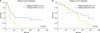
The staining score of 0 was defined as ARID1A-negative. Other staining scores were defined as ARID1A-positive. The prognosis was compared between ARID1A-negative and ARID1A-positive patients. There were 34 ARID1A-negative patients and 77 ARID1A-positive patients. PFS and OS did not differ significantly between ARID1A-negative and ARID1A-positive patients when all disease stages were included in the analysis (data not shown). On the other hand, there was a significant difference in PFS between ARID1A-negative (n=11) and ARID1A-positive (n=35) patients only in the disease stage III, IV (p<0.05) (Fig. 2A), whereas no significant difference was observed when OS was compared between the same 2 groups (Fig. 2B).
DISCUSSION
The results of the present study indicate that tumors with no or weak ARID1A expression are resistant to anticancer drugs. All tumors used in the present study had been treated with TC as the primary therapy. Paclitaxel exerts its effect as an anticancer drug by affecting microtubules [20]. Microtubules are cytoskeletal proteins formed by the polymerization of tubulin. Microtubules have a long tubular structure with the diameter of 25 nm. During cell division, microtubules are required for segregation of chromosomes. After DNA is copied and tightly packed into chromosomes, microtubules segregate the DNA into 2 cells. Cell division can be interrupted by inhibiting the formation of microtubules from tubulins. Paclitaxel achieves its antitumor effect by interrupting the function of microtubules [20]. ARID1A is involved in DNA repair. Inhibition of ARID1A expression is believed to cause inhibition of cell division. In other words, the expression level of microtubules may also be inhibited. When the ARID1A expression is lost or decreased, the tumor cells become static; thus, the efficacy of paclitaxel probably decreases because there is no action site for paclitaxel. For ovarian cancer with no or weak ARID1A expression, a new therapeutic regimen may need to be established in order to replace paclitaxel therapy.
In the present study, among the patients with stage III, IV cancer, the expression level of ARID1A was lower in patients who relapsed after achieving CR than in patients who did not relapse. In addition, when the staining score of 0 was defined as ARID1A-negative and other staining scores were defined as ARID1A-positive, there was a significant difference in PFS between ARID1A-negative and ARID1A-positive patients only in the disease stage III, IV. Katagiri et al. [18] studied the relationship between the expression level of ARID1A and prognosis in 60 patients with ovarian clear cell adenocarcinoma and reported that disease-free survival was significantly short in tumors with no ARID1A expression. There is no known biomarker for the risk of relapse of ovarian cancer. The result of the present study suggests that decreased ARID1A expression can be an important predictive factor for cancer relapse not only in clear cell adenocarcinoma but also in all other types of epithelial ovarian cancer.
When different tissue types were compared, the expression level of ARID1A was significantly lower in clear cell adenocarcinoma than in other tissue types. This result is consistent with the results of many other studies [7,14]. The result of the present study also indicates that some of clear cell adenocarcinoma overexpressed, downregulated or mutated ARID1A. Moreover, the expression level of ARID1A was not correlated with either the clinical stage, retroperitoneal lymph node metastasis, or the residual tumor size. In other words, ARID1A is unlikely to be related to angiogenesis or cell growth factors involved in tumor progression and metastasis. When all stages were included in the analysis and patients with no ARID1A expression (ARID1A-negative patients) and those with ARID1A expression (ARID1A-positive patients) were compared, no significant difference was observed in OS. The same result was obtained when only patients with advanced disease were compared. With respect to the clinical significance of ARID1A, our study suggests that decreased ARID1A expression is possibly associated with the risk of developing resistance to anticancer drugs and also that ARID1A is a predictive factor for the risk of relapse after achieving CR. The reason why no significant difference was observed in OS between ARID1A-negative and ARID1A-positive patients is probably because the effect of decreased ARID1A expression was masked by the use of various therapeutic regimens, including surgery, radiation therapy, and second-line agents such as liposomal doxorubicin, gemcitabine and topoisomerase inhibitors, in relapsed patients. In order to determine whether the expression level of ARID1A has any effect on OS, we need to conduct a study with a prospective design. Furthermore, because ovarian cancers as a whole are heterogeneous, composed of different histologic types with different molecular pathogenetic pathways, it is better to stratify ovarian cancers into different histologic groups rather than lump them all together when doing statistical analysis. Thus, the importance of a specific factor may be masked.
ARID1A mutations are reported to be consistent with the loss of ARID1A protein expression in immunohistochemical staining [9]. Thus, the biological property of a tumor can be estimated by examining the expression level of ARID1A using immunostaining, while at the same time determining the tissue type using Hematoxylin and Eosin staining. However, one of the obstacles is that the criteria for ARID1A-negative/ARID1A-positive need to be established. Various definitions/criteria have been reported. For example, one of the definitions for ARID1A-negative is that the proportion of ARID1A-positive cells is 5% or less of the epithelial cells [14]. The other definition uses the level of intranuclear expression to grade ARID1A expression as negative, weakly positive, or strongly positive [18]. In this study, in order to objectively evaluate the expression level of ARID1A, the mean score was obtained from 3 sites in each tumor by multiplying the staining intensity of the nuclei by the stain-positive area. However, we were not able to establish a concrete cutoff score that can be used to diagnose anticancer drug resistance. In the future, the criteria for the ARID1A expression (i.e., ARID1A-negative and ARID1A-positive) need to be established, and then a retrospective study should be conducted to accumulate clinical cases. Moreover, a new therapeutic strategy leading to tailored ovarian cancer therapy may be generated by conducting a prospective study of anticancer drugs selected in accordance with expression levels of ARID1A.

 XML Download