

 PDF
PDF ePub
ePub Citation
 Print
Print


INTRODUCTION
Cancer-related mortality in women has significantly decreased during the last decade due to advancements in early diagnostic tests, effective medical treatments, and surgical techniques as well as increased public awareness attributed to successful press and media communications regarding primary care and preventive strategies [1]. Unfortunately, these successful treatment strategies give rise to other challenging problems, especially in young female patients receiving chemotherapy, as most chemotherapeutic agents decrease the number of primordial and larger growing follicles, leading to premature menopause and infertility [2].
Cisplatin, a platinum-based anticancer agent, is widely used for a variety of cancers such as sarcoma, small cell lung cancer, ovarian cancer, cervical cancer, and lymphomas. The mechanism of its antitumor activity relies on the formation of a platinum complex inside of a cell, leading to DNA cross-linking, the arrest of cells in the G2 phase, and apoptosis [3]. Moreover, cisplatin is thought to induce the peroxidation of cell membranes [4], mitochondrial dysfunction [5], and inhibition of protein synthesis [6]. These effects on dividing cancer cells also affect healthy dividing cells in the body, such as the granulosa cells of the follicles, causing acute and chronic ovarian failure resulting in infertility.
Therapeutic options for preventing ovarian damage in female patients desiring future fertility have been previously studied; however, currently available methods remain limited and include fertility-sparing surgery in gynecologic cancers and hormonal protection with gonadotropin releasing hormone analogues and antagonists [7]. Recently, stem cell studies have begun to emerge in reproductive biology. The generation of male and female gametes from mouse embryonic stem cells [8,9] and the development of oocyte-like cells from porcine skin and rat pancreatic stem cells [10,11] have been described. Studies by Lee at al. [12] and Johnson et al. [13] demonstrating follicle protection and long-term fertility preservation in female mice treated with high-dose alkylating chemotherapy followed by bone marrow transplantation have inspired researchers to hypothesize that the mobilization of endogenous bone marrow stem cells by induction with granulocyte-colony stimulating factor (G-CSF) might provide a similar protection of ovarian follicles and fertility.
G-CSF is a glycoprotein with growth factor and cytokine functions [14]. G-CSF has been successfully used in congenital neutropenia and in chemotherapy-induced neutropenia without a decrease in the efficacy of chemotherapeutic agents. With long-term use, its efficacy and safety have been proven. Although toxic and adverse events have been reported, these are not clinically troublesome and seldom necessitate stopping therapy [15]. Recently, the capacity of hematopoietic stem cells to migrate to other organs in response to acute damage to participate in tissue repair has been established in various tissues including the intestine [16], heart [17], liver [18], and kidneys [19]. In addition to its stimulating effects on bone marrow cells for transdifferentiation, G-CSF also has antiapoptotic [20] and anti-inflammatory functions [21].
The potential of G-CSF to protect against cisplatin-induced ovarian failure has not been investigated to date. Thus, here we examined whether G-CSF can reduce the ovarian injury caused by cisplatin in an experimental rat model.
MATERIALS AND METHODS
1. Animals
Twenty-one adult female Sprague-Dawley rats weighing 200 to 210 g were used in this study. Animals were housed in cages and maintained under standard conditions with a 12-hour light/dark cycle at room temperature (22℃±2℃). The rats were fed a standard pellet diet and were provided tap water ad libitum throughout the study. The protocol used in this study was approved by the Institutional Animal Care and Ethical Committee of Ege University.
2. Experimental protocol
For 14 rats, cisplatin (Sisplatin Flakon, Koçak, Farma, Turkey; 2 mg/kg/day) was administered by intraperitoneal injection twice per week for 5 weeks (total 20 mg/kg) [22]. No drug was administered to the remaining rats, which were considered a control group (n=7). The 14 rats receiving cisplatin were randomly divided into two groups: the first cisplatin group was treated with 1 mL/kg/day physiological saline (PS) (CP+PS; n=7), and the second cisplatin group was treated with 100 µg/kg/day G-CSF (Neupogen, Roche, Mannheim, Germany; 48 MU/mL=480 µg) [23,24]. Injections were administered intraperitoneal every day for 5 weeks. None of the rats died in the study.
All rats were euthanized at the end of the treatment period. Both ovaries from all animals were recovered, fixed in 10% formalin, and stored at 4℃ for paraffin sectioning. Blood samples were collected by cardiac puncture and stored at -30℃ for hormone assays.
3. Histological examination
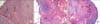
Formalin-preserved ovarian tissues were embedded in paraffin. The ovaries were sectioned at a thickness of 4 µm using a microtome, and the sections were mounted onto glass slides and stained with hematoxylin and eosin. An Olympus C-5050 digital camera (Olympus, Tokyo, Japan) mounted on an Olympus BX51 microscope was used to photograph all sections. Morphological analysis of the ovaries was performed using a computerized image analysis system (Image-ProExpress 1.4.5, Media Cybernetics Inc., Rockville, MD, USA) on 10 fields of view per section at a magnification of 20×; the observer was blinded to the study groups. Ovarian follicles at different developmental stages were categorized according to standards established by Oktay et al. [25] (Fig. 1). Primordial follicles are localized immediately below the cortex and contain a single layer of squamous granulosa cells. Primary follicles consist of a single layer of cuboidal granulosa cells. Secondary follicles include multiple layers of granulosa cells, and tertiary follicles are characterized by a stratum granulosum and a fluid filled antral space.
4. Measurement of anti-Müllerian hormone levels
Plasma anti-Müllerian hormone (AMH) levels were measured using a commercially available enzyme-linked immunosorbent assay kit (USCN Life Science Inc., Hubei, China). Samples from each animal were measured in duplicate according to the manufacturer's instructions. The detection range was 0.156 to 10 ng/mL.
5. Statistical analysis
Data analyses were performed using SPSS ver. 15.0 (SPSS Inc., Chicago, IL, USA). The groups of parametric variables were evaluated by an analysis of variance (ANOVA) followed by the post hoc Tukey honest significant difference test. The results are presented as the mean±standard error. A value of p<0.05 was considered to be statistically significant.
RESULTS
Histological examination of the ovaries demonstrated significant alterations in cisplatin+PS group including decreased follicle numbers and follicular maturation whereas control group exhibited normal ovarian morphology. However, treatment of the rats with G-CSF significantly lessened the follicular impairment and enhanced the immature follicles (Fig. 1).
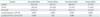
The follicle counts of the groups are summarized in Table 1. ANOVA results showed significant differences between the study groups (p=0.001 for primordial follicles, p=0.01 for primary follicles, p<0.001 for secondary follicles, and p=0.005 for tertiary follicles). The number of primordial follicles showed a significant reduction in the cisplatin+PS group compared to the control group (2.8±0.4 vs. 18.2±0.8, respectively; p<0.001). Treatment of rats with G-CSF considerably improved the number of primordial follicles (14.5±1.4, p<0.001). Similarly, primary and secondary follicle numbers of the cisplatin+PS group were significantly different compared to control group (8.3±0.5 vs. 14.6±1.1 and 6.7±0.5 vs. 10.8±0.9; p<0.001). Treatment of rats with G-CSF effectively prevented the cisplatin-induced damage in primary and secondary follicles (15.1±2.1 and 10.3±1.3; p=0.02 and p=0.004, respectively). Tertiary follicle counts were 4.7±0.2 in the control group, 1.2±0.3 in the cisplatin+PS group, and 3.5±0.7 in the cisplatin+G-CSF group. The differences between the groups were also statistically significant (p<0.001 and p=0.01, respectively).
Plasma AMH levels were measured to determine the effects of G-CSF on follicle pool of cisplatin-treated rats. Table 2 shows the alterations in AMH levels of the study groups. Compared to the control group, plasma AMH levels were significantly decreased in cisplatin+PS group (2.38±0.17 ng/mL vs. 0.41±0.02 ng/mL, respectively; p<0.001). G-CSF treatment significantly improved the AMH levels compared to cisplatin+PS group (1.56±0.247 ng/mL, p=0.006) (Table 2).
DISCUSSION
We examined the effects of G-CSF on ovarian reserves by measuring follicle counts and serum AMH levels after cisplatin therapy. Treatment without G-CSF caused a significant decrease in the follicle number and serum AMH levels, whereas these measures were significantly increased when G-CSF was added to the treatment.
Although the specific protective mechanisms of G-CSF remain unclear, we present several hypotheses. One possible explanation is that G-CSF has anti-apoptotic properties that occur through the Jak/Stat, PI3K/AKT, and MAPK/AKT pathways [26,27]. AKT is an important antiapoptotic protein that balances cell survival and cell death through the phosphorylation of Bad, which is a proapoptotic member of the Bcl-2 family. In its unphosphorylated form, Bad binds to Bcl-XL (an antiapoptotic protein) and thus blocks cell survival. However, the activation of AKT leads to Bad phosphorylation and inhibits its proapoptotic activity [28]. G-CSF also induces the expression of Bcl-2 and decreases Bax translocation to the mitochondria, leading to apoptosis inhibition and cell survival [20]. Another important antiapoptotic mechanism of G-CSF acts through the JAK/Stat signaling pathway and leads to subsequent activation of Bcl-2, which protects against cell death [29]. Although further work is needed to better understand the molecular mechanisms, it is likely that G-CSF protects against apoptosis.
A second possible explanation of the protective mechanism of G-CSF is that G-CSF has anti-inflammatory properties. In several studies, a decreased production of pro-inflammatory cytokines, such as interleukin 6 (IL-6), tumor necrosis factor-α (TNF-α), nuclear factor-κB, and IL-1β, upon G-CSF administration has been demonstrated [20,26,30], leading to reduced inflammation and an amelioration of a potentially destructive inflammatory response. Accumulating evidence also indicates that elevated plasma levels of IL-6 and TNF-α are commonly observed in animals treated with cisplatin, which might contribute to germ cell apoptosis [31]. Thus, G-CSF might contribute to the survival of follicles by suppressing the production of proinflammatory cytokines.
A third potential protective mechanism is that G-CSF plays a role in neoangiogenesis. In a study by Lee et al. [32], a 3-day treatment with G-CSF (50 µg/kg/day) after occlusion of the middle cerebral artery enhanced angiogenesis in the rat brain. An up-regulation of intracellular adhesion molecule 1 by G-CSF was also reported to facilitate the migration of monocytes that engage in angiogenesis [33]. In an acute myocardial infarction experimental rat model, Kocher et al. [34] injected G-CSF intravenously and observed significantly increased neovascularization at the site of the myocardial infarction. In a recent study by Skaznik-Wikiel et al. [35], high-dose cyclophosphamide and busulfan chemotherapy caused a significant reduction in ovarian microvessel density compared to vehicle-treated controls. In contrast, the microvessel density in mice treated with chemotherapeutic agents and G-CSF was similar to that observed in vehicle treated control mice.
G-CSF is known to mobilize hematopoietic stem cells from bone marrow into peripheral blood. These undifferentiated cells are thought to commit to a differentiation program, integrate into tissue, and replace damaged cells to reconstruct a damaged cell mass. This transdifferentiation has been reported in many studies [36]. However, it was recently reported that cell fusion, rather than transdifferentiation, is the major mechanism underlying organ repair [37]. Thus, it remains controversial whether fusion or the transdifferentiation of bone marrow cells into other tissues aids organ repair. In a study by Yannaki et al. [38], G-CSF administration ameliorated histological damage and accelerated the regeneration process in both an acute and chronic liver injury model. However, the acceleration of recovery after chemical injury and G-CSF treatment was mainly mediated by an increased proliferation of host hepatocytes with less support from cells of bone marrow origin. Another study demonstrated that in ex vivo perfused rat hearts subjected to ischemia-reperfusion injury, the injury was ameliorated by G-CSF without any bone marrow effect [29]. Based on these findings, we propose that G-CSF may have non-hematopoietic effects and function as a growth factor for tissue repair and regeneration.
Another explanation for the protective effects of G-CSF is that G-CSF as a growth factor to initiate or trigger endogenous repair mechanisms. In a study by Hess et al. [39], bone marrow-derived cells were found to reverse hyperglycemia in streptozotocin-treated mice by inducing endogenous pancreatic tissue repair through possible interactions with endothelial cells. In addition a study by Ledee et al. [40] reported that follicular fluid G-CSF influenced future embryo implantation, suggesting that G-CSF plays an autocrine/paracrine role in the reproductive system.
A final hypothesis is that the protective mechanism of G-CSF is via the beneficial effects of G-CSF in neutralizing the effects of reactive oxygen species (ROS), which are strongly induced in tissues after chemotherapy treatment [41]. In many studies, G-CSF was found to induce the synthesis of endogenous nitric oxide, which is a powerful scavenger of excessive ROS, by either activating the Akt/eNOS pathway [42] or by shifting the balance between the pro- and anti-inflammatory cytokines towards a more favorable anti-inflammatory net effect [20,26,30].
Despite the interesting findings, our study had certain limitations. The number of subjects included in the subgroups was small; hence, a larger sample size is necessary to enhance the power of our study. Moreover, we did not examine the exact molecular mechanisms by which G-CSF ameliorates follicle loss after cisplatin treatment. Detailed studies are necessary to evaluate the optimal timing and dosage of G-CSF relative to chemotherapy.
In summary, our results showed that G-CSF is beneficial in decreasing follicle loss in an experimental rat model of cisplatin chemotherapy. One of the main inferences of our study is that harmless effects of the cisplatin for patients desiring further fertility may be diminished with G-CSF. It is hoped that our findings will inspire others to undertake more detailed researches focused on the underlying molecular mechanisms of the observed effect of G-CSF, ultimately leading to protection of the patients from ovarian damage caused by cisplatin chemotherapy.

 XML Download