

 PDF
PDF ePub
ePub Citation
 Print
Print


INTRODUCTION
Microinvasive cervical cancer (MIC) was first introduced by Mestwerdt in 1947 [1,2]. The definition of MIC has been debated and controversial for decades. There are two most commonly used definition systems: one is the Society of Gynecologic Oncologists (SGO, USA) [2] and the other is the International Federation of Gynecology and Obstetrics (FIGO) [3]. The SGO defines MIC as one with a maximum depth of invasion of 3 mm. SGO does not place a horizontal limit but exclude any patient if vascular lymphatic space involvement was present [2]. The current FIGO system divides stage IA into two categories: stage IA1 MIC is defined as measured invasion of stroma no greater than 3.0 mm in depth and no wider than 7.0 mm. Stage IA2 MIC is defined as measured invasion of stroma greater than 3.0 mm and no greater than 5.0 mm in depth and no wider than 7.0 mm. Lympho-vascular space involvement (LVSI) was not included as part of the definition [3].
There's no unified standard treatment for MIC. Management of patients with MIC varied from conization to radical hysterectomy (RH) with or without lymphadenectomy. Stage IA1 MIC is traditionally treated with a simple hysterectomy (SH) and conization for young patients who have strong desire for fertility. Stage IA2 MIC is traditionally treated with more aggressive therapy such as radical or modified radical hysterectomy (MRH). The outcomes of patients with MIC are favorable [4,5,6,7,8,9,10]. Convincing data have shown that the risk of parametrial involvement and ovarian metastasis are extremely rare [4,6,11,12,13]. In a review of literatures, Baalbergen et al. [7] found the risk of recurrent disease was 1.5% (3/193) after conservative therapy and 3.5% (9/254) after radical therapy in patients with stage IA1 and IA2 cervical adenocarcinoma. Extensive treatment such as RH with pelvic lymph node dissection (PLND) or trachelectomy does not prevent recurrence [7]. Conservative treatment has been studied for decades to preserve fertility in patients with MIC [6,7,14,15,16,17]. Some researchers considered conization alone with careful follow-up appears to be an effective and safe treatment for patients with stage IA1 MIC regardless of resection margin status or LVSI [17]. However, some researchers considered conization is safe in patients with stage IA1 cervical cancer without LVSI and with negative conization margin [15]. Positive cone margin, LVSI, postmenopausal state, positive endocervical curettage, involvement of four quadrants and precone high risk-human papillomavirus (HPV) load ≥300 relative light units/positive control have been reported to be predictors of residual disease after conization by several researchers [18,19,20].
Management of patients with LVSI and stage IA2 cervical cancer has been controversial. Some gynecologists suggested that pelvic lymphadenectomy should be performed in patients with LVSI [7,16]. However, some researchers does not find relationship between LVSI and the lymph node status [4,21]. The very low rate of positive lymph nodes in stage IA2 patients can not justify the inclusion of lymphadenectomy as part of standardized care [9]. When exact evaluation of tumor extension and surgical margins of the cone are considered, conservative management of stage IA2 MIC is safe [22].
In the present study, we report a cohort of 324 Chinese women with MIC. The aim of the present study is to explore appropriate treatment modality of MIC and to analysis prognosis and risk factors of recurrence.
MATERIALS AND METHODS
There were 346 patients treated for MIC at the Department of Obstetrics and Gynecology at Peking Union Medical College Hospital from January, 2003 to November, 2013. In our study, the eligibility criteria included as following. (1) The pathological diagnosis were made and reviewed by two independent pathologists in our hospital. (2) The diagnosis of MIC was made by FIGO staging system [3], i.e., stage IA1 is defined as measured invasion of stroma no greater than 3.0 mm in depth and no wider than 7.0 mm. Stage IA2 is defined as measured invasion of stroma greater than 3.0 mm and no greater than 5.0 mm in depth and no wider than 7.0 mm. LVSI was not included as part of the definition, but it was recorded and analyzed. (3) Comprehensive medical and histopathology records are available. Patients were excluded from the study if they: (1) did not take further surgical treatment in our hospital if patients underwent conization in other hospital; (2) took neoadjuvant chemotherapy or radiotherapy before surgery; (3) did not have complete medical and histopathology records; and (4) did not take the histopathology slides reviewed in our hospital for consultation if they underwent conization in other hospital. Using these criteria, 22 patients were excluded from the study and the remaining 324 patients were eligible and identified.
Medical and histopathology records of the 324 patients were collected and reviewed retrospectively by searching the medical records and clinical database. The following information was taken from medical records or database: age at diagnosis, parity, telephone number, HPV and cytology result, biopsy result, type of treatment. Histopathological diagnosis were reviewed by two independent pathologists regarding depth of invasion, horizontal extension of invasion, histological subtype, margin status, parametrial involvement, nodal metastasis, depth and width of conization and presence of LVSI. The slides of patients who recurred after treatment were reviewed and reevaluated by another senior pathologist. Positive margin was diagnosed if the distance between cervical intraepithelial neoplasia (CIN) or disease that is more advanced and the resection surface was less than or equal to 1 mm at the ectocervical or endocervical margins, or at both margins. In our study, 45 patients had close margin of ≤1 mm. LVSI was defined as the presence of tumor cells within the endothelial-lined (capillary-like) spaces that are contiguous with the cervical stroma. Residual disease was defined as CIN 1, 2, 3, or MIC in the hysterectomy specimens or radical trachelectomy specimens after conization. Disease recurrence was defined as histology confirmed diagnosis of microinvasive or invasive cervical cancer.
After treatment, the patients were followed up regularly with cytology, HPV test, squamous cell carcinoma antigen (SCC-Ag), and pelvic examination. For all patients the follow-up data were available until November, 2013. Patients who lived far away from Peking were followed up regularly in the local hospital. We followed up them by telephone during October 2013 to November 2013. Follow-up duration was defined from the time of therapeutic surgery to the last follow-up visit or telephone follow-up.
Data from the present study were summarized using standard descriptive statistics (e.g., frequencies and percentages). Comparisons of frequency distributions between categorical variables were analyzed using the chi-square test or Fisher exact test. Multivariate logistic regression model was constructed to analyze risk factors of residual disease after conization. Life table was used to calculate survival. Cox regression analysis was used to analyze risk factors of recurrence in patients with stage IA1 cervical cancer. Kaplan-Meier method was used to compare progression-free survival (PFS) of stage IA1 MIC patients treated by conization and hysterectomy. Two sided p<0.05 was considered statistically significant. Statistical analysis was conducted with SPSS ver. 17.0 (SPSS Inc., Chicago, IL, USA).
RESULTS
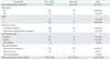
There were a total of 324 cases of MIC identified in the study. Patient characteristics are shown in Table 1. Patients with stage IA1 or IA2 diseases accounted for 86.4% (280/324) and 13.6% (44/324), respectively. The mean age at diagnosis was 42.1±8.5 years (range, 20 to 72 years). Thirty-nine women (12.0%) were nulliparous. The 91.4% of patients (296/324) underwent conization. Among them, 85.5% (253/296) underwent cold knife conization (CKC) and 14.5% (43/296) underwent loop electrosurgical excision procedure (LEEP) cone. Of 296 patients, 174 patients (58.8%) had negative margin, 113 patients (38.2%) had positive margin and in nine cases the status of margin could not be evaluated. The 91.7% of cases (297/324) was squamous cell cancer; 7.7% (25/324) was adenocarcinoma; and 0.6% (2/324) was clear cell cancer. The mean follow-up duration was 32.3 months (range, 0 to 128 months).
In patients with stage IA1 MIC, most of them (71.4%) underwent SH. Seventeen patients underwent therapeutic surgery without conization, including five cases of SH, one case of radical trachelectomy with PLND, 10 cases of RH with PLND and one case of MRH with PLND. Of the 263 patients undergoing conization, 222 cases underwent subsequent surgery within 3 months including: nine cases underwent radical trachelectomy with PLND, 195 cases underwent SH, five cases underwent MRH+PLND, and 13 cases underwent RH+PLND. Forty-one patients with stage IA1 MIC underwent conization alone. Among them, mean age at diagnosis was 42.5 years (range, 29 to 61 years) and 15 women were nulliparous. Thirty-eight patients underwent CKC and three patients underwent LEEP cone. The 87.8% of cases (36/41) was squamous cell cancer; 12.2% (5/41) was adenocarcinoma. There were 37 cases with negative cone margin and four cases with positive cone margin of CIN, in which two cases was diagnosed positive margin based on the closed margin of ≤1 mm. In patients with stage IA2 MIC, 11 patients underwent therapeutic surgery without conization, including three cases of SH and eight cases of RH with PLND. Of the 33 patients undergoing conization, 31 cases underwent subsequent surgery within 3 months including: one cases underwent radical trachelectomy with PLND, six cases underwent SH, one cases underwent MRH+PLND, and 23 cases underwent RH+PLND. Only two patients with stage IA2 MIC underwent conization alone, in which one patient underwent PLND in combination coniztion. For stage IA2 patients, most of them (70.5%) underwent RH+PLND.
Seventeen patients were treated by repeat conization, all of whom were stage IA1 MIC patients. Eleven patients chose to follow-up and six patients underwent SH. After reconization, three cases yet had positive margin and in the hysterectomy specimens, two cases had residual disease of CIN 3 and stage IA1 lesion, respectively. One patient had a residual disease of CIN 1 in spite of negative margin after reconization.
Only 25 cases (7.7%) were found to LVSI, 20 of whom were patients with stage IA1 MIC. Fourteen patients with LVSI underwent PLND, one in whom (7.1%) was found to had nodal metastasis. No parametrial involvement was noted in any of the 61 patients who underwent RH or MRH. No ovarian involvement was noted in any of the 81 bilateral salping-oophenrectomy specimens. In 73 patients who underwent PLND, we cleared away 599 lymph nodes in 39 patients of stage IA1 MIC and 748 lymph nodes in 34 patients of stage IA2 MIC. Only one staged IA2 patient with LVSI was found to have two lymph nodes metastasis in the right iliac nodes (2/13). The depth of tumor was 4 mm and the width of tumor was 7 mm. The patient opted not to receive any adjuvant therapy and was lost from follow-up 2 months later. Risk of LVSI in stage IA1 and IA2 MIC patients was 7.1% and 11.4%, respectively. Risk of node metastasis in stage IA1 and IA2 MIC patients was 0% and 2.9%, respectively. Although both risk of LVSI and lymph node metastasis in stage IA2 MIC were higher than stage IA1 MIC, no significant difference was found between them (p=0.36 and p=0.47, respectively).
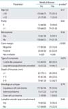
Of 324 MIC patients, 253 patients underwent subsequent surgery within 3 months after conization. Residual disease was observed in 82 cases (32.4%) including 22 cases of CIN 1, 43 cases of CIN 2-3, and 17 cases of MIC. Risk of residual disease in patients with stage IA1 and IA2 MIC was 31.5% (70/222) and 38.7% (12/31), respectively. Residual disease in postcone specimens with negative cone margin was found in 23 cases (16.4%), which included 10 cases of CIN 1 and 13 cases of CIN 2 to 3. In patients with positive margin of CIN, 17 cases (44.7%) had residual disease, including four cases of CIN 1, 10 cases of CIN 2 to 3, and three cases of MIC. In patients with positive margin of MIC, 38 cases (56.7%) had residual disease, including four cases of CIN 1, 10 cases of CIN 2 to 3, and three cases of MIC. No residual MIC lesion was found in patients with negative cone margin. Rate of residual disease in patients with negative cone margin or positive margin of CIN and MIC were 16.4%, 44.7%, and 56.7%, respectively. Negative-margin patients had significantly lower rates of residual disease than patients with positive cone margin of CIN (p<0.001) or MIC (p<0.001). However, there was no statistical difference between positive cone margin of CIN and MIC (p=0.24).
Predictors of residual disease in postcone specimen in univariate analysis are shown in Table 2. Univariate analysis showed only cone margin was significantly correlated with residual disease after conization (p<0.001), while parameters including age ≤50 years, parity, menopause, type of conization, depth of invasion, histological subtype, and LVSI status were not significantly correlated with residual disease after conization (p>0.05). We eventually selected four parameters including type of conization, age ≤50 years, parity and cone margin with p-value less than 0.5 into the multivariate logistic regression model. Multivariate logistic regression analysis showed positive cone margin was the only independent risk factor of residual disease after conization (odds ratio [OR], 4.18; 95% confidence interval [CI], 2.42 to 7.23; p<0.001).
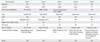
The mean follow-up duration was 32.3 months (range, 0 to 128 months). Forty-nine cases (15.1%) lost follow-up, including 37 cases with stage IA1. In 41 patients with stage IA1 disease who underwent conization alone, six patients underwent hysterectomy during follow-up (range, 5 to 14 months). Among them, four patients underwent hysterectomy due to fear of recurrence and had no disease in hysterectomy specimens. Two patients had abnormal cytology and in the subsequent hysterectomy specimen one patient had CIN 2-3 and the other case had no disease. The 2.1% of patients (5/243) with stage IA1 MIC relapsed. Clinical and pathological characteristics of patients who relapsed during follow-up were shown in Table 3. Three cases (60.0%) had LVSI. All recurrences happened in patients with depth of invasion ≥1 mm.The mean time between initial surgery and recurrence was 27.8 months (range, 12 to 59 months). No recurrence was found in FIGO stage IA2 MIC. There was no mortality in the study.
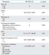
Ten-year PFS of stage IA1 MIC patients was 94%. There was no statistically significant difference in PFS between the stage IA1 MIC patients treated by conization and hysterectomy (92.3% and 98.8%, p=0.07). Table 4 demonstrated demographic and clinicopathological parameters related to predictors of recurrence in stage IA1 MIC patients by Cox regression analysis. LVSI was an independent predictor for recurrence in stage IA1 patients (OR, 12.14; 95% CI, 1.71 to 86.40; p=0.01). Age ≤50 years, parity, menopause, histological subtype, depth of invasion, and treatment modalities showed no significantly difference in recurrence of stage IA1 patients (p>0.05).
DISCUSSION
MIC is a special entity with excellent survival and prognosis. In our study 10-year PFS for stage IA1 patients was 94% and no recurrence was found in stage IA2 patients. There was no mortality in our study during 10-year follow-up time. Similar excellent outcome was also reported in recent literatures [4,5,6,7,8,9,10]. Further analysis revealed that excellent prognosis of patients with MIC may result from the extremely rare in parametrial involvement and lymph node metastasis. In our study no parametrial and ovarian involvement was found in patients with MIC which was similar as previous reports [6,11,12,13]. The risk of lymph node metastasis in patients with stage IA1 MIC ranged from 0% to 2.48% compared with 0.5% to 8.7% for the patients with stage IA2 MIC [4,9,10,20,23,24]. We found that no lymph node metastasis was detected in patients with stage IA1 MIC and only one staged IA2 patient with LVSI had lymph node metastasis. There was no significant difference in prevalence of positive lymph nodes and LVSI between stage IA1 and IA2 MIC patients. The rate of positive lymph nodes in patients with MIC was very low. However, Dedes et al. [24] reported that presence of LVSI was associated with an increased risk of lymph node involvement in patients with MIC, but some studies did not find relationship between LVSI and the lymph node status [4,21]. In a review of 1,565 patients with MIC [6], LVSI was observed in 25 of 458 cases (5.5%), but no one had positive lymph nodes, whereas positive lymph nodes were found in four of 433 cases (0.92%) without LVSI. Our result demonstrated that risk of lymph node metastasis in patients with LVSI was 7.1% (1/14) and there was no positive lymph node metastasis in patients without LVSI. Our study also demonstrated that LVSI was an independent predictor for recurrence in stage IA1 patients by multivariate analysis which was similar as Hou et al. [4] reported. Thus, special attention should be paid to the LVSI when we managed patients with MIC. PLND should be performed for evaluating the lymph node status if LVSI was present in patients with MIC, but the role of PLND in management of MIC should be further confirmed in the future.
In the present study, great majority (71.4%) of stage IA1 patients treated by SH, and most of stage IA2 patients (70.5%) underwent RH+PLND. The excellent prognosis of MIC made us heart-searching on possibility of overtreatment. Our results illustrated that patients with MIC were quite young with the mean age of 42 years at diagnosis and 12.0% of them were nulliparous. Quality of life and reproductive function are very important for those young women. For patients who desire to preserve fertility, conservative treatment such as conization has been studied [6,7,14,15,16]. Our results also demonstrated that PFS of stage IA1 MIC patients showed no statistically significant difference between conization and hysterectomy (p=0.07). Similar findings were also reported in previous studies [10,15]. A study of 3,987 women with MIC showed 5 year survival of stage IA1 was similar for conization and hysterectomy in both SCCs (95.1% and 95.6%) and adenocarcinomas (98.8% and 96.9%) [10]. Five year survival of stage IA2 was also similar for conization and hysterectomy for both SCCs (90.2% and 96.3%) and adenocarcinomas (97.8% and 98.2%) [10]. Besides, in our study no parametrial involvement was found in patients with stage IA2 MIC and there were no significant difference of LVSI, lymph node metastasis and residual disease after coniztion between stage IA1 and IA2 MIC patients. Thus, in order to avoid overtreatment in patients with MIC, correct diagnosis is essential and more conservative surgeries such as coniztion or SH may be feasible for them. However, residual disease after conization may eventually lead to persistent disease or relapse for patients treated by coniztion. Our study observed residual disease in 32.4% of cases (82/253). Compared with positive cone margin of CIN or MIC, patients with negative margin had significantly lower rates of residual disease. No residual MIC lesion was found in patients with negative cone margin. Positive cone margin has been reported by many researchers to be a predictor of residual disease [18,19,20]. In our study, positive cone margin was the only independent risk factor of residual disease after conization in multivariate analysis. Thus patients with positive cone margin should be treated subsequently. Repeat conization may be an option when the initial conization margin is positive, especially for patients desiring to preserve fertility.
Recurrence rate of stage IA1 MIC was very low ranging from 0 to 1.6 [4,5,6,7,8], compared with 0 to 5.6 for stage IA2 patients [4,5,6,8,9]. In our study, 2.1% of stage IA1 lesion recurred and no recurrence was found in stage IA2 patients. All recurrence occurred within 5 years (range, 12 to 59 months) after treatment. Late recurrences (>5 years after diagnosis) were observed in 6/28 cases (21.4%) in a study [4]. Long term follow-up should be considered for patients with MIC.
The limitation of this study is the retrospective study in nature, in which limited number of patients underwent conization and most of stage IA2 patients underwent aggressive surgery. Besides, we could not perform survival analysis due to the limited case numbers in stage IA2 MIC, which unabled us to analyze. We also had a smaller cohort of patients with microinvasive adenocarcinoma which limits the significance of our findings in these patients.
In conclusion, for MIC patients, treatment programs should be made based on a thorough histological evaluation of conization specimens including LVSI. Conization can be considered as an ideal treatment modality for stage IA1 patients with negative resection margin and no LVSI. Repeat conization may be an option for patients with positive cone margin. With no parametrial involvement and no significant difference of LVSI, lymph node metastasis and residual disease after coniztion between stage IA1 and IA2 MIC patients in our study, more conservative surgery such as SH may be considered for stage IA2 patients with or without LVSI. Long-term follow-up is essential for patients after treatment. Further studies such as prospective multicenter clinical trials are also needed to confirm the role of PLND in the treatment of patients with LVSI and the feasibility of conservative surgery in patients with stage IA2 MIC.
 XML Download