

 PDF
PDF ePub
ePub Citation
 Print
Print


INTRODUCTION
Cervical cancer is the second most common form of cancer among females worldwide [1] and the most common form of gynecological cancer in some developing countries [2] because of lack of screening programs. The Papanicolaou test (called Pap test) is a screening test for the detection of potentially precancerous and cancerous processes in the endocervical canal of the female reproductive system. The most frequent abnormal results of Pap test comprises atypical squamous cell of undetermined significance (ASC-US, typically 2% to 5%) and low-grade squamous intraepithelial lesion (LSIL, about 2%) [3], which indicates infection with human papillomavirus (HPV). Persistent infection with high-risk HPV (HR-HPV) is the main cause of cervical cancer and its precursor lesion, cervical intraepithelial neoplasia (CIN) [4,5]. Patients with positive findings for ASC-US or LSIL are frequently asked to undergo HPV screening [6]. Thus, carcinogenic HPV testing has been widely employed for the screening of primary cervical cancer in women aged 30 years and above [7,8,9].
Several studies have shown that testing for HPV DNA has high sensitivity but low specificity [9,10,11]. It is, therefore, a challenge to discriminate between transient and persistent infections, necessitating the requirement of a test with improved specificity for the detection of the presence of HPV. HPV has a small, double-stranded, circular DNA that encodes 8 genes. Some of the genes such as E6 and E7 are known to act as oncogenes that promote tumor growth and malignant transformation [12]. E6 and E7 oncogenes are overexpressed in the malignant phenotype. Moreover, the oncogenic potential of HPV infection is associated with the expression of E6 and E7 oncoproteins. The presence of HR-HPV DNA does not always lead to the disease, but the detection of this transient infection invariably points to an increased risk for disease development in women. Recent studies [13,14] have demonstrated that the detection of E6/E7 mRNAs may facilitate the screening for malignant lesions. Also, the specificity of detecting high-grade lesions is higher by using HPV mRNA-based assay than by using HPV DNA-based assay. The APTIMA HPV (AHPV) assay has been used for the detection of HPV E6/E7 mRNA in all 14 HR-HPV types and shows greater accuracy than HPV DNA-based tests in triage settings [14,15].
To the best of our knowledge, the present study is the first to compare AHPV with HC2 in a referral population in China. The performance of both assays was evaluated in the whole cohort, which was further stratified according to cytology.
MATERIALS AND METHODS
1. Patients and specimen collection
This study was performed at the Third Hospital of Peking University from February 5, 2013, to August 27, 2013. The referred women were eligible if they were not pregnant and had no history of CIN diagnosis and hysterectomy. Before referral, all women underwent liquid-based cytology test, as recommended by gynecologists, by using smears that were stored at the Third Hospital of Peking University. All women signed a written consent before sample collection. The procedures were approved by the Ethics Committee for Medical Science Research at Third Hospital of Peking University. The results obtained for 600 cases were reviewed and classified according to the 2001 Bethesda system. If the result was normal, ASC could not rule out high-grade SIL, high-grade SIL, atypical glandular cells, or cancer, and the data were excluded. A total of 411 women (median age, 34 years; range, 21 to 69 years) were enrolled, of which 231 women had ASC-US and 180 women had LSIL. Upon enrollment, a single cervical specimen was collected from all participants by using a Cervex broom-type brush (Rovers Medical Devices, Oss, Netherlands), according to the manufacturer's instructions. The median interval time from the initial cytological diagnosis and enrollment to the colposcopy referral visit was 27 days (from 5 to 90 days).
2. Colposcopy and histology
Colposcopy was performed by two experienced gynecologists. Colposcopically directed punch biopsy was performed on the day of patient enrollment, according to the standard of care. Cervical histology was read and reviewed independently by experienced pathologists; the result was used as the disease endpoint for the purpose of the study. The pathologists were blinded to HPV results.
3. APTIMA HPV assay
AHPV assay (Gen-Probe Inc., San Diego, CA, USA) was designed to detect HPV E6/E7 mRNA from liquid cervical specimens of 14 high-risk oncogenic types (HPV-16, -18, -31, -33, -35, -39, -45, -51, -52, -56, -58, -59, -66, and -68). The procedure was carried out according to the manufacturer's instructions. Briefly, a 1-mL aliquot of each of the PreservCyt sample was transferred to 2.9 mL of buffered detergent solution. A 400-µL aliquot of this mix was then tested on a semiautomated Direct Tube Sampling system (Gen-Probe). Assay results were determined on the basis of the signal-to-cutoff ratio (S/CO) for the analyte. Specimens with an S/CO value of ≥1.0 were considered positive.
4. HC2 assay
HC2 (Qiagen, Mississauga, ON, Canada) assay was used to detect the DNA of 13 high-risk oncogenic types (HPV-16, -18, -31, -33, -35, -39, -45, -51, -52, -56, -58, -59, and -68). RNA-DNA hybrids were prepared using a RNA probe cocktail and the target DNA. The hybrids were then captured by specific antibodies and detected by chemiluminescence substrate. This test was performed with 4 mL of PreservCyt sample in conversion buffer as per the manufacturer's instructions. Specimens with a relative light unit/cutoff ratio of ≥1.0 were considered positive.
5. Statistical analysis
The clinical performance of the HPV test was established by comparing the test result against the disease endpoint founded on histologically confirmed CIN 2 or greater (CIN 2+) and CIN 3 or greater (CIN 3+) samples. Sensitivity and specificity were calculated using the conventional contingency tables. The 95% confidence intervals (CIs) were computed using the binomial method. McNemar's chi-square test was used to test for the difference between their sensitivities and specificities, as they were both calculated using the same set of samples. The accuracy of AHPV and HC2 assays was calculated as the percentage of correct results from one HPV test in comparison to the results of histological analysis. A significance level of 0.05 was used to compare performance characteristics. No adjustments were made for multiple comparisons. All statistical analyses were carried out using the SPSS ver. 13.0 (SPSS Inc., Chicago, IL, USA).
RESULTS
1. Prevalence of cervical disease
A total of 411 subjects were tested using histology, cytology, AHPV, and HC2 techniques, and the results were analyzed. Histology results revealed that 339 participants were normal or were classified as CIN 1 (<CIN 2), while 55 and 17 subjects were classified as CIN 2 and CIN 3, respectively. Thus, the prevalence of cervical disease (CIN 2+) in the referred population was 17.5% (95% CI, 13.8 to 21.2).
2. Detection of cervical disease by AHPV assay, HC2 test, and cytology
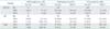
The performance of AHPV and HC2 assays were determined and compared with the histology results. The results are presented in Table 1. For AHPV assay, the positive results increased with the severity of histological abnormality (from normal to CIN 3). The positive detection rate of AHPV assay was 67.5% (95% CI, 61.5 to 73.6) in ASC-US specimens and 75% (95% CI, 68.0 to 81.1) in LSIL specimens, which was significantly lower than that of HC2 test (95.2% in ASC-US and 94.4% in LSIL, p<0.001). In all 411 participants, the positive detection rate of AHPV assay (70.8%) was significantly lower than that of HC2 test (94.9%; p<0.001).
3. Sensitivity and specificity of AHPV assay and HC2 test in the screening of cervical disease
According to the results of histological and cytological analyses, 30 specimens of the 231 ASC-US subjects (13.0%) were identified as CIN 2+ and 42 specimens of the 180 LSIL subjects (23.3%) were classified as CIN 2+. Based on a total of 411 cases, the sensitivity of AHPV assay for the detection of CIN 2+ and CIN 3 was 87.5% and 100%, respectively, while the sensitivity of HC2 test was 100% for both CIN 2+ and CIN 3 subjects (Table 2). The specificity of AHPV assay for the detection of CIN 2+ and CIN 3 specimens was 32.7% and 30.2%, respectively, while the specificity of HC2 test was 6.2% for CIN 2+ and 5.3% for CIN 3. Thus, AHPV assay has a significantly higher specificity than the HC2 test (p<0.001).
4. False-negative cases by AHPV assay
In the referred cohort of 120 AHPV-negative cases, only nine CIN 2+ cases were detected (one CIN 3 and eight CIN 2) (Table 3). In case of HC2 test, all 55 CIN 3 and 17 CIN 2 cases were HC2-positive and no false negative case was observed.
DISCUSSION
The present study, to the best of our knowledge, is the first clinical experiment in a referred cohort to evaluate the performance of the AHPV assay for cervical cancer screening in China. We evaluated the clinical performance of the assays in 411 subjects with ASC-US/LSIL. This study demonstrated that both assays had high sensitivity (>86%) for the detection of high-grade cervical lesions (CIN 2+). HC2 assay identified more CIN 2 cases than AHPV assay, which is consistent with other studies [15,16,17].
Few CIN 2 cases spontaneously regress and do not progress into CIN 3 or cancer [18,19,20] but the CIN 3 patients develop invasive cervical cancer if left untreated [21]. Timely detection of CIN 3 is thus crucial for treatment. This study was conducted to evaluate the clinical performance of AHPV assay in screening CIN 3 specimens. It was concluded that AHPV has good sensitivity and specificity in screening of CIN 3 cases. In addition, the possibility of the presence of CIN 3 in AHPV-negative specimens was as less as 0.8% (Table 3). These data indicate that AHPV is a reliable assay for screening precancerous and cancerous cervical specimens.
HC2 assay is the first FDA-approved and widely used procedure for HPV testing based on quantitative detection of L1 in 13 HR-HPV types [22]. The HC2 test has good sensitivity, but it cross-reacts with some low-risk HPV types [22,23], resulting in relatively low clinical specificity for high-grade cervical lesions [7,24,25,26]. Hence, an improvement in specificity is desired to reduce the referral rate of colposcopy.
The expression of viral oncogene E6 and E7 mRNA in HPV-transformed basal keratinocytes is essential for the development and progression of cervical cancer. These genes are mildly expressed during transient infections [5,27]. Unearthing biomarkers may assist the generation of a more specific assay for the detection of viral DNA that can accelerate the progression of abnormalities to cervical cancer [28,29].
AHPV assay is a procedure targeting E6/E7 mRNA of 14 HR-HPV types. It does not cross-react with the low-risk HPV types [30]. Some previous studies [26,31] have demonstrated that AHPV has good sensitivity along with significantly higher specificity for the detection of CIN 2+ and CIN 3+ as compared to HC2 test; our data verified this piece of information.
In this study, fewer specimens were tested positive by AHPV assay than that by HC2 test, especially in cases with normal or CIN 1 histology. This finding can be explained by the fact that the HC2 test is very sensitive to the presence of HPV DNA, and consequently, it cannot be used as a factor for evaluating the risk of cervical precancerous cells progressing to cervical cancer. The AHPV assay, on the other hand, is expected to detect fewer CIN 2 cases than HC2 assay, because some CIN 2 lesions are more likely to be transient and spontaneously regress. Low sensitivity of AHPV assay in detecting CIN 2 can be attributed to regional factor or race factor. These results indicate that the application of AHPV assay in China has good potential to further triage ASC-US/LSIL specimens and can obviously reduce a patient's burden and health care-related cost due to unnecessary colposcopy.
This study has several advantages. First, the data were obtained from a well-designed clinical experiment. Second, the biopsy samples were used as the gold standard. Biopsies were taken from the women referred to colposcopy. Colposcopic impression was also normal. However, there are few limitations of this study. First, it is a cross-sectional study and not a prospective, randomized controlled trial. Second, the study did not include HPV genotyping data, which might partly explain the difference in clinical specificity between AHPV and HC2. Third, it should be noted that women in this cohort were referred for colposcopy because of a previously abnormal Pap test. Some HC2-negative cases had been excluded. Fourth, the number of CIN 2/3 specimens was not large; therefore, further evaluation is necessary to establish the clinical performance of AHPV assay in a larger population size. Moreover, a CIN 3 specimen was misdiagnosed by AHPV assay; therefore, the procedure used for AHPV should be improved and additional methods for screening cervical disease should be discovered for future studies.
Results from this referral study show that the AHPV assay offers high sensitivity for high-grade disease and has a significantly higher specificity compared to the HC2 test. The greater performance of the AHPV assay provides supporting data for its application in cervical cancer screening as an ASC-US triage test, which may improve patient management and reduce health care costs. Further work is, however, essential to confirm these findings in a primary cervical disease screening setting.


 XML Download