

 PDF
PDF ePub
ePub Citation
 Print
Print


INTRODUCTION
In 2011 an estimated 529,800 cases of cervical cancer were diagnosed worldwide, with 275,100 deaths [1]. This global burden is attributable to the disproportionately high incidence of cervical cancer in developing, resource poor countries lacking adequate health care infrastructure and screening programs. Regionally, in the United States, an estimated 12,360 cases will be diagnosed in 2014, with 4,020 deaths; it is anticipated that this number will continue to decline as human papilloma virus (HPV) vaccination rates rise, and the focus shifts to primary prevention [2]. Despite advances in screening, vaccination and treatment of early stage disease, a proportion of patients will be diagnosed with advanced stage (stage IVB), recurrent or persistent cervical cancer. Specifically, patients with International Federation of Obstetrics and Gynecology (FIGO) stage IB-IIA disease have a recurrence risk ranging from 10% to 20% despite primary chemo-radiation, while those patients diagnosed FIGO stage IIB-IVA have a 50% to 70% chance of disease recurrence [3]. For this subset of patients, systemic chemotherapy remains the standard treatment.
In the setting of advanced stage or recurrent disease cure is exceedingly rare, and goals of care are centered on palliation of symptoms, and control of disease burden [4]. Since the publication of the initial studies exploring single agent cisplatin in the treatment of cervical cancer, a number of effective drugs have been identified, including paclitaxel, ifosfamide and topotecan, although none have exhibited significant gains with respect to overall survival (OS) [5,6,7,8,9,10,11,12,13,14,15,16]. Ultimately, various combination-based regimens were investigated, with modest gains in response rate, which analogously failed to translate into a meaningful OS advantage [17,18,19,20,21]. Importantly, the combination regimen of cisplatin and paclitaxel has been established as the backbone for future chemotherapy trials, although OS approaches only 13 months [5]. Additionally, responses to this regimen are uniformly temporary, and effective second and third line therapeutic regimens are lacking.
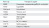
The poor oncologic outcome in this patient population represents and unmet clinical need, and catalyzed the exploration of novel treatment paradigms. Most recently, the results of Gynecologic Oncology Group (GOG) protocol 240 were presented and published, illustrating a 3.7 months improvement in OS with the incorporation of the antiangiogenic agent bevacizumab to a chemotherapy backbone [22]. In an era of molecular medicine, the development of additional biologic therapies, to be used solely or in conjunction with cytotoxic chemotherapy, is implicit. Currently, a number of biologic agents targeting various molecular pathways including epidermal growth factor receptor (EGFR), histone deacetylase, matrix metalloproteinase, check point inhibitors, Notch pathway, and mammalian target of rapamycin (mTOR) are under clinical development (Table 1). This article will explore molecularly targeted drugs developed for the treatment of cervical cancer.
NOVEL NONANGIOGENIC TARGETED THERAPIES IN CERVICAL CANCER
1. Targeting EGFR and Her2
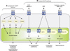
The EGFR is a trans membrane protein involved in signaling pathways critical for cell survival (Fig. 1) [23]. EGFR overexpression has been shown to correlate with resistance to cytotoxic therapy and radiation in squamous cell cancers [24,25,26,27]. Additionally, the majority of cervical cancer patients (54% to 71%) exhibit EGFR expression, with correlative studies associating increased expression with prognosis and tumor aggressiveness [28]. More importantly, the addition of cetuximab to radiotherapy in the treatment of head and neck squamous cancers resulted in a statistically significant prolongation in OS and locoregional control at 2 years, providing the biologic rational for study in cervical cancer [29].
To date 2 anti-EGFR antibodies have been developed and studied in cervical cancer: cetuximab and matuzumab [28]. Cetuximab, is a chimeric immunoglobulin G2 monoclonal antibody that targets the extracellular domain of the EGFR. Alternatively, matuzumab is a humanized immunoglobulin G1 monoclonal anti-EGFR antibody.
A total of four studies were completed, reporting on the safety an efficacy of cetuximab in the treatment of cervical cancer. Two studies used single agent cetuximab [30,31], while one combined the monoclonal antibody with cisplatin [32], and the final study combined cetuximab with cisplatin and topotecan [33].
Unfortunately, response rates were less than anticipated during trial design. When used as a single agent, cetuximab failed to result in a clinical response [30]. The median progression free survival (PFS) and OS were 1.9 and 6.7 months, respectively. Combining cetuximab with cisplatin did not translate into improved outcomes. Farley et al. [32] investigated cetuximab at a loading dose of 400 mg/m2 followed by 250 mg/m2 on days 1, 8, and 15 every 21 days in combination with cisplatin 30 mg/m2 on day 1 and 8. Sixty-nine patients with advanced, persistent or recurrent cervical cancer were eligible and evaluable, and the clinical objective response rate was 11%. The authors concluded that there was little or no evidence that cetuximab was beneficial in this patient population beyond single agent cisplatin.
Lastly, a 3-drug combination of cetuximab, cisplatin, and topotecan was studied in patients with advanced cervical cancer not amenable to curative treatment [33]. This study was terminated early, after only 19 subjects enrolled, due to unacceptable toxicity, with three treatment related deaths.
Matuzumab has also been studied in patients with cervical cancer progressing after treatment with platinum-based chemotherapy, with data presented in abstract form at the 2005 ASCO Annual Meeting. Amongst 38 evaluable patients, there were two partial responses and nine patients with stable disease. Reported grade 3/4 AEs included hepatotoxicity, diarrhea, fainting, anorexia, fatigue, abdominal pain, and pancreatitis (1 of each) [28].
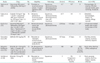
Analogous to receptor tyrosine kinase inhibition in the angiogenic cascade, investigators explored anti-EGFR tyrosine-kinase inhibitors (TKIs) in the treatment of cervical cancer (Table 2) [30,32,33,34,35,36]. EGFR signal transduction is dependent on receptor auto-phosphorylation followed by signal transduction. Inhibition via small molecule TKIs results in inhibition of phosphorylation and interruption of signal transduction.
Three trials have reported outcomes in patients with cervical cancer treated using anti-EGFR TKI's. Two, phase 2 trials investigated the use of oral, single agent gefitinib and erlotinib in patients with recurrent, persistent or metastatic cervical cancer, receiving at least one prior cytotoxic chemotherapy regimen [34,36]. In both studies, there was a lack of an observed objective response, signaling that these small molecule TKIs were unlikely to exhibit clinical activity as single agents in the advanced/recurrent setting. An additional phase 1 study, explored the combination of erlotinib orally with concurrent cisplatin+external beam radiation and brachytherapy. A total of 15 patients with clinical stage 2B-3B squamous cell carcinoma of the cervix were enrolled on trial. The authors reported a 91.7% complete response rate and an 8.3% partial response rate. No data on long-term outcome was provided. The most commonly reported grade 3/4 AEs associated with this class of targeted agents include: rash, nausea, anorexia, diarrhea, anemia, fatigue, and infection.
The only published study to date exploring Her2/neu inhibition in the treatment of cervical cancer was previously reviewed [37] (Table 2). Amongst the human EGFR (HER) family, HER2 is unique in that it exists in a constitutively active form. Dimerization results in receptor activation, phosphorylation and down stream signaling with oncogenic gene transcription. Prior studies indicated an association between increased Her2 expression and improved prognosis in cervical cancer patients [38]. Unfortunately, lapatinib was found to have only modest activity, with a 5% objective response rate, and was associated with an up to 13% rate of grade 3/4 AEs.
2. Antifolate agents
More recently, a novel chemotherapeutic combination regimen of pemetrexed (an anti-folate, which disrupts folate-dependent metabolic processes) and cisplatin was investigated in patients with cervical carcinoma. Initial phase I trials of single agent pemetrexed completed in South Africa indicated a 21% response rate in chemotherapy naïve cervical cancer patients. Recently, GOG 127T, a phase II trial evaluating pemetrexed in the treatment of recurrent cervical carcinoma in patients who failed prior chemotherapy, completed accrual. It was well tolerated and demonstrated a 15% response rate.
Given the above, protocol 77GG was developed, exploring the combination regimen of cisplatin 50 mg/m2+pemetrexed 500 mg/m2 in patients with recurrent, metastatic cervical cancer. The results were presented at the ASCO Annual Meeting in June 2013. Patients had received no prior therapeutic chemotherapy aside from concurrent with primary radiation therapy. From September 2008 to November 2011, five GOG member institutions enrolled 55 patients. Fourteen of the enrolled subjects (29%) received greater than nine cycles. The most common grade two toxicities included neutropenia (35%), leukopenia (28%), and metabolic (28%). There were one complete and 16 partial responses (RR 31%). Median response duration was 7 months and survival 12 months.
3. Histone deacetylase inhibitors in cervical cancer
Over the last several years significant interest in epigenetic modifications resulting in alteration of tumor suppressor gene expression has catalyzed the investigation of novel treatment strategies using histone deacetylase inhibitors (HDACIs). In its transcriptionally silent state, chromatin is composed of nucleosomes in which the histones have low level acetylation [39]. Acetylation of histone proteins subsequently results in relative charge neutrality, allowing for DNA unfolding and transcriptional access. HDACs remove the acetyl groups, returning an overall positive charge, which is thought to inhibit transcription of tumor suppressor genes in various malignancies [39,40,41].
The most extensively studied HDACI in cervical carcinoma is a drug commonly used in the treatment of epilepsy, bipolar disorder, and major depression, valproic acid. Valproic acid acts as a specific inhibitor of class I HDACs and induces proteosomal degradation of HDAC2, leading to cell differentiation, growth arrest and death in vitro and in vivo [42]. To date, four studies have reported on the effects of HDACI on oncologic outcome in patients with cervical cancer.
In the primary setting, Chavez-Blanco et al. [43] conducted a phase I study exploring the impact of magnesium valproate use on histone acetylation in 12 patients with stage 2B to 4B cervical carcinoma. All subjects were treated with magnesium valproate after a baseline tumor biopsy and blood sampling at the following dose levels (four patients each): 20, 30, or 40 mg/kg for 5 days via oral route. At day 6, tumor and blood sampling were repeated and the study protocol ended. Tumor acetylation of H3 and H4 histones and HDAC activity were evaluated by Western blot and colorimetric HDAC assay respectively. Blood levels of valproic acid were determined at day 6 once the steady state was reached.
Ten patients were evaluated for H3 and H4 acetylation and HDAC activity. After treatment, investigators observed hyper-acetylation of H3 and H4 in the tumors of nine and seven patients, respectively, whereas 6 patients demonstrated hyperacetylation of both histones. Serum levels of valproic acid ranged from 73.6 to 170.49 mg/mL. Tumor deacetylase activity decreased in eight patients (80%), whereas two had either no change or a mild increase. There was a statistically significant difference between pre- and posttreatment values of HDAC activity (mean, 0.36 vs. 0.21; two-tailed t-test p<0.0264). There was no correlation between H3 and H4 tumor hyperacetylation with serum levels of valproic acid. The authors concluded that magnesium valproate at a dose between 20 and 40 mg/kg inhibited deacetylase activity and hyperacetylated histones in tumor tissues.
The combined use of hydralazine, a DNA methyltransferase inhibitor, and valproic acid has also been studied in a double-blind randomized phase 3 trial [44]. DNA demethylation results in reactivation and expression of tumor suppressor genes, which was hypothesized to synergize with HDAC inhibition. Patients received hydralazine at 182 mg for rapid, or 83 mg for slow acetylators, and valproate at 30 mg/kg, beginning a week before chemotherapy and continuing until disease progression. A total of 36 patients were enrolled, 17 treated with hydralazine and valproic acid (HV) and 19 with placebo (PLA), both groups receiving combination topotecan and cisplatin. The median number of cycles was 6. There were four partial responses in the HV arm, and one in the PLA arm. At a median follow-up time of 7 months, the median PFS was 6 months for the PLA arm and 10 months for the HV arm (p=0.0384, two tailed). Molecular correlates with response and survival from this trial are yet to be analyzed.
The same combination was assessed in the up front setting in patients with stage 3B squamous and adenosquamous cervical cancer [45]. A total of 22 patients received weekly cisplatin 40 mg/m2 + pelvic radiation, in combination with hydralazine 30 mg/kg administered three times daily until completion of intracavitary radiation therapy. The reported response rate was 100%, although delay in brachytherapy administration precluded assessment of the impact of epigenetic
therapy.
4. mTOR in cervical cancer
mTOR plays an integral role in angiogenesis, cell growth, proliferation, and survival. Activation of the phosphoinositide 3-kinase (PI3K)/Akt/mTOR pathway begins with growth factor receptor tyrosine kinase ligand binding, resulting in activation of PI3K. The primary role of activated PI3K is to convert phosphatidylinositol-4,5-bis-phosphate to phosphatidylinositol-3,4,5-triphosphate (PIP3) [46]. Accumulation of PIP3 at the cell surface then results in phosphorylation and activation of Akt, a protein serine-threonine kinase. In the absence of PTEN inhibition, Akt phosphorylates and inhibits the tuberous sclerosis complex (TSC), leading to mTOR activation. Activated mTOR subsequently forms 2 different multiprotein complexes, mTOR complex 1 and mTOR complex 2, associated with the regulatory associated protein of mTOR (raptor) [47]. Ultimately, phosphorylation and activation of 2 separate down stream signaling molecules, eukaryotic translation initiation factor 4E binding protein and ribosomal protein S6 kinase 1, promotes the translation of proteins involved in cell growth, angiogenesis, proliferation and survival [46,48,49,50].
High risk HPV related E6 has been shown to cause the rapid degradation of TSC2, resulting in TORC1 activation and downstream mTOR signaling. Additionally, HeLa cells are defective in the tumor suppressor LKB1, which inhibits mTOR via TSC2 stimulation.
The sole mTOR inhibitor studied in the treatment of cervical cancer is temsirolimus. In a combined phase 1 study, two patients with advanced stage and recurrent squamous cell cervical cancer were included amongst 15 total subjects. One patient had stable disease, and the median time to progression was 3 months. Unfortunately, the combination regimen of topotecan+temsirolimus was not tolerated in patients with a history of pelvic radiation therapy.
5. WEE1 inhibition and mitotic catastrophe
In normal, nonmalignant cells, cell cycle progression is a carefully orchestrated process, with emphasis on mutation prevention. Normally, several checkpoints ensure genomic integrity, including G1-S transition, S-phase, and the G2-M transition [51]. Normal cells commonly repair DNA damage during the G1-arrest, with cancer cells commonly exhibiting a deficient G1-arrest, relying more on G2-arrest to facilitate DNA repair. This process is carefully regulated by a series of cyclin dependent kinases, including the protein kinase WEE1.
In normal cells, WEE1 phosphorylates, and inactivates cyclin dependent kinase CDK1 preventing premature entry into mitosis while DNA integrity is ensured. Thus, WEE1 functions as a mitotic inhibitor, regulating the G2-M transition. WEE1 has been shown to be overexpressed in various cancer types, including cervical cancer. The scientific rational for WEE1 targeting is based on data indicated a deficient G1-S checkpoint, resulting in increased DNA damage at the G2-M transition in cancer cells. Thus, abrogation of the G2-M arrest releases cells with unrecognized and unrepaired DNA damage into premature mitosis, resulting in mitotic catastrophe and apoptotic cell death [52]. Combination treatment with DNA damaging cytotixic agents and WEE1 inhibition, is therefore hypothesized to prime these cells for apoptosis.
Preclinical studies using cancer cell lines and animal models identified WEE1 as a potential therapeutic targerget using RNA interference screening. Preclinical in vitro studies illustrated inhibition of cervical cancer cell outgrowth, using the WEE1 inhibitor, PD0166285 [52]. Additionally, knockdown of WEE1 in cervical cancer cells, but not in normal human epithelial cells, in combination with adriamycin, induced apoptosis [53]. These findings were supported by in vivo experiments, in which combination carboplatin and MK1775 (WEE1 inhibitor) resulted in tumor growth reduction for HeLa-luc xenograft animal models.
The above findings led to the development of a phase 1/2 clinical trial (NCT01076400) evaluating MK1775 in combination with topotecan and cisplatin in patients with advanced stage, metastatic and recurrent cervical cancer.
6. Notch signaling, cell fate, and cervical cancer
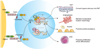
Notch genes encode transmembrane receptors that are highly conserved and are involved in cell fate decision (Fig. 2) [54,55]. Signaling via this pathway is dependent on direct contact between adjacent cells expressing the Notch receptors and ligands. Ultimately, down stream signaling results in regulation of cell fate specification, differentiation, proliferation and survival [56,57,58,59]. Notch receptors and their ligands have been shown to be overexpressed in various malignancies, including cervical cancer. The earliest studies using Notch specific antibodies, detected greater Notch gene product expression in cervical cancer specimens relative to high-grade dysplasia [60,61].
Importantly, the basal epithelial cells normally found at the ectocervical-endocervical junction are prone to metaplastic transformation, and strongly stain for Notch1, Notch2 and their ligand jagged. Specifically, squamous and columnar epithelium normally cover the ectocervix and endocervical canal, with a reserve cell population at the squamocolumnar junction. This population of reserve cells (also known as basal cells) can differentiate into squamous or columnar epithelium. HPV infection of this cell population (at the transformation zone) is thought to result in premalignant and malignant transformation, and may be related to aberrations in Notch signaling and cell fate decisions.
The relationship between cervical cancer and Notch, however, is complex, with in vitro assays indicating a requirement for both E6 and E7 oncogene expression prior to soft agar colony formation in an immortalized keratinocyte cell line [60,61]. Ultimately, regulated Notch signaling was found to be associated with cervical cancer progression, although later experimental studies indicated that Notch overexpression, beyond an undetermined threshold, results in apoptotic cell death and counteracts HPV-induced cellular transformation. A resolution of these apparent contradictory observations may emerge as we gain insight into the cancer stem cell evolutionary paradigm [62].
There are currently several clinical trials in various stages of accrual investigating the clinical utility of Notch inhibition in patients with metastatic caricnoma. An industry sponsored prospective phase 1, dose escalation, study was conducted in patients with metastatic recurrent carcinoma, using a Notch inhibitor (NCT01158404). The study is closed to accrual, with results pending.
7. Heat shock protein 90
Antiangiogenic agents have shown promise in the treatment of cervical cancer, heralded with the presentation of GOG 240 [22]. Unfortunately however, acquired resistance continues to be a clinical dilemma, as a cancer cells utilize redundant pathways to allow for continued growth and proliferation.
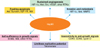
In an effort to target several proangiogenic pathways simultaneously, researchers have explored interruption of ubiquitous heat shock protein 90 (HSP90) activity. Mechanistically, HSP90 is required for proper protein folding (for its substrate molecules), and simultaneously functions as a scaffold protein, facilitating interactions between receptor tyrosine kinases and their downstream substrates. Thus, inhibition of HSP90 allows for interruption of several 'cancer signaling pathways' simultaneously, although in a nonspecific manner (Fig. 3) [63,64,65]. One of the most prominent pro-angiogenic pathways up-regulated in patients with cancer is the hypoxia inducible factor (HIF)/vascular endothelial growth factor (VEGF) cascade. In a hypoxic environment, there is up-regulation of HIF, with subsequent activity, resulting in VEGF activation, and angiogenic signaling. Importantly, several key mediators of this pathway including HIF, VEGF receptor, tumor growth factor alpha (TGF-α), and EGFR are dependent on HSP90 function. Given this interdependence, inhibition of HSP90 is hypothesized to result in impaired signaling along these parallel pathways, suppressing tumor angiogenesis [66].
In patients with cervical cancer, correlative clinical studies conducted as early as 1986, indicated an association between increased HSP expression and carcinogenesis, tumor size and cellular proliferation [64]. Additionally, HSP70 expression was associated with poor outcomes in patients with cervical cancer [67].
Bisht et al. [68] investigated the impact of the HSP90 inhibitor, Geldanamycin, on two cervical cancer cell lines (HeLa and SiHa) in conjunction with radiation. HSP90 inhibition resulted in significant tumor cytotoxicity and radiosensitization, suggesting a potential therapeutic utility. Additionally, VEGF expression was down regulated in cells treated with the geldanamycin analog, 17-allylamino-17-demethoxy (17-AAG). There are no current active clinical trials investigating the use of HSP90 inhibition in patients with cervical cancer.
8. PARP inhibition in the treatment of cervical cancer
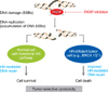
Poly ADP ribose polymerase (PARP) catalyzes the poly ADP-ribosylation of proteins involved in DNA repair [69]. Currently, this class of antineoplastic therapy is being extensively studied in patients with epithelial ovarian cancer, with an emphasis on patients with germline breast cancer susceptibility gene (BRCA) mutations. Deficiency in homologous recombination (HR) results in enhanced cell death with exposure to PARP inhibitors. Biologically, PARP inhibitors result in the creation of large numbers of single stand breaks in DNA, that when left unrepaired, lead to double strand breaks (DSBs) at replication forks (Fig. 4) [69,70]. In vitro assays indicated that cells deficient in HR (BRCA mutants) are unable to maintain genomic integrity in the contexts of large numbers of DNA DSBs, and are sensitive to PARP inhibition.
Importantly, it was discovered that the activity of PARP inhibitors is not limited to BRCA mutation carriers, indicating that PARP inhibition may result in synthetic lethality when alternate DNA repair genes are deficient. This finding subsequently opened the door to the investigation of PARP inhibitors in cancers with deficiencies in HR, aside from known BRCA mutants.
The use of PARP inhibitors in cervical cancer is in its infancy. In a study of HeLa cell lines resistant to cisplatin with constitutively hyperactivated PARP1, Michels et al. [71] were able to illustrate the cytotoxic effects of PARP inhibition. Interestingly, these investigators were also able to show that elevated levels of poly ADP-ribose (PAR) predicted response to PARP inhibition in vitro and in vivo more than PARP1 expression itself [69]. A phase 1 trial is presently enrolling patients with cervical cancer along with other gynecologic malignancies to investigate the combination of olaparib with carboplatin in refractory or recurrent disease (NCT01237067). Another phase 1/2 trial is investigating the use of veliparib with cisplatin and paclitaxel in advanced, persistent, or recurrent cervical cancer (NCT01281852). The results of the above studies are anxiously anticipated as we try and identify alternate therapeutic options in a population of patients where few effective therapies exist.
CONCLUSIONS
In summary, several nonangiogenic molecular pathways have been identified as potential therapeutic targets in the treatment of cervical cancer. To date, none has been studied in a prospective phase 3 trial, although promising phase 2 data is emerging. Despite the above, several questions remain including long-term toxicity with use of the above agents, identifying the appropriate regimen (i.e., in combination with cytotoxic agents, as single agents or in the maintenance setting), using translational end points to help predict response, and ultimately understanding acquired resistance. It is evident that the field of molecular therapeutics is in its infancy within the cervical cancer domain, and we anxiously await the growth of this field and the identification of effective therapeutic agents.
 XML Download