

 PDF
PDF ePub
ePub Citation
 Print
Print


INTRODUCTION
Radiotherapy and concurrent cisplatin-based chemosensitization has become the standard treatment for locally advanced cervical cancer. Previous randomized studies [1-3] showed that a combined approach, including cisplatin with and without 5-fluorouracil (FU) chemotherapy and radiotherapy, improved survival, progression-free survival and recurrence rates in patients with locally advanced cervical cancer. The 5-year disease-free survival is diminished because cervical cancer often metastasizes in an orderly fashion, initially involving the low pelvis and progressing to high pelvic lymph nodes and para-aortic nodes (PAN) and finally to the supraclavicular lymph nodes [4]. It has long been recognized that many patients with locally advanced cervical carcinoma harbor occult para-aortic metastases [5]. Berman et al. [5] showed a clear correlation between the incidence of nodal involvement and advancing tumor stage. They evaluated 621 patients and found PAN disease in 5% of stage I, 16% of stage II, and 25% of stage III patients [5].
Once cervical cancer has metastasized to the PAN, the patients have a poor prognosis [6]. It has been suggested that patients undergo extended field radiation therapy (EFRT), including the para-aortic region, with both pelvic masses and metastatic PANs [5,7,8]. A prospective and randomized study reported that para-aortic irradiation improved the overall survival and reduced distant metastases without concurrent chemotherapy [9]. However, there is inherent toxicity in the treatment of the pelvis and the para-aortic nodal regions. The Radiation Therapy Oncology Group (RTOG) 7920 reported an 8% risk of grade 4 to 5 toxicity with extended field treatment compared with 4% in the pelvic-only arm, which approached statistical significance (p=0.06) [9]. In the update of the RTOG 90-01 trial, 12% of the patients treated with extended field radiotherapy had late grade 3 to 4 toxicity [10]. Conflicting evidence exists regarding the toxicity of concurrent chemoradiation with EFRT. Some studies report substantial toxicities [11]. Sood et al. [12] reported that 77.5% of patients treated with EFRT and concurrent chemoradiation suffered from grade 3 to 4 hematologic toxicity. On the contrary, other studies have reported acceptable side effect profiles [13]. The different results reflect that the use of concurrent chemotherapy and EFRT has remained controversial.
Intensity-modulated radiotherapy (IMRT) has been shown to decrease the incidence of acute and late gastrointestinal toxicities. Jensen et al. [14] demonstrated that extended-fielded IMRT was associated with low rates of acute gastrointestinal (GI) toxicities, late toxicity and locoregional failure.
In the current study, we retrospectively evaluated toxicity among patients with International Federation of Obstetrics and Gynecology (FIGO) stage IB2-IIIB cervical cancer treated with extended field IMRT (EF-IMRT) and concurrent cisplatin chemotherapy. We began by exploring the feasibility of reducing ovarian toxicity and evaluated the variation of ovarian function in patients with ovarian transposition.
MATERIALS AND METHODS
1. Patients
Between 2006 and 2010, 171 patients with disease classified as FIGO stage IB2-IIIB treated at the Department of Radiation Oncology, Qilu Hospital of Shandong University were reviewed. Women who were treated with EF-IMRT with concomitant cisplatin chemosensitization were identified and included in this analysis. The initial evaluation of all patients included a history and physical exam, chest radiography, complete blood count, and measurements of liver and renal function. All patients underwent exams under anesthesia for staging purposes by two gynecologic oncologists, and additional modalities, including computed tomography (CT), magnetic resonance imaging (MRI), and biopsy, were utilized to identify nodal disease. Sites suspected of metastatic disease were either biopsied or judged to be metastatic based on radiographic criteria. Lymph nodes that were positive by radiographic assessment were defined as greater than 1 cm or reported as pathologically enlarged by an attending physician on the final radiology report. Forty-five patients underwent EF-IMRT as a result of suspicious pelvic or para-aortic lymph nodes or excessive local pelvic tumor burden. To preserve ovarian function, 7 patients underwent ovarian transposition by an abdominal procedure or a laparoscopic procedure prior to radiotherapy. The local ethics committee at Qilu Hospital of Shandong University approved the study.
2. Radiotherapy and chemotherapy
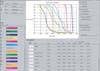
All patients underwent an initial CT simulation in the supine position with their arms on their chests, using intravenous contrast agents and free breathing. According to the consensus of our institution, we determine the dose-volume parameters and target volume in this study. The clinical target volume (CTV) included all areas of gross and potentially microscopic disease and included the upper half of the vagina, parametria, uterus, and regional lymph node regions (obturator, common, internal and external iliacs, presacral region and para-aortic regions) (Fig. 1A, B). The CTV in the pelvis consisted of a 2 cm margin around the cervix, uterus, parametria, presacral space, and vagina. Considering the unreliability of current imaging techniques to determine involvement of the parametria [15] and the possibility of movement during the course of radiotherapy, all pelvic tissue lateral to the cervix and upper vagina were considered to be part of the parametria. The vaginal volume consisted of 4 cm of vagina distal to the lower extent of the tumor (defined by imaging of tumor or marker seeds). The external, internal, and common iliac nodal volume was based on the contrast-enhanced vessels with a 0.7-1 cm circumferential margin. The CTV in the para-aortic region was contiguous with the pelvic lymph node stations and generously encompassed the aorta and inferior vena cava with at least a 1.5 cm margin (Fig. 1C). The superior CTV border was usually at the level of T12-L1 junction. When required, the superior aspect of the CTV was modified laterally for kidney sparing and anteriorly for small bowel sparing. The presacral region was included to the level of S3 to ensure coverage of the presacral lymph nodes and the uterosacral ligament. Accounting for patient motion and set-up uncertainty in our institution, the CTV was expanded 0.8-1 cm non-uniformly to create the planned target volume. Seven patients with ovarian transposition underwent separate ovarian contouring based on clips left by the surgeon. The involved nodes were also contoured separately and were either based on clips left by surgeon or on the abnormal appearance of nodal regions on the MRI or CT images. These areas were treated with a simultaneous integrated boost using a margin of up to 0.5 cm depending on the proximity of the small bowel or other critical structures. The entire extended field and pelvic field were prescribed 50.4 Gy in 28 fractions, whereas the involved nodes were treated with a total dose of 57.6-59.4 Gy. There was a dose-volume limitation with no more than 50% of the ovarian volume receiving 7 Gy (Fig. 1D). The dose-volume limitation was applied to other organs at risk. The rectum received a dose of V45≤50%; the bladder received a dose of V45≤50%; the small intestine received a dose of V35≤45%; the kidney received a dose of V25≤33%; the liver received a dose of V30≤30%; the bone marrow received a dose of V10≤90% and V35≤45%; and the spinal cord received a dose of V40≤0.1 cubic centimeters (Fig. 2). The inverse treatment planning for IMRT was performed with the sliding window technique using the Philips Pinnacle3 Planning System (Andover, MA, USA). All plans used seven coplanar beams. All patients were treated with 6-MV photons (Fig. 1E). All patients underwent high-dose-rate (HDR) intracavitary brachytherapy. Four or five fractions of six Gy each were delivered to point A. These treatments were delivered once or twice weekly, with no EF-IMRT treatment on the day of the intracavitary HDR treatment. Six patients received a parametrial boost as determined by the physician, depending on the cancer stage. Parametrial boosts of 55.8-59.4 Gy were delivered using parallel-opposed anteroposterior fields. Combining the external beam radiation doses with brachytherapy doses, the cumulative linear quadratic equivalent doses (EQD2) delivered to point A (defined as 2 cm lateral and 2 cm superior to the cervix) were 82 GyEQD2 (α/β=10 Gy)-98 GyEQD2 (α/β=10 Gy).
All patients received weekly cisplatin at a dose of 40 mg/m2 during the course of external beam radiotherapy (EBRT). During EBRT, or before the initiation of chemotherapy, weekly physical examinations, complete blood counts, and liver and renal function tests were performed. If the absolute neutrophil count was <1,000/mm3 or the platelet count was <100,000/mm3, chemotherapy was delayed or interrupted until the patient recovered.
3. Follow-up evaluation and statistical analysis
After RT completion, all patients were evaluated by a radiation oncologist and gynecologic oncologist after 1 month, followed by evaluations at 3-month intervals for 2 years and every 6 months thereafter. Radiologic studies and blood chemistries were ordered at the discretion of the treating oncologists. Ovarian function was evaluated by the presence or absence of postmenopausal symptoms and by the measurement of follicle-stimulating hormone (FSH) and estrogen (E2) levels. We checked the FSH levels frequently when patients returned for cancer status follow-up during the 3-month and 6-month intervals. Transient ovarian failure may last for a long time, and we defined ovarian failure as two elevated (>40 U/L) FSH levels measured at least 3-6 months apart, over 2 years of follow-up after the completion of the cancer treatment. Survival was measured from the date of diagnosis to the date of death or to the date of the most recent follow-up. Time to recurrence was measured from the date of diagnosis to the date of the first failure. Acute toxicities, measured from the initiation of treatment to 90 days after completion, were graded according to the National Cancer Institute Common Terminology Criteria for Adverse Events ver. 3.0 (CTCAE 3.0). Late toxicities, experienced more than 90 days after completion of therapy, were graded according RTOG late toxicity scale. Toxicities are reported as counts with percentages. The overall survival and disease-free survival rates were estimated with the Kaplan-Meier method using SPSS ver. 13.0 (SPSS Inc., Chicago, IL, USA).
RESULTS
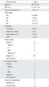
Forty-five patients were treated with EF-IMRT and concurrent cisplatin chemotherapy. The clinical and pathologic characteristics of these patients are shown in Table 1. The median age was 45 years (range, 25 to 74 years). The total median treatment length was 48 days (range, 37 to 64 days). No delay in radiation therapy was observed.
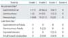
The treatments were well-tolerated. The acute toxicities are summarized in Table 2. Diarrhea and cystitis were the most common toxicities, with most patients reporting at least grade 1 and 2 toxicities. Twenty-eight patients (62.2%) and three patients (6.7%) were diagnosed with grade 2 and 3 acute gastrointestinal toxicities, respectively. Leukopenia was the most common hematologic toxicity. Chemotherapy for 5 patients was delayed due to a low neutrophil count. The median number of cisplatin cycles received per patient was six (range, 5 to 6 cycles).
There were three patients (6.7%) who suffered from RTOG grade 3 late toxicities. Two patients had a small bowel obstruction at 10 months that required a partial enterectomy. One patient developed sigmoid stenosis at 13 months, which required surgical intervention. Two patients suffered from RTOG grade 2 late toxicities and underwent medical therapy. No patients experienced grade 4 toxicity, and there were no treatment-related deaths. Seven patients underwent ovarian transposition by abdominal or laparoscopic procedure prior to radiotherapy. The ovaries were transposed as high or as lateral as possible, with metallic clips applied to each transposed ovary that were subsequently identified by CT simulation localization. The mean ovarian dose was 2.5-3.4 Gy. Five patients (71%) maintained ovarian function. Two patients, aged 43 and 44, were classified as having ovarian failure. Their serum levels of FSH and luteinizing hormone rose progressively, and their serum E2 levels declined over 4 weeks after radiation. The lengths of time when their FSH levels were first elevated to more than 40 U/L (67 U/L and 81 U/L, respectively) were 89 and 104 days after treatment, respectively.
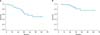
Forty-two patients had a complete response, and three had a persistent disease as confirmed by clinical examination and imaging studies. The median follow-up time was 28 months (range, 5 to 62 months). Thirty-eight patients (84.4%) were alive at the last follow-up. Twenty-seven patients (64.3%) had no evidence of disease, and 15 patients (35.7%) had recurrence that was documented clinically or by imaging at last the follow-up (Table 3). The most common sites of out-of-target failure were supraclavicular nodes (10 patients) and inguinal nodes (3 patients). The 28-month disease-free survival, and the overall survival rates for the entire cohort were 69.3% and 85.3%, respectively (Fig. 3).
DISCUSSION
Due to the high incidence of occult PAN metastases in patients with advanced cervical cancer [5], EFRT has been used as both a prophylactic and a curative treatment [7,9,16]. Several studies has demonstrated that EFRT improved survival in selected cervical cancer patients [6,9], and the European Organisation for Research and Treatment of Cancer (EORTC) showed that the incidence of para-aortic and distant metastases was significantly higher in patients receiving pelvic irradiation alone, although there was no statistically significant difference in local control, overall distant metastases and survival with no evidence of disease between the EFRT and pelvic RT [17]. There is inherent toxicity in the treatment of the pelvis and the PAN regions. The addition of concurrent chemotherapy to EFRT exacerbates the acute toxicity. Prospective phase II cooperative group trials [18] have reported grade 3 to 4 acute bowel toxicity in 49% of patients treated with concomitant chemotherapy and extended field radiotherapy. In comparison, grade 3 acute gastrointestinal toxicity was experienced by 6.7% of the patients in the current study. This result is superior to those reported in previous studies [19,20], which showed grade 3 gastrointestinal toxicity among 20% and 19.4% of patients. The use of IMRT greatly assisted in the conformality of dose distribution, confined the high-dose portions of radiation fields and reduced the absorbed dose and volume in critical organs, resulting in reduced overall toxicity. Gerszten et al. [21] found a significant reduction in critical organ irradiation with EF-IMRT and proposed that this treatment may reduce both acute and late treatment-related side effects.
To reduce myelotoxicity, we used the bone marrow-sparing IMRT approach. This technique has been shown to dosimetrically reduce the volume of bone marrow irradiated [22] and can be clinically correlated with a decreased rate of grade 2 leucopenia [23]. In our study, the volume of marrow receiving 10 Gy was reduced to ≤90%, and the volume receiving 35 Gy was reduced to ≤45%. Acute grade 3 hematologic toxicity was observed in 20% of our patients. This finding is similar to the hematologic toxicity observed in the study by Gerszten et al. [21] and is superior to the Japan study [24]. The incidence of hematologic toxicity in our series was slightly higher than the reported rates in both the Gynecologic Oncology Group (GOG) study (15%) [18] and the study by Chung et al. (10%) [25] of EFRT and concurrent chemotherapy. This difference may be related to the use of weekly chemotherapy, suggesting that the IMRT technique may not have a significant impact on this toxicity. In addition, other risk factors may account for these differences in toxicity, including low body mass index (BMI), smoking, and other causes of microvascular disease.
RTOG 90-01 [10] reported a late toxicity rate of 12% in patients treated with EFRT without concurrent chemotherapy. In previous studies with EFRT and concurrent chemotherapy, the reported late toxicities rates were 0%-14% [12,18-20,25,26]. Our result was 6.7%, which compares favorably with those results, although our follow-up is relatively limited. Fistula formation and bowel obstruction were significant late toxicities. HDR intracavitary brachytherapy was safely used with EFRT and concurrent chemotherapy [12,25]. We observed that HDR did not significantly increase late complications, which is similar to the findings of trials in which HDR brachytherapy was combined with pelvic RT and concurrent chemotherapy [27]. Jensen et al. [14] showed that the 2-year cumulative incidence of late grade ≥3 genitourinary toxicity was 4.8%, and no patients experienced late grade ≥3 gastrointestinal toxicity. It is unclear whether these toxicities were directly related to the addition of EFRT. In the present study, we found the overall survival rate to be 84.4%, which is similar to those reported in previous studies [12,19,25]. EF-IMRT concomitant with chemotherapy was effective in preventing in-target failure. The locoregional in-target control rate was good, with distant sites being the most common sites of failure. Only two patients (4.8%) developed in-target failures, and 13 patients (30.9%) had out-of-target recurrence. This result is similar to the results of two other studies [14,28]. The locoregional in-target control rate of our study compares favorably with the result reported by Kodaira et al. [24], showing a good distant control rate with high-intensity concurrent chemotherapy.
The degree and persistence of ovarian damage and suppression of ovarian function is related to the patient's age and the dose of radiation delivered to the ovaries [29]. The importance of the dose of radiation is clear because a low dose can save many follicles and repair the damage induced in some of them. For over three decades, ovarian function has been maintained by transposing the ovaries out of the field of irradiation, which reduces the ovarian dose. Bidzinski et al. [30] confirmed that ovarian function was preserved when the ovaries were transposed at least 3 cm from the upper border of the field. In our study, 7 patients underwent ovarian transposition to a position as high or as lateral as possible by abdominal or laparoscopic procedure prior to radiotherapy. Five patients (71%) maintained ovarian function; 4 of these patients had ovarian transposition at 3-3.5 cm above the level of L5-S1 (above the iliac crest), and one of these patients had ovarian transposition at 1.5 cm from the radiation field edge. The median age of the five patients was 32 years (range, 25 to 41 years). We used a dose-volume limitation with no more than 50% of the ovarian volume receiving 7 Gy for EF-IMRT. The median ovarian dose of the five patients who preserved ovarian function was 2.8 Gy (range, 2.5 to 3.1 Gy) and the ovarian dose of the other two patients was 3.2 Gy and 3.4 Gy, respectively. Two patients (29%) experienced ovarian failure; one was 44 years old with ovarian transposition at 3-3.5 cm from the upper border of the field, and the other was 43 years old with ovarian transposition at 1.5 cm from the radiation field edge. As previously described in the literature, ovarian transposition is of limited value in patients who are older than 40 because they have an intrinsically reduced fertilization potential and an increased risk of ovarian failure despite transposition [31].
This retrospective study is limited by a small sample size, which makes it difficult to evaluate toxicity, particularly grade 1 and 2 toxicities, as these symptoms may not routinely be recorded by clinicians in daily clinical practice. Large randomized multi-institutional trials are needed to verify the effectiveness of EF-IMRT and concurrent chemotherapy for patients who had disease classified as FIGO stage IB2-IIIB. One strength of this study lies in the fact that seven patients experienced ovarian transposition before radiotherapy, and ovarian function was evaluated before and after EF-IMRT.
In conclusion, cisplatin chemotherapy with EF-IMRT for local advanced cervical cancer is safe and well-tolerated. The acute and late toxicities are acceptable. The locoregional control rates are promising and similar to those reported in previous studies, although distant metastases continue to be the predominant mode of failure. EF-IMRT provides an opportunity to preserve endocrine function for patients with ovarian transposition.


 XML Download