

 PDF
PDF ePub
ePub Citation
 Print
Print


INTRODUCTION
Obesity is the main risk factor for endometrial cancer [1-3]. Both excess estrogen and insulin resistance (hyperinsulinemia) contribute to the development of endometrial cancer in obese women [3,4]. Our recent study confirmed the association of hyperinsulinemia and endometrial cancer [5]. In addition, adipose tissue may directly mediate the obesity-related endometrial cancer risk by acting as an endocrine organ [6-8]. It can increase inflammatory cytokines, adipokines and surplus free fatty acids, or provide a sustained source of estrogen by aromatizing androstenedione to estrogen [8].
Adipokines are bioactive substances produced by adipose tissue. They play a crucial role in metabolism, and the development of metabolic syndrome and related disease [9,10]. Increased serum leptin or decreased adiponectin are reliable markers for obesity and insulin resistance [9,10]. The serum leptin-to-adiponectin (L/A) ratio is believed to be a useful, surrogate marker for insulin resistance since increased adipocytes produce more leptin and less adiponectin [11]. Both adipokines and L/A ratio are associated with increased risks of obesity-related cancers [6,7]. Several studies [12-21] have investigated the direct link between serum leptin, adiponectin or L/A ratio and endometrial cancer. However, these studies did not have biologic measures of insulin resistance. More clinical data are required to investigate the association between serum leptin or adiponectin, and endometrial cancer.
This case-control study aims to investigate the relationships between serum leptin, adiponectin, or L/A ratio, and the risk of endometrial carcinoma in combination with various metabolic factors in Chinese women.
MATERIALS AND METHODS
1. Study population
We investigated 206 patients (mean age, 53.2 years [range, 26 to 81 years]; mean parity, 2.3 [range, 0 to 6]) with histologically confirmed endometrial cancer, between August 2008 and December 2010, from the Women's Hospital, Zhejiang University School of Medicine, China. Controls (n=310; mean age, 53.3 years [range, 27 to 82 years]; mean parity, 2.2 [range, 0 to 5]) were randomly selected from lists of healthy women undergoing routine physical examinations during the same period. The cases and controls were individually age-matched (at 5-year intervals) and had an approximate response rate of 1:1.5. Women with a history of hysterectomy, bilateral oophorectomy, or Lynch syndrome were not included in the study. Each subject has signed the informed consent according to an institutional review board. The cases and controls were included in our recent study [5].
We reviewed clinical details from the medical records. The available data included body weight and height for body mass index (BMI) calculation, menopause, reproductive history, cancer history, and preoperative serum fasting total cholesterol (TC), triglycerides (TG), high-density lipoprotein cholesterol (HDL-C), and glucose (GLU). BMI was categorized into normal (18.5 to 24.9 kg/m2), overweight (25.0 to 29.9 kg/m2) and obese (≥30.0 kg/m2). These data were also included in our previous study [5].
2. Blood sampling
Blood samples were drawn from patients and controls before surgery. Fasting serum was obtained and centrifuged at 4℃ in the blood collection tube containing a specific separation gel for biochemical tests (BD Biosciences, San Jose, CA, USA). The serum was kept in -80℃ freezer until use.
3. Laboratory assays
Assays of serum leptin, adiponectin, and insulin were performed blind to the case-control status of the samples. Serum leptin and adiponectin concentrations were analyzed by commercial, double-antibody, sandwich ELISA kit (Leptin: Enzo Life Sciences, Shanghai, China; Adiponectin: Bender MedSystems GmbH, Vienna, Austria) as per the manufacturer's procedures. Concentrations were calculated according to the standard concentrations and the corresponding optical density value at 450 nm. Serum insulin was automatically measured by a chemilluminescent, microparticle immunoassay (the ARCHITECT Insulin Assay, Abbott Laboratory Inc., Abbott Park, IL, USA) in the ARCHITECT i system.
4. Statistical analysis
SPSS ver. 13.0 (SPSS Inc., Chicago, IL, USA) was applied for statistical analysis. Correlations between different measures or the descriptive results between cases and controls were analyzed by the Spearman's correlations or one-way ANOVA, respectively. Measures including the L/A ratios were categorized into tertiles based on the values in cases and controls for univariate and multivariate analysis. Women with missing data were removed in the statistical analysis including body weight or height in 8 cases; previous hormone replacement therapy use in 1 case; TC, TG, and HDL in 2 cases and 6 controls; and GLU in 1 case and 3 controls. There are 197 cases and 304 controls left for multiple logistic regression analyses after exclusion of all missing data. Odds ratio (OR) and its 95% confidence interval (CI) were calculated to estimate the relative risks for endometrial cancer. The relative cancer risks of serological factors were calculated as women in intermediate or top tertiles (T2 or T3) versus the bottom tertile (T1). The threshold of statistical significance was 0.05 (2-tailed). Subgroup analysis stratified by menopausal status, reproductive history (as categorical variables), or hormone replacement therapy use was conducted to determine whether these factors influenced cancer risks of adiponectin, leptin, L/A ratios and insulin. An unconditional, logistic regression model was used to estimate the ORs of serum adiponectin, leptin, and L/A ratios, adjusted by age at 5-year intervals, and, different metabolic variables including BMI, GLU, TC, TG, HDL-C, insulin, adiponectin (for leptin) and leptin (for adiponectin). The variables were removed from the final models if they did not change risk estimates with more than 10% (removal probability=0.10).
RESULTS
We found that patients with endometrial carcinoma had a higher BMI, serum insulin, GLU, TG, and TC than controls (p<0.001, data not shown). Endometrial cancer patients were more frequently associated with nulliparous status (cases vs. controls: 26/206, 12.6% vs. 9/310, 2.9%; p<0.001) and history of type 2 diabetes mellitus (cases vs. controls: 18/206, 8.7% vs. 11/310, 3.5%; p<0.05). The data mostly overlapped with those in our recent study [5].They had significantly higher serum leptin, insulin, and L/A ratios than controls (mean±SD, cases vs. controls: leptin 28.8±2.2 ug/L vs. 19.8±1.4 ug/L, p<0.001; L/A ratios 0.018±0.050 vs. 0.013±0.014, p<0.001; insulin 15.8±1.4 mU/L, vs. 8.1±0.5 mU/L, p<0.001). Serum adiponectin was not significantly lower in patients than in controls (mean±SD, cases vs. controls: adiponectin 2,330.7±180.5 ug/L vs. 2,583.9±147.2 ug/L; p=0.078). We also noted that serum leptin or adiponectin was positively or negatively correlated with both BMI and fasting insulin level, respectively (Spearman correlation coefficients: leptin-BMI 0.516, p<0.01; leptin-insulin 0.236, p<0.01; adiponectin-BMI -0.222, p<0.01; adiponectin-BMI -0.113, p<0.01). There was also a mildly negative correlation between serum leptin and adiponectin (Spearman correlation coefficients: -0.185, p<0.05).
The associations between clinical or serological variables and endometrial cancer were included in our recent study [5]. There was an increased endometrial cancer risk in women with diabetes versus women without diabetes, and, in women with obesity versus those with normal BMI (p<0.01). Women aged >50 years, or with menopause, primary hypertension or a history of hormone replace therapy did not show significantly altered endometrial cancer risk as compared with the baseline (p>0.05). Women with high serum insulin, GLU, TG, or TC had an elevated risk for endometrial cancer while women with high serum HDL-C showed a decreased cancer risk (p<0.05, data not shown).
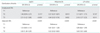
We found a dose-dependent relationship between serum leptin, adiponectin concentration, L/A ratios, and endometrial cancer (Table 1). Stratification by menopause, parity, hormone replacement therapy, or type 2 diabetes, etc, did not appreciably influence on these relationships. The multiple logistic regression model further demonstrated that serum leptin, adiponectin and L/A ratios were significantly associated with endometrial cancer. The logistic regression model-derived ORs and 95% CIs were shown in Table 1. The effect of serum letpin and adiponectin on endometrial cancer was given in Table 2. Women with high serum (T3) leptin and low adiponectin (T1) showed the highest endometrial cancer risks among various groups.
DISCUSSION
The current study is the first one to investigate the relationship between serum leptin, adiponectin and endometrial cancer in Chinese women by recruiting a relatively large number of cases. It has been noted that the prevalence of endometrial cancer in China increased approximately three times in the past two decades [22]. In concordance with most previous studies [12-21], our study demonstrates that high levels of serum leptin, low levels of adiponectin, and high L/A ratio are associated with endometrial cancer. Moreover, in this study, we have obtained clinical details on each sample including BMI, reproductive history, use of hormone replacement therapy, and measurements on serum insulin, GLU, TG, etc. Evaluation of these variables permits a comprehensive analysis of metabolic factors on the potential associations between adipokines and endometrial cancer.
Several previous studies showed that serum leptin was positively correlated with endometrial cancer [12-14,23,24]. The correlation between serum leptin and endometrial cancer risk disappeared in most studies, when adjusted by BMI. The current study indicates that adjustment for BMI, insulin and other potential cofounders does not significantly alleviate the relationship between leptin and endometrial cancer. Luhn et al. [24] also showed the presence of a positive association in women with no menopausal hormone therapy after adjustment for estradiol and BMI. BMI is a common parameter for measuring insulin resistance, and serum insulin itself is a direct marker and may be more helpful than other anthropometric measures [5]. Friedenreich et al. [25] carried out a similar case-control study, but they had somewhat different results. Their age-adjusted analysis confirmed that high levels of serum leptin or L/A ratio was associated with increased or decreased endometrial cancer risk, respectively. These associations disappeared in multi-variable analysis. However, in that study, only postoperative blood samples were available in a portion of cases. The presence of cancer, cancer treatment and perioperative metabolic response potentially influenced the measurements of serum leptin and adiopnectin. In our study, preoperative fasting blood samples (generally 2-day before diagnosis) were obtained in all cases. We think that the blood draw time might be the main factor contributing to the inconsistent results between these two studies.
We suggest that elevated serum leptin may participate in the development of obesity-related endometrial cancer by other mechanism in addition to insulin resistance. For example, leptin can contribute to hyperestrogenemia by activating aromatase, which catalyzes the conversion of androstenedione to estrogen in adipose tissue [26]. Leptin can also stimulate cell growth, migration, invasion, angiogenesis and anti-apoptosis in vitro via activating different signaling pathways [27-29].
Adiponectin functions as an insulin sensitizer, and low serum adiponectin reversely correlates with insulin resistance [10]. Moreover, adiponectin can inhibit cell proliferation, invasiveness and angiogenesis in vitro by suppression of estrogen receptor α and vascular endothelium growth factor [30-32]. Serum adiponectin would be a protective factor for endometrial cancer. As expected, we found that levels of serum adiponectin were inversely associated with the risk of endometrial cancer in Chinese women. There are five retrospective and four prospective studies that have investigated this association [15-20,23-25]. Most studies confirm a negative association between serum adiponectin and endometrial cancer, but two prospective studies showed no association [20,23]. The relatively small sample size and the short time interval between blood draw and endomtetrial cancer diagnosis may be the major factors that limited the ability to detect the association in these studies.
There are increased levels of serum leptin and decreased adiponectin in endometrial cancer; therefore, the L/A ratio might be a better indicator to characterize the endometrial cancer risk than leptin or adiponectin alone. A recent study of Japanese women supported this hypothesis [21]. However, inconsistent result was reported in a recent case-control study [25]. We have mentioned the possible cause of this inconsistent result earlier in this manuscript. Our study is generally consistent with the Japanese report. However, in our study, the estimated cancer risk of L/A ratio is not higher than leptin alone. This subtle difference might be explained by population or lifestyle differences. In women with endometrial carcinoma, the frequency of obesity (BMI≥30 kg/m2) in our study (40.3%) is similar to that previously from Western countries [15,16,18] and much higher than that from the Japanese report (8.9%) [19].
There are several limitations in our study. First, this is a retrospective study in which potential bias may inevitably occur, and, the sample size is somewhat limited. Second, we measured serum leptin, adiponectin and other variables from one preoperative fasting blood sample. The 2-day interval between blood draw and cancer diagnosis may provide an accurate reflection of insulin resistance at diagnosis, but failed to determine the time-effect of insulin resistance on endometrial cancer. Finally, we did not assess the reduced endometrial cancer risk, which was associated with decreasing insulin resistance via weight loss, exercise and other intervention.
In conclusion, our study showed the significant relationships between serum leptin or adiponectin and endometrial cancer. Serum leptin and adiponectin play important roles in the development of obesity (insulin resistance)-related endometrial cancer. However, a multi-center, long-term prospective study is necessary to disentangle some of these relationships with the risk of endometrial cancer.

 XML Download