

 PDF
PDF ePub
ePub Citation
 Print
Print


INTRODUCTION
Adenocarcinoma (ADC) of the uterine cervix is a relatively uncommon histological subtype of cervical cancer, while have been increasing recently in many areas [1]. Several reports have shown that ADC is more aggressive and exhibits more distant metastases, resulting in a lower survival rate than squamous cell carcinoma (SCC) [2,3]. Our incomplete understanding of the prognostic factors [4,5] and optimal treatment [6,7] of ADC may account for its poor outcome. Indeed, there has been no uniformly accepted form of management for ADC. As with SCC, patients with International Federation of Obstetrics and Gynecology (FIGO) stage IB1-IIB cervical ADC are treated by radical hysterectomy (RH). The prognosis of patients with ADC after RH remains unclear and conventional adjuvant therapy in high-risk group after primary surgery or salvage therapy of recurrent ADC seems generally ineffective [6]. Several reports found that patients with ADC have poorer prognosis than do those with SCC [4,5,8-10], whereas others found no differences in prognosis [11-15]. Therefore, the prognosis after RH and the optimal management of ADC are still a matter of debate.
Thus, the aim of this study was to clarify the treatment outcomes and prognostic factors after RH in patients with FIGO stage IB1-IIB ADC of uterine cervix, and to postulate the optimal management of patients with early-stage ADC of uterine cervix.
MATERIALS AND METHODS
1. Patients
After approval by the Institutional Review Board of Hokkaido University Hospital, we searched the cancer registry and computerized database of our institution for patients with 1) FIGO stage IB1-IIB cervical ADC, 2) who underwent RH with removal of a vaginal cuff of at least 2 cm, total resection of parametrial tissue and systematic lymphadenectomy (LND). This operation is a nerve-sparing modification of the Okabayashi operation [16]. The nerve-sparing procedure was further refined by introducing the preservation of vesical branches of pelvic plexus since 1997. As the preferred treatment in our institution for patients with FIGO stage IB1-IIB cervical cancer is RH, almost all patients with FIGO stage IB1-IIB cervical cancer underwent RH and only a small number of patients who were not eligible for radical surgery because of severe medical co-morbidity received radiotherapy (RT) or concurrent chemoradiotherapy (CCRT). Medical records were retrospectively reviewed, and the following parameters were collected: age, FIGO stage, tumor size, deep stromal invasion (DSI, >2/3 thickness), parametrial invasion (PI), lymphovascular space invasion (LVSI), lymph node metastasis (LNM), corpus invasion, vaginal metastasis, ovarian metastasis, adjuvant therapy, and date of death or last follow-up. Pathologic slides were reviewed by two experienced pathologists at our institution.
2. Adjuvant therapy
Since there is no definitive evidence that adjuvant RT is more effective than adjuvant chemotherapy for cervical ADC undergoing RH, we have used more frequent adjuvant chemotherapy after surgery since the year 2000 [16]. Patients with risk factors for recurrence, including DSI, LVSI, PI, LNM, and/or bulky tumor, received whole pelvic irradiation or systemic platinum-based chemotherapy as postsurgical adjuvant therapy. RT consisted of whole pelvic external irradiation by four-field technique with 50 Gy for 25 fractions beginning four weeks after surgery. Chemotherapy was given at least three courses at four-week intervals beginning approximate 3 weeks after surgery. Chemotherapeutic regimens used were previously described [17]. Briefly, IEP (ifosphamide: 1.5 g/m2, day 1-3; epirubicin: 40 mg/m2, day 1; cisplatin: 14 mg/m2, day 1-5) was used for eleven patients, CAP (cyclophosphamide: 500 mg/m2, day 1; adriamycin: 50 mg/m2, day 1; cisplatin: 50 mg/m2, day 1) for five, MEP (mitomycin C: 10 mg/body, day 1; cisplatin: 70 mg/m2, day 1; etoposide: 100 mg/m2, day 1-3) for two, BOMP (bleomycin: 7 mg/body, day 1-5; vincristine: 0.7 mg/m2, day 5; mitomycin C: 7 mg/m2, day 5; cisplatin: 14 mg/m2, day 1-5) for one, TC (paclitaxel: 175 mg/m2, day 1; carboplatin: AUC5, day 1) for one, TP (paclitaxel: 135 mg/m2, day 1; cisplatin: 50 mg/m2, day 2) for one, ITP (ifosphamide: 1.5 g/m2, day 1-3; paclitaxel: 175 mg/m2, day 1; cisplatin: 14 mg/m2, day 1-5) for one, and intra-arterial infusion of cisplatin (100 mg/body) for one, and cisplatin-based chemotherapy (unknown) for seven.
3. Statistical analysis
Categorical variables were analyzed using the chi-square test or Fisher's exact test. We used the Kaplan-Meier method, log-rank test for survival analysis and the Cox hazard method for prognostic analysis. A result was considered significant when the p-value was less than 0.05.
RESULTS
1. Patients' characteristics
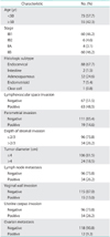
The clinicopathological characteristics of 130 patients with cervical ADC are summarized in Table 1. Among 130 patients with cervical ADC, sixty had clinical stage IB1, six had stage IB2, four had stage IIA, and sixty had stage IIB. Histologic subtypes include eighty-eight cases of endocervical type, two of intestinal type, thirty-two of adenosquamous carcinoma, seven of endometrioid type, and one of clear cell carcinoma. Median age of the patients was 47 years (range, 26 to 69 years). Median follow-up period was 72 months (range, 4 to 120 months). Among 130 patients, seventy-one patients did not receive adjuvant therapy, because 53 patients showed no risk factors for recurrence, and 18 patients refused adjuvant therapy due to personal reasons. Fifty-nine patients with risk factors for recurrence received adjuvant therapy, including 29 cases of RT and 30 cases of systemic platinum-based chemotherapy.
2. Ovarian metastasis in adenocarcinoma of the uterine cervix
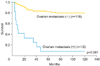
All cases of ovarian metastasis had stage IIB disease. Ovarian metastasis was found in 12 of 130 cases (9%), and 11 of 12 cases died within five years. Ovarian metastasis is significantly related to other pathological risk factors including LN metastasis, LVSI, parametrial invasion, DSI, vaginal invasion, and corpus invasion (p<0.001 for all 6 risk factors), which should explain why the patients with ovarian metastasis showed extremely poor survival (Table 1). We, therefore, excluded ovarian metastasis for multivariate survival analysis in this study (Fig. 1) (Table 2).
3. Multivariate survival analysis
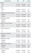
An estimated five-year survival rate of stage IB1 was 96.6%, 75.0% in stage IB2, 100% in stage IIA, and 52.8% in stage IIB. Among the clinicopathological factors analyzed in this study, all risk factors except for age are significantly related to poor survival by univariate analysis (Table 3). Multivariate analysis revealed that LNM (hazard ratio [HR], 4.4, 95% confidence interval [CI], 1.7 to 11.4; p=0.002), LVSI (HR, 4.0; 95% CI, 1.1 to 14.1; p=0.03), and PI (HR, 4.6; 95% CI, 1.8 to 11.5; p=0.001) were independent prognostic factors in cervical ADC treated with RH and systematic LND (Table 3).
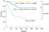
Survival of the patients with cervical ADC could be stratified into three groups by the combination of three independent prognosticators. An estimated five-year survival rate for the patients without three independent risk factors (group A, n=59) was 98%, that for the patients with LVSI and/or PI without LNM (group B, n=37) was 75%, and that for the patients with LNM irrespective of the presence of LVSI/PI (group C, n=34) was 37%. There is statistically significant difference of disease-specific survival among each group (p=0.004 for group A vs. group B, p<0.001 for group B vs. group C, p<0.001 for group A vs. group C) (Fig. 2).
4. Impact of LNM on the survival of ADC of the uterine cervix
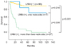
Since multivariate analysis has shown that LNM was one of the most important prognostic factors in cervical ADC, we analyzed its impact on the survival in cervical ADC according to the number of positive-node sites. Estimated five-year survival rate of the patients without LNM (group A, n=96) was 89%. Estimated five-year survival rate with one positive-node site (group B, n=7), and with more than two positive-node sites (group C, n=27) was 86% and 23%, respectively (Fig. 3). There was statistically significant difference of survival between group A and C (p<0.001), and between group B and C (p=0.009). However, there was no statistically significant difference of survival between group A and B (p=0.29).
DISCUSSION
In this retrospective analysis, we demonstrated that LNM, LVSI, and PI were independent prognostic factors for ADC of the uterine cervix, and their survival was stratified by the combination of three independent prognostic factors. Postoperative treatment and follow-up modality could be individualized according to the independent risk factors for survival.
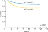
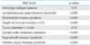
Several reports found that patients with ADC have poorer prognosis than do those with SCC [4,5,8-10], whereas others found no differences in prognosis [11-15]. When we compared the frequency of clinicopathologic risk factors and survival between ADC and SCC (260 cases) at our institution during the same study period, we found no significant difference of frequency of clinicopathologic risk factors according to the histologic subtype (Table 4). However, overall survival of ADC was significantly worse than that of SCC (Fig. 4), suggesting that adjuvant therapy might be less effective for ADC than SCC.
Currently, standard adjuvant therapy was not fully established after RH and LND for cervical ADC, but RT is widely employed as a standard adjuvant therapy as well as for SCC. However, there is no definitive evidence that RT is more beneficial than chemotherapy after radical surgery for cervical cancer. Thus, adjuvant chemotherapy combined with RH and systematic LND may also provide a survival benefit. However, there are no randomized controlled studies comparing the clinical efficacy of RT and chemotherapy after surgery so far. We need to analyze the survival difference according to type of adjuvant therapy in other patients' cohort and in randomized trials in the future.
LNM have been shown to be the most important prognosticator for ADC of the uterine cervix. However, it is still unclear whether number of LNM sites affect differential survival of cervical ADC treated with RH and pelvic lymph node dissection. We, therefore, analyzed overall survival according to the number of positive-node sites and demonstrated that survival with one positive-node site was not significantly worse than that with negative-node (Fig. 3), which is similar to our previous report [18]. We speculate that single LNM site might be a local disease, and can be curable by our current treatment strategy consisting of extensive surgery and adjuvant therapy. This result is similar to that in node-positive endometrial cancer as we previously reported [19].
In contrast to node-negative patients, prognosis of patients with multiple positive-node sites was significantly worse than that with no and one positive-node site, indicating that most appropriate treatment strategy for patients with multiple positive-node sites remains to be established. If we can accurately predict LNM preoperatively, we can choose CCRT instead of radical surgery as a primary treatment for patients with multiple positive-node sites. Among risk factors, which can be assessed preoperatively, tumor size is supposed to be a good predictive factor for LNM. However, its prognostic value remains controversial [10,15]. In fact, tumor size was not an independent prognostic factor in our patient cohort. One possible preoperative assessment to predict LNM might be use of the combination of serum tumor markers, because we have previously shown that preoperative serum SCC and CA-125 levels strongly associated with number of positive pelvic nodes, site of positive-node in cervical SCC [20]. Alternatively, it is worth to utilize new imaging technique to efficiently detect LNM. Magnetic resonance/positron emission tomography (PET) has been reported to be more accurate than PET-computed tomography (CT) to predict LNM in cervical cancer [21].
In summary, we found three independent prognostic factors for patients with ADC of the uterine cervix who underwent RH and systematic LND. Prospective study is necessary to establish standard primary treatment (CCRT or RH) or standard adjuvant therapy (RT or CCRT or chemotherapy) after RH and systematic LND for early-stage ADC of the uterine cervix with multiple-node sites to improve their survival.

 XML Download