

 PDF
PDF ePub
ePub Citation
 Print
Print


INTRODUCTION
In women, cervical cancer is the third most commonly diagnosed cancer and the fourth leading cause of cancer death in females worldwide, accounting for 9% (529,800) of the total new cancer cases and 8% (275,100) of the total cancer deaths among females in 2008 [1].
Treatment of cervical cancer depends on FIGO staging. FIGO criteria is important for pretreatment staging and choice of appropriate treatment planning and treatment [2]. The inclusion of CT or MRI scans in the staging work-up has been advocated in order to improve accuracy for cervical carcinoma [2,3]. Cystoscopy and sigmoidoscopy are necessary to confirm bladder and rectal invasion in cervical cancer [4].
Traditional pretreatment evaluations of patients with cervical cancer include physical examination, chest radiography, cystoscopy, intravenous urography, sigmoidoscopy, and barium enema [2,3]. However, imaging modalities (CT, MRI scan) and endoscopy (cystoscopy, sigmoidoscopy) are not included in the recent FIGO guidelines for routine pretreatment staging of cervical cancer [5]. Recent FIGO staging system for the cervix encourages the use of CT and MRI scans, but cystoscopy and sigmoidoscopy were classified as optional modalities and are not recommended as mandatory examinations. However, there are no recommendations regarding which patients should receive endoscopy as an alternative examination.
We carried out a retrospective analysis of cervical cancer patients who underwent imaging work-up and endoscopy before radiotherapy to determine the clinical utility of CT or MRI scans for ruling out bladder or rectal invasion, and the indications of endoscopy for patients with cervical cancer.
MATERIALS AND METHODS
Between January 1997 and December 2010, 1,610 patients with biopsy-confirmed cervical cancer were treated by the radiation oncology department of Samsung Medical Center, Seoul, Korea. Among these patients, we retrospectively reviewed the records of 769 patients who underwent imaging work-ups such as CT (503 patients) or MRI (749 patients) scans, and 473 patients who underwent both CT and MRI in addition to the standard FIGO staging work-up. Among them, endoscopies such as cystoscopy or sigmoidoscopy were done in 590 patients and 735 patients, respectively. In Samsung Medical Center, if there is no contraindication, it is routine practice to conduct imaging and endoscopy for all patients with cervical cancer. We analyzed data regarding age, tumor size, stage, lymph node involvement, menopause status, and squamous cell carcinoma antigen (SCC-Ag).
Considering endoscopy as the standard reference investigation, the sensitivity, specificity, positive predictive value (PPV), negative predictive value (NPV), and accuracy of CT and MRI scans for bladder and rectal invasion were determined.
Each patient underwent pretreatment imaging work-up of the abdominopelvic area and endoscopy of the bladder or rectum. CT (Lightspeed VCT* XTe, GE Healthcare, Buckinghamshire, UK) scans were performed using contiguous axial, sagittal, and coronal 5-mm thickness slices after administration of contrast medium. MRI scans were performed using a 1.5 T unit (Achieva, Phillips Medical System, Eindhoven, Netherland; Signa HDe, GE Healthcare). The cardiac or torso coil was used in the supine position from the pelvis to the abdomen. The CT scan criteria for bladder or rectal invasion included the focal loss of the periorgan fat plane between the bladder or rectum and the growth, accompanied by asymmetrical wall thickening, nodular indentations along the bladder or rectal wall, and intraluminal tumor masses. MRI findings of wall irregularity with heterogeneous signal, enhancement with thickening and nodularity, loss of fat plane or mass protruding into the bladder or rectal lumen were interpreted as positive invasion.
Twenty five out of 65 patients (38.5%) and 7 out of 16 patients (43.8%) were pathologically confirmed by cystoscopy or sigmoidoscopy when the bladder or rectum invasion suspected on image work-up. Endoscopy was considered the gold standard for determining the presence of bladder or rectal invasion. In all cases, cystoscopy and sigmoidoscopy were performed by an urologist and gastroenterologist, respectively. Flexible or rigid cystoscopy was used for bladder investigation, and fiberoptic sigmoidoscopy was used for rectal examination with biopsy for pathological confirmation. Endoscopically directed biopsy specimens were taken from all areas in the bladder and rectum that were suspected of cancer development.
Seven hundred fifty three patients received radiotherapy. Most of these patients (603 patients, 80.1%) received 5,040 cGy of external beam radiotherapy, and 359 patients (47.7%) underwent additional brachytherapy. A total of 450 patients underwent combined chemotherapy with radiotherapy. Sixteen patients who did not receive radiotherapy were treated with palliative aim. 424 patients did not receive brachytherapy of patients with radiotherapy. Among these patients who did not received brachytherapy, most patients underwent postoperative adjuvant radiotherapy or palliative radiotherapy.
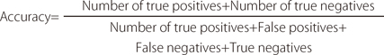
We evaluated the sensitivity, specificity, PPV, NPV, and accuracy of CT and MRI findings for the diagnosis of bladder or rectal invasion, by comparing the frequencies of each imaging work-up with the final endoscopic biopsy. The sensitivity, specificity, PPV, NPV, and accuracy of CT scans for bladder or rectal invasion were calculated using the following formulae:


RESULTS
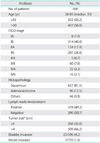
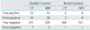
Seven hundred sixty nine are reviewed and their characteristics are given in Table 1. The median age of the patients was 50 years (range, 20 to 85 years) and the median follow-up was 37 months (range, 1 to 162 months). Of 769 patients, 29 (3.8%), 50 (6.5%), and 25 patients (3.3%) had bladder invasion on CT, MRI scan, and cystoscopy, respectively. Rectal invasion was identified in 11 (1.4%), 14 (1.8%), and 7 patients (0.9%) on CT, MRI scan, and sigmoidoscopy, respectively. The results for each of the imaging modalities are given in Tables 2 and 3, with endoscopic findings considered as the gold standard. CT and MRI scans revealed bladder invasion in 15 and 12 patients, respectively, who had endoscopically confirmed bladder invasion (true-positive for bladder invasion) and CT and MRI both revealed rectal invasion in 6 patients, respectively, who had endoscopically confirmed rectal invasion (true-positive for rectal invasion). Thus, 14 patients demonstrated bladder invasion on CT scan only and 38 patients demonstrated bladder invasion on MRI scan only (false-positive for bladder invasion). Five patients demonstrated rectal invasion on CT scan only and 8 patients demonstrated rectal invasion on MRI scan only (false-positive for rectal invasion). Seven and 3 patients who had cystoscopically confirmed invasion (false-negative bladder invasion) did not show any invasion on CT and MRI scan, respectively. For each image modality, there was one patient who showed no invasion, but had sigmoidoscopically confirmed invasion (false-negative rectal invasion). Finally, in 2 patients showing no bladder involvement on CT and MRI scan, bladder invasion was confirmed by cystoscopy. As shown in Table 3, CT scan showed a sensitivity of 68.2% and 85.7%, and a specificity of 96.4% and 98.9%, for detecting bladder and rectal invasion, respectively. CT scan had a low PPV (51.7% and 54.5%) and a high NPV (98.2% and 99.8%). MRI scan showed a sensitivity of 88.0% and 85.7%, and a specificity of 93.1% and 98.9% for detecting bladder and rectal invasion, respectively. MRI scan had a low PPV (35.6% and 42.9%) and a high NPV (99.4% and 99.7%). The accuracies of the imaging modalities in detecting the bladder and rectal invasion are given in Table 3.
DISCUSSION
In FIGO staging, some studies concluded that the use of imaging is not superior to physical examination [2,6]. In contrast, many studies have suggested that imaging is an important work-up tool and should be included in staging [3,7-11]. Cystoscopy and sigmoidoscopy, previously categorized as mandatory investigations, were reclassified as optional investigations in a recent revision of FIGO staging [5]. Since the 2009 revision of FIGO staging, a few studies have explored the identification of patients who will need endoscopy [12-14]. The present study therefore had two objectives. The primary objective was to establish how to identify patients who required cystoscopy or sigmoidoscopy according to the revised FIGO staging. The secondary objective was to demonstrate the accuracy of CT and MRI scans for pretreatment diagnosis of bladder and rectum invasion.
Based on previous studies, the sensitivities, specificities, PPVs, NPVs, and accuracies of CT and MRI scanning for bladder or rectal invasion were about 40-100%, 92-100%, 40-100%, 85-100%, and 86-98%, respectively [4,12,14-17]. The results of this study correspond well with those of previous studies (Table 4), with the exception of a few differences in sensitivity. In this study, the NPV was high enough so that additional invasive endoscopy was not necessary for patients who presented without invasion in imaging work-up (CT, MRI scan). Endoscopy should be considered as a tool for confirming invasion when patients were positive for invasion based on imaging work-up even though CT and MRI scans are not as effective for the purpose of diagnosis due to low sensitivity and PPV. These results suggest that there are advantages to using imaging modalities for the physician and patients, and that imaging offers additional cost benefits. There is no evidence that the low sensitivity of imaging can be increased by simultaneously using CT and MRI scans as shown in Table 3. MRI scans may be instrumental as a diagnostic tool to evaluate myometrial invasion or lymph node involvement, but it has no additional benefit as part of the confirmation process for bladder or rectal invasion.
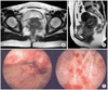
Fig. 1 shows a false-negative image from an MRI scan and endoscopy. There is no evidence of bladder invasion on the CT and MRI scan, but cystoscopy revealed that the patient was positive for invasion. False negative in CT or MRI is a rare case in this study and in previous studies which conducted similar purpose with the current study. The false negative finding in this case may have been the result of poor image quality or focal invasion in between the CT slices, so additional invasive endoscopy is not recommended to check the invasion for patients without invasion on imaging work-up.
The findings of this study are significant because they are based on a larger patient sample than those of previous studies. In addition, we analyzed the accuracy and indications of two imaging modalities, CT and MRI, for the diagnosis of bladder and rectal invasion.
There are some limitations to this single institution retro-spective study. First is that the number of patients in a specific group, such as stage IV, was too small for analysis and stage IIA was too large to bring about selection bias. Second is that this study does not include cervical cancer patients treated with surgery or chemotherapy only. Another limitation is that endoscopy was not performed in all patients, but was more likely to be used in patients who were suspected of having bladder or rectal invasion based on imaging work-up or physical examination.
In conclusion, if there is no evidence of invasion on imaging work-up, endoscopy is not necessary as an invasive diagnostic modality. However, if there is any evidence of invasion on imaging work-up, endoscopy is necessary to obtain an accurate prognosis for appropriate treatment. Patients prefer non-invasive diagnostic methods, which have fewer side effects. Therefore, future work should focus on the use of CT virtual endoscopy, which can be used in place of invasive endoscopy.


 XML Download