Journal List > J Gynecol Oncol > v.23(4) > 1079015

Kang, Lee, Lee, Kim, Cho, Kim, Park, Park, and Kim: How low is low enough? Evaluation of various risk-assessment models for lymph node metastasis in endometrial cancer: a Korean multicenter study

Abstract

Objective

The aim of this study was to identify a standard for the evaluation of future models for prediction of lymph node metastasis in endometrial cancer through estimation of performance of well-known surgicopathological models.

Methods

Using the medical records of 947 patients with endometrial cancer who underwent surgical management with lymphadenectomy, we retrospectively assessed the predictive performances of nodal metastasis of currently available models.

Results

We evaluated three models included: 1) a model modified from the Gynecologic Oncology Group (GOG) pilot study; 2) one from the GOG-33 data; and 3) one from Mayo Clinic data. The three models showed similar negative predictive values ranging from 97.1% to 97.4%. Using Bayes' theorem, this can be translated into 2% of negative post-test probability when 10% of prevalence of lymph node metastasis was assumed. In addition, although the negative predictive value was similar among these models, the proportion that was classified as low-risk was significantly different between the studies (56.4%, 44.8%, and 30.5%, respectively; p<0.001).

Conclusion

The current study suggests that a false negativity of 2% or less should be a goal for determining clinical usefulness of preoperative or intraoperative prediction models for low-risk of nodal metastasis.

INTRODUCTION

Despite recently published randomized trials suggesting no survival benefit for routine lymphadenectomy in endometrial cancer [1,2], full pelvic and paraaortic lymphadenectomy is still recommended by many gynecologic oncologic societies and guideline committees [3-5]. However, although there is ongoing controversy concerning the benefit of routine lymphadenectomy [6-8], the general consensus is that there is a certain subset of patients in which the omission of routine lymphadenectomy may be justified [9-11].
For several decades, researchers have proposed several models to predict patients at low-risk for nodal metastasis [12-15]. Most of these prediction models were designed using surgicopathological parameters, such as depth of myometrial invasion or pathological grade [13,14,16,17]. Therefore, it has been frequently challenged whether we can apply frozen section results in these models due to the inaccuracy of frozen section examination [18,19], and many have claimed that routine lymphadenectomy is unavoidable [20].
Although many researchers have suggested several methods to identify the low-risk group of nodal metastasis before lymphadenectomy [21-25], many gynecologic oncologists are still skeptical about these results [26]. Moreover, there has been no consensus about the desirable performance of a prediction method. The American Society of Breast Surgeons recommended a false-negative rate of 5% or less in order to abandon axillary dissection [27]. Then, how should an acceptable false-negative rate of lymph node metastasis be determined in endometrial cancer?
To answer these questions, we began a multi-institutional, retrospective study. If we are able to estimate the innate false-negative rate of the final pathology-based models, we may also use that as a tool for determining clinical usefulness of pre- or intra-operative prediction models which are in development.

MATERIALS AND METHODS

1. Patient selection

Using data from eight independent institutions, we retrospectively reviewed the medical records and pathological findings of patients surgically treated for endometrial cancer between 2000 and 2006. A total of 1,298 patients were identified after approval from the institutional review board. A part of the dataset has been used in previous reports; eligibility for the study and treatment strategy have been described previously [28]. Briefly, patients with histologically confirmed endometrial cancer who underwent surgical management, including hysterectomy, were enrolled in the study. At all institutions, patients were consecutively enrolled and defined using the selection criteria. Exclusion criteria were as follows: histologic diagnosis of sarcoma including carcinosarcoma, double primary tumor, or other metastatic cancer. Our study was designed and analyzed as recommended by the Standards for Reporting Diagnostic Accuracy Steering Group [29].
As an index test, three models predicting low-risk groups based on pathologic data were used. These included the following: 1) criteria modified from the GOG pilot study suggested by Boronow et al. [12,13] (Model A) ; 2) criteria modified from the GOG-33 data suggested by Creasman et al. [14] (Model B) ; and 3) the Mayo clinic criteria suggested by Mariani et al. [15,21] (Model C) . Detailed descriptions of these models are summarized in Table 1.
The reference standard was defined as the final pathologic diagnosis of the harvested lymph nodes. Central pathologic review was not performed, as pathologists from each participating center assessed lymph node status. No restriction of harvested lymph nodes was applied if one or more lymph nodes were harvested. Instead, we categorized optimal and suboptimal lymphadenectomy based on the number of harvested lymph nodes. Optimal lymphadenectomy was arbitrarily defined as more than ten harvested nodes and four or more harvested paraaortic nodes [30,31].

2. Statistical analysis

All statistical analyses were performed using STATA ver. 11.0 (STATA, College Station, TX, USA). To estimate continuous variables, Student's t-test and the Wilcoxon rank-sum test were used. For categorical variables, chi-square and Fisher exact tests were used. All p-values presented are two-sided, and associations are considered significant if the p-value is <0.05.
To assess the performance of models predicting low-risk groups for lymph node metastasis, we selected the negative likelihood ratio (LR) as a primary endpoint [32,33]. We concluded that the negative predictive value was not an adequate endpoint, as negative predictive value is vulnerable to the prevalence of events. Using Bayes' theorem, the negative post-test probability (PTP) was derived from the negative LR based on the assumed pre-test probability of lymph node metastasis as 10%. PTP was calculated as: post-test odds/(post-test odds+1), where post-test odds is calculated as: prevalence/(1-prevalence)×sensitivity/(1-specificity).

RESULTS

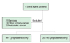
The records of 1,298 patients who received surgical management for uterine cancer were reviewed (Fig. 1). Of the 1,298 patients, 58 patients were excluded because of a diagnosis of non-epithelial cancer including carcinosarcoma, double primary tumor, or other metastatic cancer. Furthermore, 293 patients who did not undergo lymph node dissection were excluded. The characteristics of the remaining 947 patients are summarized in Table 2. As expected, the distribution of stage, tumor grade, myometrial invasion, lymphovascular space invasion (LVSI), and extra-uterine involvement were significantly different between the lymphadenectomy versus non-lymphadenectomy groups, representing the tendency to avoid lymphadenectomy in cases with fewer risk factors.
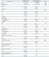
The negative predictive values (NPVs) and negative LRs were not statistically different among the three models (Table 3). However, the proportion of patients classified as low-risk group was significantly different among the models. Model A, which included LVSI information, identified the largest number of patients as a low-risk group (56.4%) without hampering the negative predictive value. Model C identified the smallest low-risk group (30.5%), although its predictive performance was similar to that of other models. In addition, using Bayes' theorem, the negative PTP could be calculated at the 10% of assumed prevalence of lymph node metastasis (Table 3). All models indicated that false negative rate might be 2% when the prevalence of lymph node metastasis was 10%.

DISCUSSION

In the current study, we compared the predictive performance of various prediction models to identify a low-risk group in a large cohort of patients with endometrial cancer. Several clinical implications suggested by our data are as follows.
First, our study revealed that three models based on surgical pathology showed similar negative predictive powers. Our data suggest that the low-risk group can be identified with a false negativity rate of 2% by final pathologic data (Table 3), regardless of the choice of prediction model. Second, although the false negativity of these models was similar, the model from the Gynecologic Oncology Group (GOG) pilot study [12,13], which included LVSI as a predictor, was able to identify the largest number of patients (56%) as a low-risk group. The proportions of patients in the low-risk group identified using those two models (A and C) were significantly different.
In summary, even with final pathologic data, the currently available prediction identifying the low-risk group of lymph node metastasis in endometrial cancer has a false negative rate about 2% at 10% of the assumed prevalence. Therefore, future pre-/intra-operative prediction models may be regarded as clinically useful if the model shows a false negative rate less than 2%, when the prevalence of nodal metastasis was assumed as 10%.

Figures and Tables

Fig. 1
Flowchart of patient selection process.
jgo-23-251-g001
Table 1
Description of three surgicopathological prediction models for the low-risk group of lymph node metastasis in endometrial cancer
jgo-23-251-i001
Table 2
Characteristics of the 1,240 patients included in the analysis
jgo-23-251-i002

Values are presented as number (%) or median (range).

NA, not available.

Table 3
Comparison of model performance in predicting a low-risk group of nodal metastasis
jgo-23-251-i003

Values are presented as percentage (95% confidence interval).

*Comparison with model B and C yielded p<0.001 for both comparisons (chi-square test).

ACKNOWLEDGMENTS

This study was funded by National Cancer Center, Korea (grant no. 1210200).

Notes

Presented at the 2012 Annual Meeting of Society of Gynecologic Oncology.

No potential conflict of interests relevant to this article was reported.

References

1. Benedetti Panici P, Basile S, Maneschi F, Alberto Lissoni A, Signorelli M, Scambia G, et al. Systematic pelvic lymphadenectomy vs. no lymphadenectomy in early-stage endometrial carcinoma: randomized clinical trial. J Natl Cancer Inst. 2008. 100:1707–1716.
2. ASTEC study group. Kitchener H, Swart AM, Qian Q, Amos C, Parmar MK. Efficacy of systematic pelvic lymphadenectomy in endometrial cancer (MRC ASTEC trial): a randomised study. Lancet. 2009. 373:125–136.
3. Creasman WT, Odicino F, Maisonneuve P, Quinn MA, Beller U, Benedet JL, et al. Carcinoma of the corpus uteri: FIGO 26th Annual Report on the Results of Treatment in Gynecological Cancer. Int J Gynaecol Obstet. 2006. 95:Suppl 1. S105–S143.
4. Greer BE, Koh WJ, Abu-Rustum N, Bookman MA, Bristow RE, Campos SM, et al. Uterine neoplasms: clinical practice guidelines in oncology. J Natl Compr Canc Netw. 2009. 7:498–531.
5. American College of Obstetricians and Gynecologists. ACOG practice bulletin, clinical management guidelines for obstetrician-gynecologists, number 65, August 2005: management of endometrial cancer. Obstet Gynecol. 2005. 106:413–425.
6. Kitchener HC. To stage or not to stage? That is the question: (with apologies to Shakespeare). Int J Gynecol Cancer. 2010. 20:S55–S56.
7. Walsh CS, Karlan BY. Lymphadenectomy's role in early endometrial cancer: prognostic or therapeutic? J Natl Cancer Inst. 2008. 100:1660–1661.
8. Uccella S, Podratz KC, Aletti GD, Mariani A. Re: Systematic pelvic lymphadenectomy vs no lymphadenectomy in early-stage endometrial carcinoma: randomized clinical trial. J Natl Cancer Inst. 2009. 101:897–898.
9. Chan JK, Wu H, Cheung MK, Shin JY, Osann K, Kapp DS. The outcomes of 27,063 women with unstaged endometrioid uterine cancer. Gynecol Oncol. 2007. 106:282–288.
10. Trimble EL, Kosary C, Park RC. Lymph node sampling and survival in endometrial cancer. Gynecol Oncol. 1998. 71:340–343.
11. Lee TS, Kim JW, Kim SH, Seong SJ, Song ES, Kim JH, et al. Surgical practice patterns in endometrial cancer: results of the Korean Gynecologic Oncology Group survey. J Gynecol Oncol. 2009. 20:107–112.
12. Boronow RC. Surgical staging of endometrial cancer: evolution, evaluation, and responsible challenge: a personal perspective. Gynecol Oncol. 1997. 66:179–189.
13. Boronow RC, Morrow CP, Creasman WT, Disaia PJ, Silverberg SG, Miller A, et al. Surgical staging in endometrial cancer: clinical-pathologic findings of a prospective study. Obstet Gynecol. 1984. 63:825–832.
14. Creasman WT, Morrow CP, Bundy BN, Homesley HD, Graham JE, Heller PB. Surgical pathologic spread patterns of endometrial cancer: a Gynecologic Oncology Group Study. Cancer. 1987. 60:2035–2041.
15. Mariani A, Webb MJ, Keeney GL, Haddock MG, Calori G, Podratz KC. Low-risk corpus cancer: is lymphadenectomy or radiotherapy necessary? Am J Obstet Gynecol. 2000. 182:1506–1519.
16. Schink JC, Lurain JR, Wallemark CB, Chmiel JS. Tumor size in endometrial cancer: a prognostic factor for lymph node metastasis. Obstet Gynecol. 1987. 70:216–219.
17. Morrow CP, Bundy BN, Kurman RJ, Creasman WT, Heller P, Homesley HD, et al. Relationship between surgical-pathological risk factors and outcome in clinical stage I and II carcinoma of the endometrium: a Gynecologic Oncology Group study. Gynecol Oncol. 1991. 40:55–65.
18. Case AS, Rocconi RP, Straughn JM Jr, Conner M, Novak L, Wang W, et al. A prospective blinded evaluation of the accuracy of frozen section for the surgical management of endometrial cancer. Obstet Gynecol. 2006. 108:1375–1379.
19. Neubauer NL, Havrilesky LJ, Calingaert B, Bulusu A, Bernardini MQ, Fleming ND, et al. The role of lymphadenectomy in the management of preoperative grade 1 endometrial carcinoma. Gynecol Oncol. 2009. 112:511–516.
20. Chan JK, Kapp DS. Role of complete lymphadenectomy in endometrioid uterine cancer. Lancet Oncol. 2007. 8:831–841.
21. Mariani A, Dowdy SC, Cliby WA, Gostout BS, Jones MB, Wilson TO, et al. Prospective assessment of lymphatic dissemination in endometrial cancer: a paradigm shift in surgical staging. Gynecol Oncol. 2008. 109:11–18.
22. Todo Y, Okamoto K, Hayashi M, Minobe S, Nomura E, Hareyama H, et al. A validation study of a scoring system to estimate the risk of lymph node metastasis for patients with endometrial cancer for tailoring the indication of lymphadenectomy. Gynecol Oncol. 2007. 104:623–628.
23. Ballester M, Dubernard G, Lecuru F, Heitz D, Mathevet P, Marret H, et al. Detection rate and diagnostic accuracy of sentinel-node biopsy in early stage endometrial cancer: a prospective multicentre study (SENTI-ENDO). Lancet Oncol. 2011. 12:469–476.
24. Suh DH, Kim K, Kim JW. Major clinical research advances in gynecologic cancer in 2011. J Gynecol Oncol. 2012. 23:53–64.
25. Kang S, Kang WD, Chung HH, Jeong DH, Seo SS, Lee JM, et al. Preoperative identification of a low-risk group for lymph node metastasis in endometrial cancer: a Korean gynecologic oncology group study. J Clin Oncol. 2012. 30:1329–1334.
26. Soliman PT, Frumovitz M, Spannuth W, Greer MJ, Sharma S, Schmeler KM, et al. Lymphadenectomy during endometrial cancer staging: practice patterns among gynecologic oncologists. Gynecol Oncol. 2010. 119:291–294.
27. Lyman GH, Giuliano AE, Somerfield MR, Benson AB 3rd, Bodurka DC, Burstein HJ, et al. American Society of Clinical Oncology guideline recommendations for sentinel lymph node biopsy in early-stage breast cancer. J Clin Oncol. 2005. 23:7703–7720.
28. Lee KB, Ki KD, Lee JM, Lee JK, Kim JW, Cho CH, et al. The risk of lymph node metastasis based on myometrial invasion and tumor grade in endometrioid uterine cancers: a multicenter, retrospective Korean study. Ann Surg Oncol. 2009. 16:2882–2887.
29. Bossuyt PM, Reitsma JB, Bruns DE, Gatsonis CA, Glasziou PP, Irwig LM, et al. Towards complete and accurate reporting of studies of diagnostic accuracy: the STARD initiative: Standards for Reporting of Diagnostic Accuracy. Clin Chem. 2003. 49:1–6.
30. Chan JK, Kapp DS, Cheung MK, Osann K, Shin JY, Cohn D, et al. The impact of the absolute number and ratio of positive lymph nodes on survival of endometrioid uterine cancer patients. Br J Cancer. 2007. 97:605–611.
31. Chi DS, Barakat RR, Palayekar MJ, Levine DA, Sonoda Y, Alektiar K, et al. The incidence of pelvic lymph node metastasis by FIGO staging for patients with adequately surgically staged endometrial adenocarcinoma of endometrioid histology. Int J Gynecol Cancer. 2008. 18:269–273.
32. Jaeschke R, Guyatt GH, Sackett DL. The Evidence-Based Medicine Working Group. Users' guides to the medical literature. III. How to use an article about a diagnostic test. B. What are the results and will they help me in caring for my patients? JAMA. 1994. 271:703–707.
33. Jaeschke R, Guyatt G, Sackett DL. Evidence-Based Medicine Working Group. Users' guides to the medical literature. III. How to use an article about a diagnostic test. A. Are the results of the study valid? JAMA. 1994. 271:389–391.
TOOLS
ORCID iDs

Sokbom Kang
https://orcid.org/http://orcid.org/0000-0002-8428-9555

Jae-Kwan Lee
https://orcid.org/http://orcid.org/0000-0003-3101-6403

Jae Weon Kim
https://orcid.org/http://orcid.org/0000-0003-1835-9436

Similar articles