

 PDF
PDF ePub
ePub Citation
 Print
Print


INTRODUCTION
Epithelial ovarian cancer (EOC) is still the leading cause of death among gynecologic malignancies because it is usually diagnosed in advanced-stage of the disease. Although EOCs are highly responsive to initial combination chemotherapy with taxane-carboplatin, successful management of advanced or recurrent gynecological malignancies is often difficult because of resistance of cancer cells to cytotoxic drugs [1,2]. Therefore, new strategies that may be effective in improving survival and enhancing the response rate to chemotherapy are needed for these patients.
Accumulating epidemiological and translational studies suggest that selenium protects against the development of various cancers including prostate, colon, esophagus, lung, and stomach [3-6]. Especially, numerous case-control studies have demonstrated an inverse relationship between selenium status and prostate cancer risk [7-11]. A randomized controlled study of selenium as a chemopreventive agent demonstrated that supplementation of people with selenized yeast is capable of reducing the overall cancer morbidity by nearly 50% [3].
Generally, selenium compounds fall into two categories, organic and inorganic forms. Organic selenium exists in the form of selenoproteins such as glutathione peroxidases and thioredoxin reductases [12] which regulate cellular redox balance, and together with several low-molecular weight selenium compounds have been shown to play an important role in protection of mammalian cells against various forms of environmental stress as well as against tumor development [13]. Sodium selenite is a major inorganic form of selenium which is often used in chemopreventive studies [14]. Several studies have shown that sodium selenite inhibits growth of cancer cell lines including prostate and liver cancer, malignant melanoma or various hematologic malignancies via inducing apoptosis via different mechanisms involving oxidative stress, mitogen-activated protein kinases, p53-dependent signaling and mitochondria [15-18]. Although there is some evidence for selective selenium toxicity to cancer cells, there is relatively few data on specific effects cytotoxic of selenium and as a supportive element during chemotherapy in EOC. In this study, we tried to evaluate the in vitro and in vivo effect of inorganic selenium (sodium selenite) on tumor growth and food intake in EOC using an animal model.
MATERIALS AND METHODS
1. Chemical and cell culture
Sodium selenite was obtained from Sigma-Aldrich Inc. (St. Louis, MO, USA). Human EOC cell lines (HeyA8, A2780, and SKOV3ip1) were obtained from the American Type Culture Collection (ATCC, Manassas, VA, USA). All of ovarian cancer cells were maintained in RPMI 1640 supplemented with 10% fetal bovine serum (FBS) and 0.1% gentamicin sulfate (Gemini Bioproducts, Calabasas, CA, USA) in 5% CO2 at 37℃.
2. Cell proliferation assay
Cells were plated in culture medium in a 96-well plate at 3×10³ cells/well. After 24 hours, cells were treated to the sodium selenite and the assay was performed at 48 hours and 72 hours. For proliferation assays, cells were stained with 3-(4,5-Dimethylthiazol-2-yl)-2,5-diphenyltetrazolium bromide MTT (Amresco, Solon, OH, USA), after 4 hours of additional incubation, the medium was discarded, 100 µL of acidic isopropanol (0.1 N HCL in absolute sopropanol) was added, and the plate was shaken gently. Absorbance was measured on an enzyme linked immunosorbent assay (ELISA) reader at a test wavelength of 540 nm.
3. Apoptosis assay
The relative percentage of apoptotic cells was assessed at 24 hours using the Annexin V-FITC apoptosis Detection Kit-1 (BD Pharmingen, San Diego, CA, USA) according to the manufacturer's protocol. Briefly, SKOV3ip1 cells were washed twice in phosphate buffered saline (PBS), and the pellet was resuspended in annexin V binding buffer at a concentration of 106 cells/mL. Annexin V FITC and propidium iodide (PI) were added (5 µL to each per 105 cells). Samples were mixed gently and incubated for 15 minutes at room temperature in the dark before fluorescence activated cell sorter (FACS) analysis.
4. Animals and in vivo treatment
Female BALB/c nude mice were purchased from Orient Bio (Seongnam, Korea). This study was reviewed and approved by the Institutional Animal Care and Use Committee (IACUC) of the Samsung Biomedical Research Institute (SBRI). SBRI is an Association for Assessment and Accreditation of Laboratory Animal Care International (AAALAC International, protocol No. H-A9-003) accredited facility and abides by the Institute of Laboratory Animal Resources (ILAR) guide. To generate tumors, SKOV3ip1 (1.0×10⁶ cells/0.2 mL HBSS) were injected into the peritoneal cavity of BALB/c nude mice [19,20], that were 8 to 12 weeks old. Paclitaxel (100 µg) or PBS was injected i.p. once weekly; sodium selenite (1.5 mg/kg) was injected 3 times weekly i.p. in 200 µL volume [21]. Mice (n=10 per group) were monitored for adverse effects, and tumors were harvested after 4 weeks of therapy or when any of the mice began to appear moribund. Mice (n=10 per group) were monitored daily for tumor development and postoperative complications and were sacrificed on days 35 to 40, or if mice seemed moribund. Total body weight, tumor weight, tumor incidence, and the number of tumor nodules were recorded. To assess the effect of survival and food intake in mice, we performed separate animal experiments and measured the oral intake for every week until death.
5. Statistical analysis
Continuous variables were compared with the Student's t-test or ANOVA if normally distributed and the Mann-Whitney rank sum test if distributions were nonparametric using GraphPad Prism 4 for Windows ver. 4.0 (GraphPad Software Inc., La Jolla, CA, USA). A p<0.05 was considered statistically significant.
RESULTS
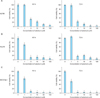
1. Selenium inhibits cell survival of ovarian cancer cells in vitro
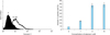
Ovarian cancer cells were treated with sodium selenite for 48 hours and 72 hours. Selenium significantly inhibited cell survival in A2780, HeyA8, and SKOV3ip1 ovarian cancer cells with a dose-dependent manner (Fig. 1). All cells types showed inhibited cell survival over 50% when treated with more than 5 µM of selenium. Moreover, we assessed the effect on apoptosis of selenium using the SKOV3ip1 cell. Apoptosis was significantly increased in these cells dose-dependently by adding selenium (Fig. 2).
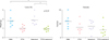
2. Selenium does not have addictive effect with paclitaxel in inhibiting tumor growth of SKOV3ip1 animal model
The SKOV3ip1 ovarian cancer cells were implanted into the peritoneal cavity of nude mice for experiments designed to test the therapeutic potential of selenium administration. From seven days after cell injection, therapy was started according to the four treatment groups; control, paclitaxel, selenium and co-treatment of paclitaxel and selenium. The animals were sacrificed after 4 weeks of therapy and a necropsy was done. The data for the effects of these therapies are summarized in Fig. 3. Selenium alone was not effective in inhibiting tumor growth compared with controls treated with PBS. Treatment with paclitaxel alone or paclitaxel plus selenium was effective in inhibiting tumor growth compared with controls. However, there was no significant difference between paclitaxel alone and paclitaxel plus selenium groups. This result suggests that adding selenium into tumor bearing mice did not seem to have an addictive effect in inhibiting tumor growth. Daily monitoring of the animals throughout the course of therapy showed acceptable tolerability with no untoward side effects, such as changes in body weight, mobility, posture, and feeding habits between all groups.
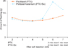
3. Selenium supplementation exhibited slightly increased food oral intake during paclitaxel chemotherapy
To evaluate the effect of selenium as a supportive element during the chemotherapy in EOC, we checked the survival, food and water intake of mice receiving paclitaxel with or without selenium supplementation. Survival and water intake of both groups did not show significant difference according to selenium supplementation. However, we found that the mean food intake was slightly increased in the selenium-treated group, especially 5 to 7weeks after cell injection (Fig. 4).
DISCUSSION
In this study, we found that selenium treatment significantly decreased cell survival in a dose-dependent manner in A2780, HeyA8, and SKOV3ip1 ovarian cancer cells. However, adding selenium into tumor bearing mice did not seem to have an addictive effect in inhibiting tumor growth in the SKOV3ip1 ovarian cancer model. In addition, the combination of selenium and paclitaxel showed slightly increased food intake compared with paclitaxel alone.
The anti-cancer mechanisms of selenium are still not fully understood. Several mechanisms have been proposed, which include maintenance of glutathione peroxidase activity to protect against oxidative damage, detoxification of intermediate metabolites of chemical carcinogens, stimulation of the immune system, induction of cell cycle arrest and apoptosis, and inhibition of angiogenesis [13,22-24]. Selenium has been shown to have potent anti-proliferative and cytotoxic properties and several in vitro studies carried on different cancer cell lines further demonstrated its ability to induce apoptosis in target cells, mostly via increased oxidative stress and DNA damaging [25,26]. In colon cancer cells, selenium inhibits the growth and proliferation in a time- and dose-dependent manner, and selenite-induced toxicity may be related to p53 and dependent signaling [27]. Studies have shown that selenium induced prostate cancer cell apoptosis and cell cycle arrest, processes that have been postulated to be critical for cancer chemoprevention by selenium [28-30].
Several studies have showed the in vivo effects of selenium as therapy to treat mice bearing human cancers. Cao et al. [31] reported that selenium is a highly effective modulator of the therapeutic efficacy and selectivity of anticancer drugs in nude mice bearing human tumor xenografts of colon carcinoma and squamous cell carcinoma of the head and neck. Corcoran et al. [32] reported that high dose dietary supplementation of inorganic selenium inhibits the progression of hormone refractory prostate cancer, which is due at least in part to a decrease in angiogenesis. In addition, selenite treatment inhibits tumor volume by 58% after 42 days of treatment [33] in a xenograft model of human prostate cancer. Moreover, Cafrey and Frenkel [21] reported that when either selenite or selenomethionine were administered close to the time of the initial cisplatin treatment, the induction of resistance was prevented in human ovarian tumors in vivo. They suggested that selenium compounds can prevent the induction by cisplatin of drug resistance [21]. However, this study did not show any addictive in vivo effects of selenium for inhibiting tumor growth. We could not suggest exactly the possible explanation for the disparity of this study between the in vitro and in vivo results, but our results that used a partially immunodeficient model suggest other mechanisms by which selenium can modulate the tumor microenvironment. Further experiments with other chemotherapeutic agent rather than paclitaxel or use of organic selenium should be required to confirm this result.
There have been several few clinical trials investigating the effect of selenium supplementation during cancer chemotherapy. Forty-one patients with solid tumors undergoing cisplatin chemotherapy were randomized into two groups, and the group that received selenium showed significantly higher WBC counts on day 14 after initiation of chemotherapy [34]. Furthermore, consumption of granulocyte colony-stimulating factor and volumes of blood transfusion were significantly less in the selenium-supplemented group. An ovarian cancer study was done with 62 women undergoing cisplatin and cyclophosphamide combination chemotherapy and half of the patients received selenium supplementation [35]. The group that received selenium showed significantly reduced neutropenia as well as increased WBCs from the second to third chemotherapy cycle. The authors also report that with selenium supplementation, there seemed to be a significant decrease in all cited side effects: nausea, vomiting, hair loss, etc. In this study, we found slightly increased food intake in selenium-treated mice during the chemotherapy. Further evaluation could be needed to identify these effects in human trial.
In conclusion, we found that selenium treatment significantly decreased cell survival in a dose-dependent manner in several ovarian cancer cells. However, adding selenium into tumor bearing mice did not seem to have any addictive effect in inhibiting tumor growth in SKOV3ip1 ovarian cancer model. In addition, the combination of selenium and paclitaxel showed slightly increased food intake compared with paclitaxel alone.
 XML Download