

 PDF
PDF ePub
ePub Citation
 Print
Print


INTRODUCTION
Borderline ovarian tumors constitute about 4-14% of all ovarian cancers. In contrast, fallopian tubal or paratubal borderline tumors are extremely rare and have not been well defined [1,2]. To our knowledge, only 10 cases of fallopian tubal serous borderline tumors have been described since 1965. In addition, only one case of paratubal serous borderline tumor has been reported in the literature [2], although paratubal cysts are common incidental findings during gynecologic operations [3]. Therefore, the clinicopathologic characteristics and treatments of these tumors are unclear. We describe here a patient with a paratubal serous borderline tumor, which should be differentiated from an ovarian or fallopian tubal serous borderline tumor, along with a review of the literature.
CASE REPORT
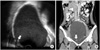
A 27-year-old, nulliparous (0-0-0-0), married woman was referred to our institution with a huge pelvic mass and left flank pain. She had no history of surgical or medical disease. Transvaginal ultrasonography revealed a 16 cm right adnexal cyst with intramural nodules and thickened cyst wall (Fig. 1). Abdomino-pelvic computed tomography (CT) scan showed a 16 cm cystic mass with enhancing solid intramural nodules in the pelvic cavity (Fig. 1). The CT scan showed no evidence of ascites, peritoneal seeding, omental cake, or pelvic or para-aortic lymph node involvement. It looked like an epithelial ovarian cancer because the CT scan showed delayed enhancement. Her serum cancer antigen 125 (CA-125) concentration was 19.3 U/mL (reference value, <35 U/mL), and the results of other preoperative work-up tests were unremarkable.
We performed an exploratory laparotomy through a low midline abdominal incision, which revealed a 16 cm right paratubal cyst completely differentiated from the right ovary and fallopian tube. Paratubal cystectomy was performed without rupture of the cyst. Frozen section analysis of the specimen showed that it was a paratubal serous borderline tumor. Exploration of the entire peritoneal cavity showed no evidence of intraperitoneal tumor spread. Her fallopian tubes, ovaries and uterus were not involved. Washing cytology and multiple peritoneal biopsies were taken. Because the patient and her husband had a strong desire to preserve fertility, her uterus and both adnexae were preserved.
Grossly, the tumor had a homogeneous, purplish white inner surface and multiple papillary projections up to 2.1 cm in greatest dimension (Fig. 2). The external surface of the tumor was homogeneously purplish white and smooth, with multifocal hemorrhage. However, there was no evidence of surface involvement. The final pathology results were identical to the frozen biopsy results indicating that the mass was a paratubal serous borderline tumor (Fig. 2). The results of the peritoneal washing cytology and multiple biopsies were negative.
Her postoperative course was uneventful and she was discharged 5 days after surgery. Regular check-ups at the outpatient clinic include serum CA-125 measurements, transvaginal ultrasonography, and/or abdomino-pelvic CT. She delivered a healthy baby at term 13 months after surgery. Twenty months after surgery, she is doing well, without evidence of disease.
DISCUSSION
Paratubal cysts, also known as hydatid cysts of Morgagni, are common incidental findings during surgery or autopsy [3]. These small round cysts are attached by a pedicle to the fimbriated end of the fallopian tube. Paratubal cysts are of paramesonephric (müllerian) origin and are lined by a single tubal-type ciliated epithelium [3]. Most paratubal cysts are unilateral, asymptomatic and benign, but they may occasionally give rise to clinical problems due to enlargement or torsion. Paratubal serous borderline tumors are very rare, with only one previous case report in a 26 year-old woman who presented with torsion of the cyst [2]. This patient was successfully managed with right salpingo-oophorectomy and fertility-sparing comprehensive surgical staging procedures [2]. Our patient underwent a paratubal cystectomy and fertility-sparing comprehensive surgical staging procedures. After surgery, she achieved a successful pregnancy and delivered a normal healthy baby.
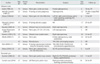
Borderline tumors, also called tumors of low malignant potential, are characterized pathologically by features of malignant tumors, including cellular proliferation, stratification of the epithelial lining of the papillae, nuclear atypia, and elevated mitotic activity, but without destructive stromal invasion [2]. Borderline epithelial ovarian tumors are not uncommon and have been well characterized. In contrast, fallopian tubal and paratubal borderline tumors are much rarer and therefore not well defined. To our knowledge, only 11 cases of serous borderline tumors of the fallopian tube and paratubal cyst have been reported in the English language literature [1,2,4-12]. The clinico-pathologic features of these patients, as well as of the patient described here, are summarized in Table 1 [1,2,4-12]. The patient's age ranged from 19 to 47 years, with a mean age of about 31 years. Most tumors occurred in young women, although a few occurred in postmenopausal women. Seven of the 13 women presented with abdominal pain, whereas the others had asymptomatic masses that were discovered during routine pelvic examination. Two patients showed elevated serum CA-125, as high as 108 and 43.6 U/mL, respectively [7,10]. All tumors were unilateral, measuring 1.7 to 16 cm in diameter, with the largest to date in the patient described here (16 cm right paratubal cyst). None of the patients showed evidence of peritoneal seeding. Nine of the 13 patients underwent conservative surgery. None of the 13 had metastasis or recurrence after follow-up periods of 11-72 months. Two patients, including the one described here, successfully delivered healthy babies at term [6].
Because fallopian tubal and paratubal borderline tumors are very rare, optimal management of patients with these tumors must be extrapolated from their ovarian counterparts [2,10]. Since most of these tumors are unilateral and confined to the fallopian tube or paratubal cyst and since most patients are young, fertility-sparing surgery with comprehensive surgical staging procedures is acceptable, and adjuvant chemotherapy is likely unnecessary. Acceptable fertility-sparing procedures for young patients who desire to maintain childbearing potential include salpingectomy for fallopian tubal serous borderline tumors and paratubal cystectomy for paratubal serous borderline tumors.

 XML Download