

 PDF
PDF ePub
ePub Citation
 Print
Print


INTRODUCTION
Less than 5% of patients with metastatic gestational trophoblastic neoplasia (GTN) have initial involvement of the gastrointestinal tract [1]. Also, chemotherapy is the primary treatment for metastatic GTN. Although the role of surgery in metastatic choriocarcinoma has diminished, local resection of metastases still seems to play a role in a small subset of patients [2]. Here, we present a case of chemo-resistant choriocarcinoma, metastatic to the colon, which was salvaged by surgery.
CASE REPORT
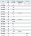
In September 2000, a 48-year-old woman, gravid 4 para 2, presented with acute abdominal pain and hypovolemic shock. Her initial blood pressure was 82/56 mm Hg, pulse rate was 112 beats/minute, and initial hemoglobin was 7.6 g/dL. She underwent an emergency laparotomy, which showed uterine perforation and severe intractable hemorrhage. But there was no seeding nodules or mass, except for the uterus. Total hysterectomy and bilateral salpingo-oophorectomy showed an invasive choriocarcinoma. The postoperative human chorionic gonadotropin (hCG) level was 34 mIU/mL and was normalized during the four cycles of methotrexate and folic acid rescue (MTX-CF) after the operation (Table 1).
In January 2002, the hCG level was elevated (64.9 mIU/mL) and abdomen-pelvis magnetic resonance imaging (MRI) showed a soft tissue nodular mass adherent to the inferoposterior wall of the sigmoid colon. There was no evidence of metastases in other sites on the abdomen-pelvis MRI, chest computed tomography (CT), and brain CT. Four cycles of combination chemotherapy with EMACO (weekly alternating etoposide, methotrexate, actinomycin D/vincristine, and cyclophosphamide) were administered, and the hCG level was normalized following the 2nd cycle. Since 2002, she had been lost for follow-up.
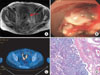
In August 2008, she was hospitalized again for rectal bleeding. Her hemoglobin value was 4.7 g/dL, and the hCG level was 154,380 mIU/mL. Diffuse irregular wall thickening of the rectosigmoid colon with regional lymphadenopathy was identified in the abdomen-pelvis MRI (Fig. 1A). Sigmoidoscopy displayed a lobulating metastatic choriocarcinoma proven by biopsy (Fig. 1B). In the chest CT, pleural thickening with minimal amount of effusion was seen, which was suggestive of metastases. Under the impression of recurrent choriocarcinoma, EMACO chemotherapy had been administered seven times. The hCG level normalized following 4th cycle of chemotherapy.
Three months after the chemotherapy, the hCG level had again risen up to 657 mIU/mL. The abdomen-pelvis CT showed interval progression of the recurrent mass. The positron emission tomography (PET) scan revealed increased fludeoxyglucose uptake (SUVmax=9.9) in the rectosigmoid colon (Fig. 1C). Sigmoidoscopic findings showed stenosis and a protruding mass 14 cm above the anal verge junction.
A secondary cytoreductive surgery for chemo-resistant choriocarcinoma was performed and a huge ulcerative mass appeared at the rectosigmoid junction. Low anterior resection with lymphadenectomy up to the level of the inferior mesenteric artery was performed with no residual mass. The histological examination confirmed metastatic choriocarcinoma involving from the mucosa to the subserosa of the sigmoid colon (Fig. 1D).
After the operation, the hCG level dropped from 6,298 mIU/mL to 2.8 mIU/mL. The patients refused further chemotherapy. However, there has been no evidence of recurrence for 13 months since the operation.
DISCUSSION
We have described a rare case of metastatic GTN responsive to surgery. In this case, the chemo-resistant GTN was treated with low anterior resection. Moreover, the patient reached complete remission shortly after removing the colon metastases.
The prognosis of choriocarcinoma has been substantially improved by the introduction of chemotherapeutic agents. GTN is extremely responsive to chemotherapy, even in its metastatic forms [3]. Therefore, the role of surgery in the treatment of patients with metastatic choriocarcinoma has diminished.
The role of surgery in the treatment of metastatic choriocarcinoma has long been debated. In general, resection of distant metastasis is unlikely to succeed if there is evidence of disseminated disease resistant to chemotherapy. In the 1960s, Lewis and his associates documented that the common indications for surgery in GTN were the need for controlling hemorrhage, removing residual disease when resistant to chemotherapy, relieving urologic obstruction, and treating infection [3,4]. Also, local resection of metastases still seems to play a significant role even in curing the disease in a small subset of patients.
Choriocarcinoma is characterized by the rapid invasion of surrounding tissue and early hematogenous metastases [5]. Common sites of metastases outside the pelvis are the lung, liver, and kidney [5]. Less than 5% of cases of metastatic GTN involve the gastrointestinal tract [1]. Although risk factor of colonic metastases is not known, it is assumed that previous uterine perforation is associated with colonic recurrence [3]. It is not known whether intestinal metastases would place patients at a higher risk of treatment failure. However, bowel resection may be beneficial in selected patients with intestinal metastases resistant to chemotherapy.
The treatment of chemo-resistant GTN should be individualized. All patients with high-risk should be treated with intensive combination chemotherapy and the selective use of radiation therapy and surgery. Recently, several authors reported favorable results with salvage surgery for chemotherapy-resistant GTN [6]. Hysterectomy may be necessary to control uterine hemorrhage or sepsis, or to resect resistant disease [6]. Thoracotomy may be performed to excise a persistent viable tumor despite intensive chemotherapy [7]. Hepatic resection may be required to manage bleeding metastases although embolization has also been utilized in this setting [5]. Craniotomy may be necessary to provide acute decompression or to control bleeding, in addition to its role in the primary resection of solitary metastatic disease [8].
Feng et al. [9] have suggested that age older than 35 years, antecedent non-molar pregnancy, distant metastasis outside of the lungs and uterus, and a preoperative serum β-hCG level greater than 10 IU/L are important clinical predictors of treatment failure, which will require following surgery. An extensive metastatic survey should be undertaken to exclude other sites of persistent tumor. A PET scan may be useful to identify occult sites of viable tumor [10].
In conclusion, although combination chemotherapy is the main treatment used in patients with high-risk GTN, surgical intervention may occasionally be required. In this case, surgery was effective for the cure of chemo-refractory choriocarcinoma, metastatic to the colon.
 XML Download