

 PDF
PDF ePub
ePub Citation
 Print
Print


INTRODUCTION
Ovarian cancer is the leading cause of death from gynecologic malignancies [1]. A retrospective study by Griffiths [2] in 1975 demonstrated a strong association between postoperative largest residual tumor and survival. Since then, initial cytoreductive (debulking) surgery and adjuvant chemotherapy has become the standard of care in advanced epithelial ovarian cancer [3-5].
Currently the treatment concept of neoadjuvant chemotherapy has been introduced. In this alternative management approach the initial treatment in some advanced ovarian carcinoma patients usually consists of 3 courses of chemotherapy followed by cytoreductive surgery (interval debulking) and additional three courses of postoperative chemotherapy. Proposed advantages of neoadjuvant chemotherapy include an increased rate of optimal residual disease, less extensive surgery, reduced blood loss, lower morbidity, shortened hospital stay and improved quality of life.
Neoadjuvant chemotherapy may also act as a mechanism to select out patients with platinum-resistant disease. While still controversial, it has been found in many retrospective and prospective studies that the outcome after the neoadjuvant approach is not inferior to that after initial cytoreductive surgery [6-9].
Various noninvasive means have been proposed to predict cytoreduction inability for the selection of patients in whom the neodjuvant approach is more appropriate. These include various computed tomography (CT) criteria [10-12] clinical and CA-125 level criteria [13,14].
A few investigations deal with the ability to predict progression free survival (PFS) and overall survival prior to interval debulking [15-18]. Most of them are by assessment of CA-125 level reduction [15-17]. Up to now no reliable prediction method has been found.
The purpose of the present study was to assess whether there is an association between improvement of CT imaging results prior to interval debulking with survival in patients treated by neoadjuvant chemotherapy.
MATERIALS AND METHODS
The records of all advanced ovarian, primary peritoneal and tubal carcinoma patients who after diagnosis had initial chemotherapy during the period 2000-2010, were abstracted after institutional review board approval. The diagnosis of malignancy in these patients was confirmed by cytology examination of ascitic fluid and/or by core biopsy. Patients were allocated to neoadjuvant chemotherapy according to Nelson's CT criteria [10] i.e., mainly in the presence of extensive disease in the upper abdomen and/or disease outside the peritoneal cavity. Their clinical and outcome data were recorded. Neoadjuvant and post-interval debulking chemotherapy consisted of intravenous paclitaxel (175 mg/m2) and carboplatin (AUC 6) for three 21-day cycles.
Results of CT imaging at diagnosis and prior to interval debulking were interpreted and compared by a certified roentgenologist (IU) who was unaware of the outcome. Two parameters where assessed: the change of the diameter and number of abnormal findings, especially in the areas considered to be non-debulkable, and the change in the amount of ascites. Evaluation of response was based on RECIST criteria [19]. In brief according to RECIST criteria complete response (CR) is defined as disappearance of all target lesions. Partial response (PR) is defined as at least a 30% decrease in the sum of diameters of target lesions, and stable disease (SD) is when there is neither sufficient shrinkage to qualify for PR nor sufficient increase to qualify for progressive disease.
In the present study the assessed variables were scored on a 0-2 scale based (but not identical) on the RECIST criteria where 0 represented no improvement (SD according to RECIST), 1 represented some improvement (PR according to RECIST), and 2 represented marked improvement i.e., a reduction of more than 50% in the number and/or the size of the CT findings and no or only minimal presence of ascitic fluid (less stringent than CR according to RECIST).
In addition the CA-125 level at diagnosis and its level prior to interval debulking were recorded. A CA-125 decrease to <35 U/mL was considered as normalization. The percent reduction of CA-125 levels from the value at diagnosis to the value prior of interval debulking was also calculated. An assessment of PFS, i.e., the time between the last course of post interval debulking chemotherapy and the time of recurrence, and an assessment of survival was made according to the change in CT imaging results, according to CA-125 normalization and according to the percent reduction of CA-125 levels. PFS and overall survival were assessed by the Kaplan-Meier method and differences by the log-rank test. Patients with recurrence were treated by a large number [4-7] and dissimilar treatment lines and by a variety of chemotherapeutic agents.
RESULTS
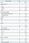
Initial chemotherapy was given to 48 patients. Of the total group 11 patients did not undergo interval debulking -2 expired prior to surgery of concurrent diseases, 3 refused surgery and 6 had progressive disease. The study group thus comprises 37 consecutive patients (29 with ovarian carcinoma, 6 with primary peritoneal carcinoma and 2 with tubal carcinoma) who were managed by the neoadjuvant approach and underwent interval debulking. All tumors were of the papillary serous type. The mean age of the patients was 64.1±10.6 years (range, 38 to 81 years). Additional selected characteristics of the patients are presented in Table 1. The largest percentage of patients was in the 60-69 age group. The majority of the patients presented with abdominal pain and/or distention, and had stage IIIC grade 3 tumors. More than half of the patients had no visible macroscopic disease after interval debulking surgery and more than half where defined as platinum sensitive i.e. recurrence occurred more than 6 months after the end of primary treatment.
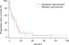
When compared to the CT at diagnosis, the CT prior to interval debulking showed no demonstrable improvement, some improvement and marked improvement in 2 (5.4%), 16 (43.2%) and 19 (51.4%) patients, respectively. Since there were only 2 patients with no improvement, they were combined with those with some improvement for the purpose of further analysis. No ascites was present at diagnosis in 7 patients. In all the remaining 30 study group patients a marked improvement in the amount of ascites was observed. Normalization of CA-125 was found in 16 (43.2%) and a reduction greater than 90% was found in 24 (64.9%) of the patients. A decrease of more than 50% of CA-125 levels was observed in all patients. The PFS according to improvement of CT findings is presented in Fig. 1. The median PFS of the study group was 7.9 months. The PFS of the patients with no/some improvement and of those with marked improvement was 7.93 and 7.23 respectively (p=0.89; 95% CI, 0.55 to 1.99). The median survival of the patients was 49.2 months (mean, 36.9±23.8 months). The median survival of those with no/some improvement and of those with marked improvement was 45.8 months and 52.5 months respectively (p=0.95; 95% CI, 0.41 to 2.09).
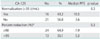
Table 2 presents the association of CA-125 response with PFS and with survival. No statistically significant difference was found between the different categories of CA-125 response.
DISCUSSION
To the best of our knowledge the association between the improvement of imaging findings prior to interval debulking surgery with survival has not been reported until now.
We found that the median PFS of patients with marked improvement in the size and number of abnormal findings was similar to those with less favorable imaging results, and that marked improvement in the amount of ascites was present in all patients. Our study therefore indicates that imaging results prior to interval debulking surgery cannot be used for prediction of PFS. This holds true for survival as well. However, we feel that it is less relevant because of the heterogeneous management of the patients subsequent to recurrence.
It is noteworthy that the PFS of our patients is of shorter duration and that the median survival is longer compared to those reported by Vergote et al. [9] (7.9 vs. 12 months and 49.2 vs. 30 months, respectively). The differences can be attributed to our small number of patients. Although the patients were informed after 3 treatment cycles that the chance of response to additional treatment is slim, they all requested and received multiple additional treatment cycles. Whether the longer survival of our patients is due to the administration of the large number (up to 7) of treatment lines, is questionable. Indeed recent evidence suggests that more number of cycles does not improve the outcome [20].
The rate of patients in our series with no residual disease after interval debulking (62%) is in the range of that reported by Vergote et al. [9] and Brun et al. [21] (50% and 73%, respectively).
Only four previous studies attempted to compare variables at diagnosis with the same variables after neoadjuvant chemotherapy and prior to interval debulking surgery in order to investigate whether improvement in these variables might predict outcome. In two studies Le et al. reported that normalization, defined as a reduction in serum CA-125 levels to less than 35 U/mL, in 16 patients with elevated levels at diagnosis [15] and that a decrease of at least 50% from baseline prior to interval debulking surgery [16] were found not to be independent predictors of either progression-free or overall survival. In contrast, in a third study, CA-125 regression coefficient was calculated and found to be a significant prognostic factor for overall survival [17]. This study was criticized [15] because the definition of the response using a CA-125 regression coefficient was not a standard one and difficult to reproduce, and because in the majority of patients significant cytoreductive surgery was not attempted. In an additional study, in vitro tumor cloning assay results regarding platinum or paclitaxel resistance of 22 ovarian cancer patients treated with neoadjuvant chemotherapy were assessed and were also found not to be predictive of PFS [18]. The results of our study, namely the lack of association of PFS and survival with CA-125 normalization and with the percent CA-125 reduction, are in line with those reported by Le et al. [15,16].
The ability to predict the outcome after neoadjuvant chemotherapy prior to interval debulking is important for two main reasons. It is of great prognostic significance and it may identify patients who might have an unfavorable prognosis thus allowing a more rational planning of further management. This could include additional treatment with different chemotherapeutic agents prior or after interval debulking or to forgo, after informed consent, interval debulking with its inherent morbidity.
It seems that neither improvement in imaging results nor CA-125 level response can predict the outcome after neoadjuvant chemotherapy prior to interval debulking. We are aware of the small size of our series (post hock power of only about 10%), and that our results should be confirmed in a larger study. Whether a larger series would yield a different result remains to be proven. Since positron emission tomography (PET) is an indicator of the metabolic state of malignancies it is possible that PET-CT could be a more accurate imaging modality to predict the outcome prior to interval debulking. However, this remains to be investigated.
Further studies are indicated in order to find a method that will enable prediction of outcome after neoadjuvant chemotherapy and prior to interval debulking.
 XML Download