

 PDF
PDF ePub
ePub Citation
 Print
Print


INTRODUCTION
Posterior pelvic exenteration (PPE) is a significant procedure to achieve complete tumor removal within the pelvis in the surgical treatment of primary advanced ovarian cancer or, less frequently, of recurrent disease. In most cases of this supralevator-type resection a low colorectal anastomosis, which is usually located 4 to 7 cm above the anocutan line, is possible because the infraperitoneal rectum is preserved from invasion by the pelvic mass. Current studies, including a review of literature, report a frequency of clinically significant anastomotic leakage in up to 10%, mostly less than 4% [1,2]. Anastomotic dehiscence is associated with an increased early and long term morbidity and mortality, mostly accompanied by a surgical re-intervention, a longer hospitalization, an impairment of anorectal function, and a delay of subsequent chemotherapy [1-5].
CASE REPORT
A 44-year old woman (gravida 2, para 1) underwent an optimal debulking surgery defined as complete tumor removal because of ovarian cancer FIGO stage IIIC characterized by a pronounced intraperitoneal tumor spread. Midline laparotomy includes the following procedures: posterior pelvic exenteration (PPE; en bloc resection of inner genitals, about 19 cm rectosigmoid and complete pelvic peritoneum), infragastric omentectomy, appendectomy, extensive deperitonealization of the right hemidiaphragm, removal of multiple peritoneal tumor deposits and, finally, a systematic pelvic and para-aortic lymph node dissection. A functioning colorectal anastomosis was realized by end-to-end circular stapling. The anastomotic doughnuts were complete and, additionally, the integrity of anastomosis was tested by insufflation of methylene blue-stained saline solution.
Histopathology revealed a serous adenocarcinoma originating from the ovaries (TNM-classification [2010]: pT3c, pN1 [5/97], pM1 LYM [1/19], L1, V0, G3).
The patient's postoperative recovery was uneventful over the next few days and her condition improved continuously. However, increased inflammation indicating values, leukocytes and C-reactive protein, decreased only gradually. After exclusion of possible foci a 'blind' antibiotic treatment with cefuroxim and metronidazol was initiated on postoperative day 5. Despite this therapy, the patient had complained of fever up to 38.5℃ every afternoon since the eighth day.
On day 11 after surgery, leukocytes and C-reactive protein increased considerably in comparison with the values of the day before (11.3 → 15.5×10⁹/L; 146.2 → 207.3 mg/L). An abdominopelvic computer tomography scan revealed infected lymph cysts surrounding the iliac vessels on both sides (right, 7×6 cm; left, 8×7 cm), but there was no suspicion of any anastomotic leak. The antibiotic treatment was changed to imipenem. Regardless of the continued high temperature during the afternoon, the patient felt relatively comfortable; her bowel function was inconspicuous.
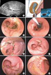
One day later, the causal diagnosis of an anastomotic dehiscence, located 5 cm above the anocutan line, was demonstrated by transvaginal ultrasound (Fig. 1A) and was verified by endoscopy. Considering the patient's condition, it was decided to attempt an Endo-SPONGE® assisted treatment (Fig. 1B; Aesculap AG, Tuttlingen, Germany). A parenteral nutrition for two weeks was initiated and during the nine-day transanal vacuum therapy the sponge was changed twice. Subsequently, a bowel rinsing was semi-weekly performed over the following two weeks (Fig. 1C-H). The anastomotic leakage healed successfully and the patient recovered uneventfully and was discharged on postoperative day 29. The first cycle of combination chemotherapy consisting of Caboplatin AUC 5 and Paclitaxel 175 mg/m2 could be applied two weeks later.
DISCUSSION
The application of transvaginal ultrasound to diagnose a leakage of colorectal anastomosis has not been previously described. The diagnostic criteria are the discontinuity of the rectal wall and an oscillating fluid flow between the bowel lumen and a surrounding hyperechogenic fluid collection, which is induced by pushing in the bowel wall by means of the ultrasound probe. In our opinion, ultrasound diagnostics should be integrated early in the diagnostic management of divergences from the expected course after any surgery.
Further points of interest are the use of protective stoma to prevent anastomotic leakage and the treatment options of this complication in patients receiving PPE for ovarian cancer. The authors of a multicenter study concluded that a temporary protective stoma should be considered only exceptionally, because the rates of "anastomotic fistula" were the same with and without stoma, respectively (8.5% vs. 8.2%) [1]. In addition, there are only a few published studies including small numbers of patients [3]. Our empiric intraoperative management is in accord with this recommendation. However, if an anastomotic dehiscence occurs, a re-operation to secondarily create a covering ileo- or colostomy or to drain the pelvic cavity in order to prevent severe septic complications is necessary in the majority of these cases [4]. The endoluminal vacuum therapy of anastomotic leakages has been increasingly applied following resections in both the upper and lower gastrointestinal tract over the last few years [4,5]. This approach can be integrated into both a surgical and conservative treatment regime, but the therapeutic effectiveness has not been verified by randomized clinical trials. Reports about the application of this method in the field of gynecologic oncology are absent.
Our case demonstrates, firstly, that anastomotic leakage of colorectal anastomosis can be obviously diagnosed by transvaginal ultrasound and, secondly, in selected cases characterized by a small leak size and a local peritonitis confined to the pelvis, an attempt of conservative management based on transanal vacuum therapy is justified and may avoid creating a secondary diverting stoma.
 XML Download