

 PDF
PDF ePub
ePub Citation
 Print
Print


INTRODUCTION
Lipoleiomyoma is a very rare tumor accounting for 0.35 to 2.1% of all uterine leiomyomas and is composed of adipocytes and smooth muscle cells.1,2 Lipoleiomyoma is most commonly located in uterine corpus although cervical, ovarian, and retroperitoneal locations were also reported.1-3 However, lipoleiomyoma located in broad ligament is extremely uncommon. A review of the English literature revealed only five cases of broad ligament lipoleiomyoma reported by this time.1,4-6
Here, we report the sixth case of lipoleiomyoma of broad ligament which was diagnosed in a postmenopausal woman who was subjected to exploratory laparotomy with a preoperative diagnosis of a solid adnexal mass suggesting an ovarian malignancy. The English literature regarding lipoleiomyoma of uterus and lipoleiomyoma of broad ligament was reviewed as well.
CASE REPORT
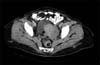
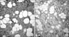
A 54-year-old, gravida 2, parity 2 woman was admitted to our clinic for lower abdominal pain. Her past medical and surgical histories were unremarkable and she experienced her last menstruation 8 years ago. Abdominal examination revealed some tenderness at left lower quadrant and a left adnexal mass was palpated on bimanual pelvic examination. Routine laboratory tests including tumor markers were in normal limits. A transvaginal ultrasonography showed a solid mass of 75×40 mm at left adnexal site. Computerized tomography of abdomen and pelvis confirmed the heterogeneous solid mass of left adnexa measuring 70×35 mm (Fig. 1). An exploratory laparotomy was performed with a preoperative diagnosis of solid adnexal mass suggesting ovarian malignancy given the age of the patient and solid nature of the mass. During laparotomy, a 7 to 8 cm mass was detected within the left broad ligament. Otherwise, the uterus and ovaries were atrophic and exploration of the whole abdomen was free of additional abnormalities. A total abdominal hysterectomy with salpingo-oophorectomy was performed. Frozen section analysis did not suggest malignancy. Final pathologic examination showed a lipoleiomyoma without mitotic figures, nuclear atypia or pleomorphism which immunohistochemically showed reactivity with desmin (Fig. 2). The patient was discharged home 3 days after surgery following an uneventful postoperative course and she is asymptomatic clinically 8 months after the surgery.
DISCUSSION
Lipoleiomyoma of the uterus is an uncommon pelvic tumor with an incidence varying from 0.03% to 0.2%.7 Finding an admixture of mature adipocytes and smooth muscle cells on microscopy is required to be able to designate a neoplasm as lipoleiomyoma. The adipocytes may be evenly distributed throughout the tumor or they may be concentrated in only focal areas. Also, adipocyte component in lipoleiomyoma may differ widely and a certain level of adipocytes was not defined to achieve the diagnosis of lipoleiomyoma. These tumors may contain microscopic foci of adipocytes resembling regular leiomyomas in gross appearance or high amounts of adipocytes may be detected resulting in yellow and lobulated cut surface.1 The tumor in our case had a yellow cut surface grossly and high amounts of adipocytes on microscopy.
The clinical features are uncertain due to its rarity. The exact histogenesis has not been explained clearly. Nevertheless, immunohistochemical studies indicated a complex histogenesis of lipoleiomyoma which might arise from immature mesenchymal cells or from transformation of smooth muscle cells into adipocytes.8 It was also demonstrated that lipoleiomyomas may be associated with some metabolic disorders including hyperlipidemia, hypothyroidism and diabetes mellitus. This suggests that changes in lipid metabolism after menopausal transition may play a role in the development of lipomatous change in leiomyomas.9 This hypothesis is consistent with advanced age of most of the patients at time of diagnosis. However, the current case did not have any of the aforementioned metabolic disorders.
By contrast with ordinary leiomyomas which tend to occur predominantly in women of reproductive age and regress after menopause, the lipoleiomyomas are frequently seen in older women. Actually, mean age of the patients in the largest two series so far was 55.4 years and almost 60% of patients were aged older than 50.1,2 Our patient was a 54-year-old postmenopausal woman as consistent with the literature.
Most patients are asymptomatic and are diagnosed incidentally, but among symptomatic ones pelvic pain, palpable mass or abnormal bleeding are the most common symptoms similar to those caused by leiomyomas. Patients with symptoms usually undergo physical exam followed by imaging modalities which reveal a solid pelvic mass.1 Although some features in different imaging modalities may suggest the possible diagnosis of these tumors, the precise diagnosis is based on pathologic examination.7
The presenting symptom was pain in our case who was found to have an adnexal mass on examination. Therefore, imaging was requested and a heterogenous solid mass located at left adnexal site which did not have an appearance of regular leiomyomas was reported on ultrasonography and computed tomography. Also, during surgery, the mass grossly did not resemble a leiomyoma which was fairly soft on palpation and located outside the uterus. The cut surface was reported to be yellow on gross pathological examination and it contained high amounts of adipocytes on microscopic evaluation. Current patient was subjected to laparotomy with a suspicion of ovarian malignancy. The presence of a pelvic mass in a postmenopausal woman is certainly not enough to consider ovarian malignancy especially if it is not associated with ascites and elevated tumor markers. However, the heterogenous solid nature of the lesion on imaging modalities and its adnexal localization may suggest a malignant neoplasm that warrants surgical exploration with frozen section analysis where available.
Although the lipoleiomyoma is most commonly located in uterine corpus, it may be found elsewhere in pelvis. However, the extrauterine location including broad ligament is the rarest site reported by this time.1-6 In fact, to best of our knowledge, only five cases of broad ligament lipoleiomyoma were reported previously.1,4-6
The long-term follow-up of patients with uterine lipoleiomyoma demonstrated that these lesions are benign without any recurrences or disease-related deaths if they are diagnosed as the unique pelvic pathology.1 On the other hand, among patients with uterine lipoleiomyoma in two largest series, 18.8% of patients were reported to have associated gynecologic malignancies which may originate from uterus, cervix or ovaries,1,2 Therefore, the patients with uterine lipoleiomyoma should be subjected to detailed clinical and pathological evaluation in order not to overlook a coexistent gynecologic malignancy.
In conclusion, lipoleiomyoma of the broad ligament may necessitate surgical intervention not only due to associated symptoms but also to be able to exclude an ovarian malignancy given its solid nature and the postmenopausal age of the affected individuals. The specimens should be examined carefully to document whether or not a coexisting gynecologic malignancy is present. The patients without an associated malignancy may be followed with routine annual gynecologic exams, because the tumor lacks the risk to recur.
 XML Download