

 PDF
PDF ePub
ePub Citation
 Print
Print


INTRODUCTION
A cellular fibroma is an ovarian stromal tumor with pure proliferation of fibroblastic cells, and is characterized by increased cellularity, with slight to moderate nuclear atypia, and three or fewer mitotic figures per ten high-power fields. Cellular fibromas are not rare, and constitute 10% of fibromatous tumors.1
Leydig cells are the primary source of androgens in the testes. In females, Leydig cell tumors of the ovary are rare and are associated with androgenic manifestations. In addition, Leydig cell components are rarely encountered in mixed tumors, which are generally in the group of Sertoli-stromal cell tumors. However, stromal tumors containing Leydig cell components are extremely rare. Only a few ovarian stromal Leydig cell tumors have been reported, and these were characterized by clusters of Leydig cells containing Reinke crystals within fibrothecomatous tissue without sex cord elements.2,3 To date, there have been no reports of a stromal tumor with cellular atypia or mitotic activity containing Leydig cells. Here, we report the first case of an ovarian cellular fibroma containing Leydig cell hyperplasia.
CASE REPORT
1. Clinical history
A 65-year-old gravida 7 para 4 postmenopausal woman presented to our gynecology outpatient clinic with lumbago. A pelvic examination revealed a palpable non-tender pelvic mass. Transvaginal ultrasonography showed a multiloculated cystic mass in the left ovary. Abdominopelvic computed tomography (CT) revealed a mass with solid and cystic components in the pelvic cavity, measuring 10 cm in diameter, with dense, amorphous calcification. The results of laboratory studies, including analysis of tumor markers, such as AFP, CA125, CA19-9, β-hCG, and inhibin A, were all within normal limits.
At laparotomy, there was no ascites and a lobulated bulging mass measuring 10×6×4 cm was observed on the left ovary. The right ovary, uterus, omentum, and pelvic and paraaortic lymph nodes were unremarkable. A frozen section of the ovarian mass revealed a sex cord-stromal tumor with nuclear atypia and mitosis. The patient underwent abdominal total hysterectomy, bilateral salpingo-oophorectomy, bilateral pelvic and paraaortic lymph node dissection, omentectomy, and appendectomy. Intraoperative pelvic washing cytology was negative and all of the resected tissues, except the left ovary, were free from tumor. Her postoperative recovery was uneventful, and no problems were noted at the 26-month follow-up examination.
2. Pathological findings
On gross examination, the left ovary was mostly replaced by a lobulated bulging solid mass, measuring 10.5×6.5×4.5 cm. It was composed partly of densely fibrotic, hyalinized, and calcified tissue, while the remainder was composed of tan, soft fleshy tissue.
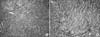
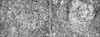
Histological examination revealed the tumor to consist of proliferating spindle cells arranged in intersecting bundles. Mild nuclear atypia and an average of 2-3 mitotic figures per ten high-power fields were present (Fig. 1). The left ovarian tumor was diagnosed as a cellular fibroma. The cellular fibroma contained multifocal nests of polygonal cells with abundant eosinophilic cytoplasm and round nuclei. These cells were considered Leydig cells, and so multifocal areas of Leydig cell hyperplasia were observed (Fig. 2).
DISCUSSION
Pure Leydig cell tumors are extremely rare, and Leydig cells in such lesions usually originate from the hilum of the ovary. These are referred to as Leydig cell tumors, hilus cell type. Rarely, Leydig cell tumors originate from the ovarian stroma, and are referred to as Leydig cell tumors, non-hilar type. While most Leydig cell tumors, non-hilar type, originate from normal stroma, the stromal-Leydig cell tumor arises in a background of neoplastic stromal proliferation. Therefore, according to the World Health Organization (WHO) classification of ovarian sex cord-stromal tumors, the stromal-Leydig cell tumor does not belong in the steroid cell tumor category, but in the Sertoli-stromal cell tumor category.1
In our case, Leydig cell hyperplasia containing polygonal cells with abundant eosinophilic cytoplasm was observed within the neoplastic stromal tumor, as in a cellular fibroma. However, it is difficult to conclude that this case was definitely a stromal-Leydig cell tumor, due to the difference in their distribution.3 Unlike a stromal-Leydig cell tumor, which is characterized by an even distribution of stromal proliferation and clusters of Leydig cells, this tumor was a cellular fibroma with small amounts of Leydig cell hyperplasia. Therefore, it was thought to belong to the theca-fibroma group rather than the stromal-Leydig cell tumor group, similar to a simple cellular fibroma.
The mechanism leading to the development of clusters of Leydig cells in the proliferating stroma is unknown. Sternberg and Roth4 hypothesized the rare non-neoplastic transformation of ovarian stromal cells into Leydig cells. Concurring with this hypothesis, Konishi et al.5 observed immature and mature Leydig cells intermingled with stromal cells in an ovarian tumor by electron microscopy, suggesting that the Leydig cells had differentiated from the ovarian stromal cells, perhaps under the influence of stretch stimuli or hormones.
Leydig cells secrete androgen hormones. Therefore, tumors containing Leydig cell are usually associated with androgenic manifestations and occasionally produce estrogenic effects. The androgenic manifestations include amenorrhea, hirsutism, breast atrophy, clitoral hypertrophy, and hoarseness, whereas the estrogenic effects include isosexual pseudoprecocity and menometrorrhagia. Hirsutism and/or virilization occurs in 75% of pure Leydig cell tumors.1 However, our patient showed no signs of androgenic or estrogenic effects. Her Leydig cell hyperplasia was thought to be non-functional.
Both pure Leydig cell tumors and stromal-Leydig cell tumors are considered benign.1 In this case, the Leydig cells showed neither atypia nor mitotic figures and the Leydig cell hyperplasia was thought to be benign. However, cellular fibromas have low malignant potential intermediate between typical fibroma and fibrosarcoma. Therefore, the treatment modality of a cellular fibroma with Leydig cell hyperplasia should follow that of a neoplasm with low malignant potential.
In summary, we reported a 65-year-old woman with a cellular fibroma including Leydig cell hyperplasia without steroid function.
 XML Download