

 PDF
PDF ePub
ePub Citation
 Print
Print


INTRODUCTION
Conization of the uterine cervix such as large loop excision of the transformation zone (LLETZ) and cold knife conization (CKC) is not only a diagnostic procedure but also an appropriate treatment for cervical intraepithelial neoplasia (CIN).1,2 However, CIN can recur, and invasive cervical carcinoma can develop, following such CIN treatment. The cumulative rate of invasion 8 years after CIN treatment is 5.8 per 1000 women, which is five times higher than for the general population.3 These findings indicate the importance of continuous and meticulous follow-up. Factors reported to be associated with persistent or recurrent cervical neoplasms after conization include menopausal status, grade of dysplasia, follow-up cervical cytology, cone diagnosis of CIN 3, cone margin status, and positive endocervical curettage. However, these factors are suboptimal predictors,4-12 and cannot be used to dictate the follow-up strategy after conization. While there is increasing evidence that testing for the presence of high risk-human papilloma virus (HR-HPV) after conization may help predict the likelihood of persistent or recurrent disease,1,13-22 no study has shown how HR-HPV testing might be integrated into post conization surveillance.
The aim of this study was to determine whether HR-HPV test after conization is a predictive factor for CIN persistence or recurrence after LLETZ or CKC of the cervix. The study also investigated whether HR-HPV test results should influence post conization surveillance.
MATERIALS AND METHODS
From March 2001 to May 2006, 754 patients underwent conization of the cervix including LLETZ and CKC for CIN or microinvasive cervical cancer at the Center for Uterine Cancer, National Cancer Center, Korea. A retrospective chart review was performed on these patients. The inclusion criteria of this study were: 1) patients whose follow-up cytology results and HR-HPV test results using the Hybrid Capture II (HC II) assay after conization were available, 2) patients whose first follow-up cytology and HR-HPV test were performed within 6 months after conization, and 3) patients whose follow-up period was longer than 12 months.
The detailed methods for cervical cytology, HR-HPV test with HC II, and conization (LLETZ and CKC) were described in our previous reports.23,24 HC II is the only HPV test approved by the United States Food and Drug Administration and is a liquid hybridization assay designed to detect 13 high-risk HPV types (HPV type 16, 18, 31, 33, 35, 39, 45, 51, 52, 56, 58, 59, and 68). In our study, a RLU/PC ratio of 1 or higher was considered a positive result. The follow-up HR-HPV test and cytology was performed at 3-6 months after conization, after which the patients were followed-up every 3-6 months.
A logistic regression model and the Kaplan-Meier method were used to identify risk factors for persistent or recurrent cytological and pathological abnormalities after conization, and to determine the relative risk of persistence or recurrence. Student's t-test and Mann-Whitney U-test were used to evaluate the differences in the mean and median values between groups, and Chi-squared test and Fisher's exact test were used to evaluate the differences in the proportions. The differences were regarded as significant when the p-value was less than 0.05 in the two-sided test. SPSS software for Windows (version 9.0; SPSS inc., Chicago, IL) was used for analysis of data.
RESULTS
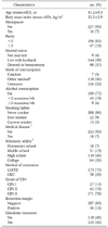
A total of 243 patients met the inclusion criteria and were included in this study. For the 243 study patients, the mean age was 41.2 years (range, 23 to 75 years), and 16 were postmenopausal. The parity was 1 or 2 in 196 patients. LLETZ was performed in 173 patients, and CKC was performed in 70 patients. Following conization, the diagnosis was CIN I in 27 patients, CIN II in 45 patients, and CIN III in 171 patients. Patient characteristics are listed in Table 1. The first follow-up visit after conization was within 6 months for all patients, and the median follow-up period was 24 months (range, 12 to 57 months).
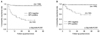
HR-HPV testing between 3 and 6 months after conization showed that 44 patients were HR-HPV positive and 199 were HR-HPV negative. Recurrent cytological abnormalities were found in 26 of the 44 HR-HPV positive patients, and in 12 of the 199 HR-HPV negative patients. Analysis showed that a positive HR-HPV result was a risk factor for recurrent cytological abnormality (p<0.001, OR=22.51, 95% CI=9.74-52.02) (Fig. 1).
The types of recurrent cytological abnormalities were ASCUS in 7 patients, ASCH in 8 patients, LSIL in 9 patients, and HSIL in 14 patients. Of these patients, 13 showed regression to normal cytology in subsequent follow-up tests, and 25 underwent colposcopy-directed biopsies of the cervix. The biopsy results of those 25 patients showed that 9 had no dysplasia, while 16 had a recurrent pathological abnormality. Recurrent pathological abnormalities were found in 12 of the 44 HR-HPV positive patients, and in 4 of the 199 HR-HPV negative patients. Analysis showed that a positive HR-HPV test result was a risk factor for recurrent pathological abnormality (p<0.001, OR=18.28, 95% CI=5.55-60.20). The types of recurrent pathological abnormalities were CIN I in 4 patients, CIN II in 2 patients, CIN III in 9 patients, and invasive carcinoma in 1 patient. Ten patients had repeat conizations, and 6 had hysterectomies. The sensitivity, specificity, negative predictive value, and positive predictive values of the HR-HPV test results were 86%, 75%, 98%, and 27%, respectively.
The resection margin was positive in 36 patients and negative in 207 patients. Recurrent cytological abnormalities were observed in 11 of 36 patients with positive resection margins, and in 27 of 207 patients with negative resection margins. Analysis showed that a positive resection margin was a risk factor for recurrent cytological abnormality (p=0.01, OR=2.93, 95% CI=1.30-6.64). Recurrent pathological abnormalities occurred in 4 of 36 patients who were resection margin positive, and in 12 of 207 who were resection margin negative. Analysis found that a positive resection margin was not a risk factor for recurrent pathological abnormality (p=0.268). There was no association between the HR-HPV test result and resection margin status (p=0.821).
Univariate analysis showed that age, body mass index, menopausal status, parity, marital status, alcohol consumption, smoking habits, medical disease, scholastic ability, method of conization, grade of dysplasia, and glandular extension were not risk factors for recurrent cytological or pathological abnormalities.
DISCUSSION
Our data showed that a positive HR-HPV test result between 3 and 6 months after conization was a significant risk factor for recurrent cytological or pathological abnormality for CIN. The study also found there was no recurrent disease 10 months after conization in HR-HPV negative patients after conization (Fig. 1). In terms of patient management, the study data suggest that HR-HPV positive patients should undergo frequent and meticulous surveillance, while HR-HPV negative patients do not require such high-level surveillance.
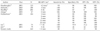
There is increasing evidence that HR-HPV testing after conization is important for detecting persistent or recurrent disease.1,13-22 The 2001 ASCCP guidelines state that HR-HPV testing is acceptable for post treatment surveillance.25 Post-conization HR-HPV testing is useful for detecting not only persistent disease but also recurrent disease. The sensitivity, specificity, positive and negative predictive values of HR-HPV testing for detecting persistent or recurrent disease after conization have been reported in several studies (Table 2).13-19 In particular, the negative predictive value was found to be very high in all studies.
Depending on the study, patients have been tested for HR-HPV at different times, including immediately after conization,21 within 6 months after conization,13-15,18,19,22 or at 6 months after conization (Table 2).16,17 Nobbenhuis et al. reported that results were similar at both 3 and 6 months after conization (Table 2).17 The 2001 American Society for Colposcopy and Cervical Pathology (ASCCP) guidelines recommend that testing be performed at least 6 months after treatment to provide sufficient time for clearance of the HPV infection, and that it can be performed at 12 months after treatment unless a patient has risk factors for persistent/recurrent CIN, such as a large lesion or endocervical extension.25 While this may be a reasonable guideline under some circumstances, such a delay in testing may have a negative impact in cases where there is residual high grade CIN or invasive carcinoma after conization. In the present study, HR-HPV tests were performed between 3 and 6 months after conization, and the median time interval from conization to recurrence was 5 months (range, 1 to 30 months).
The present study indicates that HR-HPV testing between 3 and 6 months after conization is important for predicting the risk of disease persistence or recurrence. In addition, such testing can assist in designing patient management, since HR-HPV negative patients should undergo routine surveillance, while HR-HPV positive patients should undergo frequent and meticulous surveillance.
 XML Download