

 PDF
PDF ePub
ePub Citation
 Print
Print


INTRODUCTION
Vaginal intraepithelial neoplasia (VAIN) is thought to be a precursor lesion of invasive vaginal carcinoma.1 VAIN is much less common than cervical intraepithelial neoplasia (CIN); it is estimated to account for about 1% of all lower genital tract intraepithelial neoplasias.2 However, the incidence of VAIN is expected to increase due to the wider application of cytological screening and colposcopy as well as the increased awareness of the condition. There are several different treatment options available for VAIN such as laser vaporization, radiation therapy, vaginectomy, immunotherapy, and watchful expectancy.
The risk factors for VAIN are similar to the risk factors associated with neoplasia of the cervix and vulva. A prior hysterectomy for cervical neoplasia3 is the main risk factor for VAIN. In addition, the human papillomavirus (HPV) has been implicated as a causative agent.4,5 Other risk factors include antecedent or coexistent neoplasia of the lower genital tract, a history of pelvic radiation,6,7 and immunosuppression.6 VAIN often accompanies CIN, and is thought to have a similar etiology.8 Such lesions may be extensions into the vagina from the CIN, or they may be satellite lesions occurring mainly in the upper vagina. High-grade VAIN usually occurs in association with high-grade CIN lesions, which, in approximately 3% of cases, extend into the vaginal fornices. However, primary foci of high-grade VAIN does occur.9 Because the vagina does not have a transformation zone with immature epithelial cells that can be infected by HPV, the mechanism of entry of HPV is by way of skin abrasions from coitus or tampon use. As these abrasions heal with metaplastic squamous cells, HPV may begin its growth in a manner similar to that occurring in the cervical transformation zone.
For the detection of VAIN, abnormal cytology usually initiates a diagnostic survey for cervical pathology in patients with a history of a previous hysterectomy; in the past, many cases of VAIN were detected incidentally. The utility of HPV DNA test for the diagnosis and follow-up of VAIN has not been widely used, and its value for this purpose has not been previously analyzed. The aim of this study was to evaluate the efficacy of the HPV DNA test, especially for viral load, for the diagnosis and prediction of regression or persistence of VAIN.
MATERIALS AND METHODS
A retrospective review of the medical records of 48 patients that presented with VAIN lesions between March 2001 and February 2008 was performed. The records were obtained from the database at the Department of Obstetrics and Gynecology, Guro Hospital, at the Korea University. All VAIN lesions were diagnosed by histopathological examination of specimens obtained by colposcopy guided biopsies. The grade of the histological diagnosis was determined according to the WHO accepted criteria. All patient specimens and data were obtained with Institutional Review Board approval and informed consent.
All 48 eligible women were followed for cytology, HPV DNA test, and colposcopy guided biopsies at 3- to 6-month intervals. The grade of the cytological diagnosis was determined according to the Bethesda system10 for classification of Papanicolaou smear reports. The hybrid capture 2 (HC2) assay was used to identify patients as HPV DNA positive or negative, and the viral load was determined.
Data regarding the history of previous CIN or vulvar intraepithelial neoplasia (VIN), a prior hysterectomy, and previous treatment for cervical dysplasia were confirmed by review of the medical records. Women that were previously treated for VAIN were excluded. Cytology results, HPV DNA test results and viral load, colposcopy findings, and treatment modality (vaginectomy, radiation therapy, immunotherapy, laser therapy or no treatment) were also recorded.
Among the 48 women, 11 patients were excluded from the study due to loss to follow-up. Thus, there were 37 patients studied with an average follow-up of 30 months (range, 12 to 72 months). The association of HPV DNA test results with VAIN, according to grade, was compared to the cervical cytology. In addition, the efficacy of the HPV DNA test was compared to cytology for the prediction of disease prognosis. The persistence or regression of lesions was confirmed by histopathology.
1. Liquid-based cytology
A Cervex brush (Rovers Medical Devices, Oss, Netherlands) was used to obtain samples from the cervical os and vaginal wall. The brush was immediately rinsed in a vial of PreservCyt solution (Cytyc Corporation, Marlborough, MA, USA) and the vial was placed in the ThinPrep (Cytyc Corporation) Processor.
2. HC2 assay
Vaginal scrape samples from the high end of the vaginal wall (vaginal fornix) were collected for the HC2 assay using Dacron swabs, specifically targeting areas not touched by the speculum, and the samples were placed in UCM (Digene Corporation, Gaithersburg, MD, USA).
HPV DNA test by the HC2 assay was performed according to the manufacturer's protocol (Digene Corporation); signal amplification was performed based on the production of DNA/RNA hybrids using a chemiluminescent reporter system. Results are reported as the relative light unit (RLU) ratio, a semi quantitative estimate of the viral load in the samples. This test has been validated to detect approximately 4,700 genome equivalents (or 1 pg/ml) of target HPV represented by an RLU greater than or equal to the cutoff value calculated in each run by a series of standards. Measurements below the cutoff were scored as negative. Samples were analyzed for the presence of 13 high-risk (HR) HPV types: 16, 18, 31, 33, 35, 39, 45, 51, 52, 56, 58, 59, and 68. Positive and negative controls were included in each run.
3. Statistical analysis
The data were computerized and analyzed with the SPSS ver. 12.0 (SPSS Inc., Chicago, IL, USA) statistical package. Statistical analyses were performed using the Kruskal-Wallis test, the Chi-Square test, and the Fisher Exact test. The Kruskal-Wallis test was used to assess the association between the grades of VAIN and age, parity and the HPV viral load. The Chi-Square test and the Fisher Exact test were used to evaluate the association between the grades of VAIN and the cytological results, HC2 results, previous CIN/VIN, and previous hysterectomy. In addition, these two tests were used to evaluate differentiation of the HPV DNA viral load between the regressed and persistent VAIN groups. The p-values for all tests were 2-sided and considered statistically significant when less than 0.05.
RESULTS
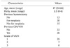
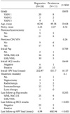
The clinical characteristics of the initial 48 patients with VAIN are shown in Table 1. The mean age at the time of diagnosis was 47 years (range, 34 to 66 years). Among the 48 patients with VAIN, 35 had VAIN 1 (72.9%), seven VAIN 2 (14.6%), and six VAIN 3 (12.5%) at the mean ages of 45.8, 55.7, and 53.3 years, respectively (Table 2). Patients with VAIN 2 and VAIN 3 were older than those with VAIN 1 (p=0.005 and 0.008, respectively).
The patients with VAIN more frequently had a history of a previous hysterectomy (p=0.016). Among 35 VAIN patients with a previous hysterectomy, a hysterectomy for neoplasia was significantly higher in the VAIN 1 group compared to the VAIN 3 group (p=0.035) and vice versa for patients with a history of hysterectomy for reasons other than neoplasia.
We also analyzed the association of the HC2 results and viral load according to the grade of VAIN. The HPV DNA load was significantly higher in patients with high grade VAIN compared to the patients with low grade VAIN (p=0.009). The patient's parity, history of CIN or VIN and Pap smear results did not show any significant difference according to the grade of VAIN.
Among the 48 patients, 37 were considered to have adequate follow-up (a mean of 30 months). Among 26 out of the 37 patients (70.3%), the VAIN lesions resolved during the follow-up visits. For the other patients (11/37) the lesions persisted. However, there was no patient with progression to invasive vaginal carcinoma (Table 3).
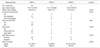
In addition, there was no difference in the grade of VAIN between the regressed and persistent groups (Table 4). The mean age of the patients in the VAIN regressed and persistent groups was 47.0 and 49.2 years, respectively; this difference was not statistically significant. The mean parity also was not significantly different between the two groups. In addition, a history of previous hysterectomy and of previous CIN or VIN did not affect the prognosis of the patients with VAIN (Table 4).
Once VAIN was diagnosed, we initiated treatment of the lesions using various modalities, for example, vaginectomy, radiation therapy, immunotherapy, laser therapy or observation. There was no difference in the disease prognosis between the two groups among the various treatment modalities. We evaluated the initial HPV DNA results and the viral load in the regressed and persistent groups and found no statistically significant difference. As previously mentioned, during the 3- to 6-months of patient follow-up, we performed both cytology and HC2 assays. The last follow-up HC2 and viral load results of the regressed and persistent groups were analyzed and compared. The results showed that the detection rate for HR HPV DNA was significantly higher in the persistent group than in the regressed group (p<0.001). Furthermore, the mean HPV DNA viral load in the regressed group was 6.99, whereas that in the persistent group it was 680.94. These findings demonstrate that the viral load was significantly higher in the persistent group compared to the regressed group (p<0.001).
In the normal or atypical squamous cells of undetermined significance (ASC-US) cytology group, 23 VAIN 1, three VAIN 2 and two VAIN 3 patients were identified. In the low-grade squamous intraepithelial lesion (LSIL) and high-grade squamous intraepithelial lesion (HSIL) cytology group, 11 VAIN 1, three VAIN 2 and four VAIN 3 patients were present. Among the 21 patients that initially presented with within normal limits (WNL) or ASC-US cytology, 16 were in the regressed group and 5 were in the persistent group; while among the 15 patients with the initial diagnosis of LSIL or HSIL, 10 regressed and 5 persisted. At the last follow-up cytology, a negative cytology was observed in 25 out of 26 patients that had regressed lesions and in 9 out of 11 patients that had persistent lesions; this difference was not statistically significant (p=0.205).
During the follow-up of the VAIN lesions, the diagnostic accuracy for the HPV DNA positive results as predictive of VAIN persistence or regression was as follows: the sensitivity was 81.8%, specificity 88.5%, positive predictive value 75.0% and negative predictive value 92.0%. By contrast, the diagnostic accuracy for the Pap smear had a sensitivity of 18.2%, specificity of 96.2%, positive predictive value of 66.7%, and a negative predictive value of 73.5%.
DISCUSSION
VAIN in almost all cases is asymptomatic, and in many patients there is no lesion visible in the vagina. Previously, the possibility of the presence of VAIN was suggested on the basis of abnormal cytology in patients that had a hysterectomy.
Why has VAIN been overlooked so far? Is it because of the difficulty of colposcopic assessment of the lesions? Perhaps because the so-called flat condyloma might have been neglected? The true reason may well be a cytological screening artifact since cytological smears are usually taken from the cervix but not from the vagina. To recognize VAIN with certainty, it is useful to look for HPV DNA in vaginal smears. The natural history of VAIN 1-3 and the risk of progression to invasive vaginal carcinoma remain poorly defined. The immediate importance of VAIN might be its role as a viral reservoir for potential sexual transmission. Therefore, it is important to recognize and monitor VAIN to control lower genital tact neoplasia.
HPV has proven to be an important etiologic agent for the development of CIN and invasive cervical cancer. In a population based study, Castle et al. found a similar distribution of HR HPV genotypes in vaginal and cervical specimens in patients without a history of gynecological malignancy.11 Although far less common than cervical neoplasia, VAIN and invasive vaginal carcinoma are also caused by HPV in the majority of cases.5,12 Infection by HR HPV genotypes in both CIN and VAIN may manifest as both high and low-grade lesions. In fact, it was due to the high percentage of LSIL lesions associated with HR HPV genotypes, that the ALTS group modified the triage algorithm in these patients.13
The purpose of follow up after treatment of VAIN was to detect regression or progression in both the short term and the long term. The rationale of adding HPV DNA test to the cytology was to increase the sensitivity of detection of HR HPV positive disease, and accordingly continue to follow these patients. Similarly, a significant number of vaginal low-grade lesions were associated with HR HPV infections. A previous study reported that 84% of VAIN 1 lesions were associated with HR HPV DNA.14 In this study, we found that 74.3% of VAIN 1, 85.7% of VAIN 2 and 100% of VAIN 3 lesions were associated with HR HPV DNA.
Previously, it was reported that malignant pathology of the vagina (intraepithelial neoplasia or vaginal cancer)3,15-17 may be more frequently identified in women that had a hysterectomy for benign disease. Hence, it was suggested that vaginal smears should continue to be performed at regular intervals, even after a hysterectomy. In this study, there were 35 patients that had a hysterectomy and also had VAIN; among this group 24 (68.6%) had neoplasia, and the others (11/35, 31.4%) were did not have neoplasia. Although the sample size was quite small, the proportion of VAIN after benign pathology cannot be ignored; we recommend that vaginal vault cytology be continued in women that previously had a hysterectomy with benign pathology.
Age was a strong demographic predictor of HR HPV infection. There was a significant association between patients with a higher degree of VAIN and older age (p=0.001). VAIN 1 was diagnosed at around 45 years of age compared to VAIN 2 and VAIN 3, which are most frequently diagnosed after 50 years of age. These results are consistent with the findings of Aho et al.18 (41 years), Benedet and Sanders8 (55 years), and Lenehan et al.19 (49 years). Audet-Lapointe et al.7 reported that patients with VAIN were on average older than those diagnosed with CIN. Other authors reported an increasing mean patient age with increasing grade of VAIN.6
As shown in Table 2, among the 28 patients with VAIN 1 that had had a hysterectomy, 21 (75.0%) had the hysterectomy for neoplasia and seven for other reasons. By contrast, among the VAIN 3 patients, those that had a hysterectomy for reasons other than neoplasia were 4-times more common than patients that had a hysterectomy for neoplasia. A possible explanation for this finding is that more attention may be paid to the individuals that had a hysterectomy for neoplasia due to the possibility of the development of VAIN, and therefore VAIN may be detected earlier.
As mentioned above, the relationship between HPV infection and the development of VAIN is well recognized. In this study, positive HPV DNA was superior to negative HPV DNA regardless of the grade of VAIN; reflecting the close relationship between HPV infection and the development of VAIN. Furthermore, this is the first study to demonstrate a positive correlation between the HPV DNA load and the severity of the VAIN grade. In contrast to HPV DNA testing, the Pap smear results did not show a positive correlation with the VAIN grade. Therefore, the results of this study showed that the HPV DNA test was a more useful predictor of the VAIN grade.
During the follow-up of patients with VAIN after treatment, we evaluated the Pap smear and HPV DNA test results as predictors of disease prognosis. As in the prediction of the VAIN grade, the HPV DNA test, by the HC2 assay, showed a significantly higher viral clearance in the regressed group than in the persistent group (p<0.001). More importantly, the HPV DNA viral load was significantly higher in the persistent group than in the regressed group (p<0.0001). These findings suggest that decrease of the HPV DNA viral load might be a marker for disease regression. Our results indicate that the HR HPV viral load was a better predictor of VAIN regression or persistence than abnormal cytology.
One study recommended including HR HPV test to monitor women initially treated for CIN 2 or 3.20 They suggested that all women should be tested at 6 and 24 months after treatment, and should only be referred for routine cervical cancer screening when these tests are negative on both visits. They evaluated the predictive value of the follow-up HPV test in patients treated for CIN 2 or 3. After monitoring patients by cytology and HR HPV testing at 3, 6, 9, 12, and 24 months after treatment for CIN, they found that a positive HR HPV DNA test performed 6 months after treatment was more predictive than abnormal cervical cytology, with sensitivities of 90% and 62%, respectively, and with similar specificity.
In this study, we did not evaluate the best treatment among all modalities. Because of the retrospective nature of this study, it was not possible to identify all of the factors that influenced the choice of treatment for all patients. Some of the factors that were considered included the location of the lesion and multifocality, as well as the patient's and physician's preference. This study was performed based on patient data in a retrospective fashion, and the sample size was small; these are significant limitations of this study. Future studies should include prospective data and a larger sample size.
In conclusion, HPV DNA test, especially the HPV DNA load was more effective than cytology for the diagnosis and prediction of regression or persistence of VAIN. Thus, HPV DNA test should be performed in addition to cytology during the follow-up of patients with VAIN after treatment.

 XML Download