

 PDF
PDF ePub
ePub Citation
 Print
Print


INTRODUCTION
Cervical cancer is the second most frequent cancer in women and the third leading cause of cancer-related death in women after breast cancer and lung cancer.1 Every year about 270,000 women die of cervical cancer. The death and severe pain and distress that accompanies the disease could be eased by improved treatment processes.2 Cervical cancer is associated with human papillomavirus (HPV) infection, smoking-related impairment of the immune resistance, long-term use of oral contraceptives, and venereal infections caused by Herpes virus and Chlamydia trachomatis. Among these, HPV accounts for over 90% of cervical cancer cases.3 Of the approximately 100 known HPV types, more than 15 species of oncogenic HPV have been shown to be directly related with cervical cancer and low-risk HPV, which causes verruca plana or condyloma, but few instances of cancer.4 Carcinogenic HPV is classified into the high-risk group that includes types 16, 18, 45, 31, 33, and 52. Among these, types 16 and 18 are associated with more than 70% of all cervical cancers.5 Types 6 and 11 are classified into the low-risk group of HPV and are found in more than 90% of cases of anogenital warts.6
The classification of HPV into the risk groups relies on base sequences of specific genes. HPV genes include an early gene (E gene) that controls viral production early in the infection period, a late gene (L gene) that is crucial in the formation of virus structure and long control region (LCR) or upper regulatory region (URR).7 L gene encodes the capsid protein of HPV; the expressed protein not only induces infection by reacting with a host accepter but becomes a target of helper T-cells.8 E genes are organized with four heritable regions that participate in virus reproduction and two other heritable regions that promote splitting of the host. LCR genes are organized with promoters controlling reproduction of virus DNA, and enhancer and base sequences that are capable of combining with gene silencers.
Among the three genes, HPV is classified according to the base sequence of the phylogenetically well-preserved L1 gene. Based on L1 gene similarities, HPV have been classified into three categories: HPV genus (<60%), HPV species (60-70%) and HPV genotype (71-89%).9 L1 gene analysis utilizes three analytical methods, namely DNA sequencing,10 restriction enzyme digestion patterns,11 and polymerase chain reaction (PCR) using primers specified for the HPV gene type.12 Since the mid-1990's, HPV types have been classified by the Southern blot method, which utilizes a probe specified for each gene type after amplifying the GP5/GP6 loci within L1 gene.13 However, this approach can be complicated by a non-specific response that arises due to the marked similarity of inner base sequences among each gene type. Very recently, a new method was devised that relies on the amplification of the LCR-E7 gene and whose HPV classification rationale is based on restriction fragment length polymorphism (RFLP).14 Subsequently, another method was developed that relies on the amplification of the L1 gene from the GP portion to the E6 portion with the same primer, and then using nested PCR with a specific inner primer.15
In this way, E gene analysis as a viral type classification has been used rather than the existing method for limited L1 gene. When we consider the extreme variation of specific characteristics of viruses and that over 100 HPV gene types have been reported, various analytical methods that key on the heritable portions should be developed. It is important that these methods identify cervical cancer-associated E gene following classification of HPV into the high-risk group.
The present study compared the L1 gene and E6/E7 gene analytical methods, which are standards for classifying gene types, to aid in the development of a refined method that minimizes HPV typing errors in the real-world of the clinic.
MATERIALS AND METHODS
1. Sample and reagent
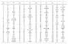
HPV positive DNA samples were acquired from Diaprobe (Seoul, Korea). A total of 104 samples were used for related PCR template DNA; all samples were stored frozen after dissolving in TE buffer. Primers used for discerning HPV positive and negative samples, and for deciding gene type are presented in Table 1. Type-specific probes designed by Jacobs et al.13 were used to membrane assay on L gene and primers designed by Sotlar et al.15 were used to nested multiplexed PCR on the E gene. A probe-based membrane assay method was applied to assess the gene type of the L1 gene amplified product. Streptavidin-peroxidase (SA-POD; Sigma-Aldrich, St. Louis, MO, USA) was also used in a reaction with 3' biotin of the amplified product. The reaction substrate was 3,3',5,5'-tetramethylbenzidine (TMB; Sigma-Aldrich, St. Louis, MO, USA).
2. L1 gene amplification via PCR and membrane assay
PCR amplification of L1 was carried out using GP5+ and GP6+ primers. PCR reagents were composed of 5 µl 10× Omni buffer (Genenmed, Daejeon, Korea), 2 mM dNTP, 10 pmol primer, 5 units Han Taq (Genenmed, Daejeon, Korea) and 5 µl sample DNA in a final reaction volume of 50 µl sterile water. PCR conditions were: 5 min pre-denaturant step at 94℃; extension step of 40 cycles and 5 min post-extension step at 72℃, with each cycle composed with 30 s denaturing step at 94℃; 1 min annealing step at 55℃; and 30 s extension step at 72℃. After completion of the reaction, electrophoresis was conducted using a 2% agarose gel with ethidium bromide. Positive and negative products were verified by demonstration of the presence and absence, respectively, of the band corresponding to L1. The PCR amplified product present in positive samples was mixed with hybridization buffer and was developed in a membrane that was clustered with a specific probe for each gene type. The amplified product was hybridized with probe and the 3' end of biotin was combined with SA-POD during membrane development. Color reaction was induced by adding the TBM substrate.
3. Nested multiplexed PCR
Primary PCR was performed using a primer set comprising E6-E7 to amplify the E gene. PCR was conducted as described above, except that the annealing temperature was lowered to 50℃. After completion of PCR, nested multiplexed PCR was processed based on the amplified product as a template. Nested multiplexed PCR consisted of four cocktails with a peculiar primer combination capable of amplifying a specific gene type added for each cocktail. Electrophoresis was then carried out using an ethidium bromide containing 2% agarose gel. The banding pattern confirmed the gene type.
RESULTS
1. HPV typing by L1 gene analysis
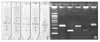
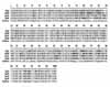
For HPV positive DNA, amplification of the PCR product using the GP5+/GP6+ primer and electrophoresis revealed a band of about 150 bp. The PCR amplified product was deployed onto a membrane with hybridization buffer and the gene type was ascertained by the result of the SA-POD-catalyzed color development (Fig. 1A). Table 2 shows the result of the 104 positive samples classified in this study. Eighty percent of the HPV types were in the high-risk group with the remainder in the low-risk group. Types 58, 16, 6, and 39 constituted 62% of all the viral samples.
2. HPV typing by E gene analysis
E gene amplification products were processed for DNA, which was confirmed with positive gene type through L1 gene amplification and membrane assay. The E6 to E7 portions were amplified and were used in the nested multiplexed PCR procedure (Fig. 1B). Gene types of the positive samples are summarized in Table 2. Eighty-two percent of the gene types were in the high-risk group with the remainder being in the low-risk group. The main viral types 39, 58, 51, and 68 constituted 52% of all sample types.
3. Comparison of HPV typing by L1 gene and E gene analyses
Greater than 65% dissimilarity was revealed between gene types classified from L1 gene analysis and from E gene analysis (Table 2). Thirty-five percent of the results of the two tests were similar but 31% of the results were not. Twenty-nine percent of the results of E gene analysis were more diversified than that of L1 gene analysis. Less than 4% of the L1 results were more diversified. In particular, type 6 of the low-risk group of L1 gene analysis was revealed as a high-risk group of types 51, 58, and 66 in E gene analysis.
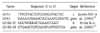
As a result of comparing internal base sequencing for gene types 6, 51, 58, and 66 classified from L gene analysis and E gene types (Fig. 2), similarity of L1 gene between low-risk type 6 and high risk types 51, 58, and 66 exceeded 67%. Diversity of internal base sequencing was very low.
DISCUSSION
As seen in Table 2, HPV typing was more diversified in the results of E6 and E7 gene analyses than for L gene analysis. This indicates that the L gene is stably preserved and that HPV gene diversity arose from the E gene.
HPV genes are structured with non-coding regions that include two parts of the L gene and six parts of the E gene. The L gene encodes L1 and L2 structural proteins and is the existing standard for classifying gene type because of its relative stability and high degree of preservation.9 As a result, commercial products based on membrane chip technology mainly target the L gene. But, carcinogenicity in cervical cancer is mainly due to the reciprocal action between the E6 and E7 genes.16 Among the E genes, E1, E2, E4, and E5 participate in virus reproduction, and E6 and E7 induce the carcinogenic process by promoting splitting of the host cell.17 In other words, classifying gene type through conserved regions is important, but a test that addresses the E gene is also very important.
Comparison of the base sequences of the low-risk group type 6 with that of high-risk group types 51, 58, and 66 (Fig. 2) revealed that gene type could be decided through base sequence based on the high similarity of L1 among the HPV types. However, clinical application of this finding is still challenging for several reasons. More than 100 diversified gene types are known; sequence-based discernment of viral types is difficult in the case of a complex infection involving various gene types. High cost and prolonged analytical time are additional limitations. Moreover, the classification of HPV gene type requires accurate analysis. The present test distinguishes high-risk group types that are implicated in cervical cancer. Thus, this test is promising in the development of an effective standard for suggesting corresponding treatment.
However, presently about 31% of total tested group revealed different results in mutually compared data of L1 and E6/E7 gene analytical methods, and a single low-risk type that appeared in the high-risk group was verified. Considering that the currently used classification method of HPV typing depends on L1 gene classification, we suggest that the nested multiplexed PCR analytical method for E6/E7 genes could be a useful supplement to the existing L1 gene analytical method until a new accurate HPV typing analytical method is developed.
 XML Download