

 PDF
PDF ePub
ePub Citation
 Print
Print


INTRODUCTION
Cadherins are a family of cell-cell adhesion molecules essential for tight connection between cells.1 E-cadherin is the major cadherin molecule expressed in epithelial cells. The cadherin-mediated cell adhesion system is known to act as an invasion suppressor system in cancer cells, since noninvasive cells can be transformed into invasive ones when treated with antibodies to block cadherin's function or with cadherin-specific antisense RNA,2,3 and transfection of human cancer cell lines with E-cadherin cDNA can reduce their invasiveness.4 Recently, immunohistochemical examination has revealed that decreased E-cadherin expression is associated with tumor dedifferentiation and progression in endometrial carcinoma5 and many other tumors.6-10
DNA methylation is one of the best-understood epigenetic changes in human cancers. DNA methylation may play roles in carcinogenesis by virtue of 3 mechanisms: 1) DNA cytosine methylation facilitates gene mutation, as 5-methyl-cytosine is deaminated to thymine; 2) aberrant DNA methylation may be associated with alleic loss; and 3) DNA methylation occurs frequently in CpG islands near regulatory regions of genes and affects the transcriptions of specific genes.11-13 CpG methylation has recently been shown to be an important mechanism in the transcriptional inactivation of E-cadherin, and CpG methylation around the promoter region of E-cadherin was found to be significantly correlated with reduced E-cadherin expression in various cancers.14 Though it has been thought that hypermethylation of the E-cadherin gene is associated with immunohistochemical expression, tumor progression, tumor grade, tumor invasion, and lymph node metastasis in various tumors, this has been rarely studied in endometrial carcinoma of Korean women. In the current study, we analyzed methylation status of the E-cadherin and E-cadherin expression in endometrial hyperplasias and endometrial carcinomas. Also, we correlated these results with various clinicopathological factors of endometrial carcinomas.
MATERIALS AND METHODS
1. Patients and tissue specimens
Samples of endometrial tissues were obtained from 50 women who had undergone hysterectomy or curettage at the Inha University Hospital, Incheon and the Hallym University Hospital, Seoul, Korea, between 1995 and 2002. Endometrial hyperplasia tissues (n=20) were taken from the endometrial curettage and diagnosed according to the system of the World Health Organization. As a result, fifteen cases were found to be simple hyperplasias and five cases were atypical endometrial hyperplasias. Endometrial carcinoma tissues (n=30) were taken from surgical resection. None of the patients had received preoperative chemotherapy.
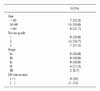
Pathologic stage, grade, and histological subtype were determined for each surgical specimen according to 1988 International Federation of Gynecology and Obstetrics (FIGO) system. Slides from each case were re-reviewed by a gynecological pathologist to verify the diagnosis, stage, grade, and histological type. The tumor grade resulted in 9 cases of tumor grade G1 (well differentiated carcinoma), 14 cases of G2 (moderately differentiated carcinoma), 7 cases of G3 (poorly differentiated carcinoma). All of the endometrial carcinomas were endometrioid endometrial carcinomas. Surgical stage resulted in 6 cases Ia, 9 cases Ib, 9 cases Ic, 4 cases II, 2 cases III (Table 1). Clinical data of patients were obtained from medical records. Tumor tissues were frozen in liquid nitrogen, and stored at -80℃ until use.
2. Immunohistochemistry (IHC)
Tissues were fixed overnight in 10% buffered formalin, dehydrated, and embedded in paraffin. Five micrometer serial sections of each sample were used. Sections were cut, floated onto albumin coated slides, dried at 56℃, deparaffinized in xylene, rehydrated, and washed with phosphate-buffered saline (PBS) for 15 minutes at room temperature. Specimens were treated in a microwave oven in 0.01 mol/L citrate buffer (pH 6.0) for 30 minutes at 100℃, slowly cooled to room temperature, and then washed with PBS for 5 minutes at room temperature. After quenching endogenous peroxidase with 3% hydrogen peroxide in PBS for 10 minutes at room temperature, the sections were incubated with a blocking solution (PBS containing 5% skimmed milk) for 60 minutes at room temperature. Then the slides were incubated overnight at 4℃ with a 1:100 dilution of anti-E-cadherin antibody (Monoclonal mouse anti E-cadherin, Zymed, CA, USA). After several washes with PBS, they were incubated with a second antibody, a 1:200 dilution of peroxidase conjugated anti-mouse immunoglobulin (LSAB kit, DAKO, Denmark), for two hours. 3.3'-diamniobenzidine tetrahydrochloride was used as a chromogen, and Mayer's hematoxylin counterstain was applied. For the negative control, the same dilution of nonimmunized mouse mmunoglobulin was used as the first antibody. Blocks of normal endometrium tissues were prepared as controls from the normal endometrium tissues of ten patients with endometrial carcinomas. In accordance with previous reports,15,16 E-cadherin expression was considered "positive" when more than 70% of tumor cells showed strong membranous staining and "negative" or "decreased" when less than 70% of tumor cells showed strong membranous staining or showed only slight membranous staining (Fig. 1).
3. Methylation-specific PCR (MSP)
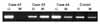
DNAs were extracted from tumor and nontumoral tissues with a genomic DNA purification kit (Promega, Madison, WI, USA). Both methylation-specific PCR (MSP) and bisulfite-PCR take advantage of the fact that unmethylated cytosine is efficiently converted to uracil after 16 hours of Na-bisulfite treatment, whereas methylated cytosines remain unchanged. To examine the DNA methylation patterns, we treated genomic DNA with sodium bisulfite, as described by Herman et al.17 In brief, 2 ug of genomic DNA was denatured by treatment with NaOH and modified 3 M sodium bisulfite for 16 hours. DNA samples were purified with Wizard DNA purification resin (Promega, Madison, WI, USA), treated with NaOH, precipitated with ethanol and resuspened in 25 ug of water. MSP was used to examine the methylation status of E-cadherin. The primer sequences of E-cadherin for the methylated and unmethylated reactions are described in Table 2. These primers were designed for the E-cadherin promoter region and contained six sites (116 bp) of the CpG promoter at genomic position of -205 for methylated-specific primers and five sites (97 bp) at genomic position of -210 for unmethylated- specific primers, respectively.17 PCR was performed in 10ug reaction volumes. PCR products underwent electrophoresis on 2% agarose gel and were then visualized under UV illumination using an ethidium bromide stain. Samples were scored as unmethylated when there was a clearly visible band on the gel with the unmethylated primers. Samples were scored as hypermethylated when there were a clearly visible band on the gel with both the methylated and the unmethylated primers (Fig. 2).
RESULTS
1. E-cadherin expression
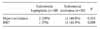
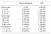
Decreased expression of E-cadherin was significantly associated with endometrial carcinomas than endometrial hyperplasias (p=0.009). Decreased expression of E-cadherin was detected 13 of 30 endometrial carcinomas (43.3%), 1 of 20 endometrial hyperplasias (5%) (Table 3). Decreased expression of E-cadherin was significantly associated with tumor dedifferentiation, advanced tumor stage, and lymph node metastasis in endometrial carcinomas (p=0.01, p=0.02, p=0.03) (Table 4).
2. Promoter methylation of E-cadherin gene
Hypermethylation of E-cadherin was more frequent in endometrial carcinomas than endometrial hyperplasias (p=0.015). Hypermethylation of E-cadherin was detected 12 of 30 endometrial carcinomas (40%), 2 of 20 endometrial hyperplasias (10%) (Table 3).
Methylation status of E-cadherin of endometrial carcinomas had no correlation with the tumor grade and lymph node metastasis. However hypermethylation of E-cadherin was found in higher frequency in above stage Ic endometrial carcinomas (p=0.025) (Table 4).
3. The correlation between hypermethylation and immunohistochemical findings of E-cadherin
There was no correlation between DNA methylation and immunohistochemical findings of E-cadherin in endometrial carcinomas. Seven of 17 samples (41.2%) with positive expression of E-cadherin showed methylation, whereas 5 of 13 samples (38.5%) with decreased expression of E-cadherin showed methylation (p>0.05).
DISCUSSION
Reduced cell-cell adhesiveness allows cancer cells to disobey the social order, resulting in destruction of the histologic structure, the morphologic hallmark of malignant tumors.18 Several reports have indicated that E-cadherin, an epithelial-specific cadherin, is a key molecule for the maintenance of epithelial integrity and of polarized states in association with alpha-, beta-, and gamma-catenin, and that the reduction of E-cadherin-mediated cell-cell adhesion favors the dispersion of cancer cells.19 Consistent with this concept, immunohistochemical studies have revealed that decreased E-cadherin expression is associated with tumor dedifferentiation and progression in endometrial carcinoma5 and other tumors.6-10
In the current study, decreased expression of E-cadherin was of a higher incidence in endometrial carcinoma than endometrial hyperplasia. Decreased expression of E-cadherin in endometrial carcinoma was associated with tumor dedifferentiation (G1<G2<G3). The expression of E-cadherin also showed a tendency to decrease in advanced surgical stages and when there was lymph node metastasis. Our results are in agreement with several previous studies.5,20 These results suggest that decreased expression of E-cadherin may play a role in carcinogenesis and be a prognostic factor for aggressive disease.
Work over the past several years has demonstrated that the silencing of tumor suppressor genes associated with promoter methylation is a common feature in human cancers.21 It was discovered that some tumor suppressor genes, including RB, VHL, p15, and p16, were inactivated as a result of reduced expression due to CpG methylation.22 As observed with these tumor suppressor genes, the E-cadherin invasion suppressor gene in human cancers is silenced by an epigenetic mechanism, promoter hypermethylation.18 But hypermethylation in the promoter region of E-cadherin has been rarely studied in endometrial carcinoma of Korean women. So we studied the promoter-region CpG island methylation of E-cadherin gene in endometrial hyperplasias and endometrial carcinomas. Hypermethylation of E-cadherin was more frequent in endometrial carcinomas than endometrial hyperplasias. The incidence was 40% in endometrial carcinomas. Saito et al.23 firstly reported methylation of the E-cadherin promoter of endometrial carcinoma. They reported promoter methylation of E-cadherin was 37.7% in endometrial carcinomas. The incidence of methylation status of E-cadherin correlated with our results. But Banno et al.24 reported that promoter hypermethylation of E-cadherin in endometrial carcinomas was detected in only 14.3%. These results suggest that DNA hypermethylation of E-cadherin may play a role in carcinogenesis. But further studies are required to show the relation between promoter hypermethylation of E-cadherin and endometrial carcinoma. Hypermethylation of the E-cadherin was detected 2 cases of endometrial hyperplasia in our study whereas Saito et al.23 reported that methylation of the E-cadherin was not detected in endometrial hyperplasia. They reported that methylation of the E-cadherin gene correlated quite well with decreased E-cadherin expression.23 However, our study showed no significant correlation between the methylation status and E-cadherin expression. This may be due to the small sample size in our study and further study for confirmation is required. The other cause of the difference may be due to different assessment of immunohistochemical findings of E-cadherin. In our study E-cadherin expression was considered "positive" when more than 70% of tumor cells showed strong membranous staining and "negative" or "decreased" when less than 70% of tumor cells showed strong membranous staining or showed only slight membranous staining.5,16 But Saito et al.23 classified E-cadherin staining patterns into three types: positive expression, heterogeneous staining pattern, and negative staining pattern. Moreover, intratumoral E-cadherin expression heterogeneity was noted in human cancers.9 Methylation status on an all-or-nothing basis may not directly predict the E-cadherin immunoreactivity, and the extent of methylated areas within a single tumor may be more relevant to the E-cadherin immunoreactivity. In addition, we noted positive E-cadherin expression in 26.7% of endometrial carcinomas. This suggests that mechanisms other than methylation are likely to be involved in impaired E-cadherin expression. Dysfunction of catenins or the effect of signals from other cell junctions might include the instability of translated E-cadherin. Saito et al.23 reported that hypermethylation in the promoter region of the E-cadherin gene correlated with tumor progression, tumor dedifferentiation, and the depth of myometrial invasion. Therefore, they suggested that methylation of the E-cadherin gene occurred in association with the acquisition of invasive capacity. But in the current study, methylation status in endometrial carcinomas had no correlation with the tumor grade and lymph node metastasis. However, DNA hypermethylation was found in higher frequency in above stage Ic endometrial carcinoma. Further study for confirmation of invasive capacity of E-cadherin in endometrial carcinoma is required.
These results indicate that hypermethylation of E-cadherin promoter region is a frequent event in endometrial carcinoma, which may play an important role in the progression of carcinogenesis. Also, the promoter methylation of E-cadherin in endometrial carcinomas was found to be significantly associated with higher stage above Ic endometrial carcinomas.


 XML Download