

 PDF
PDF ePub
ePub Citation
 Print
Print


INTRODUCTION
Vulvar cancer accounts for 3-5% of all gynecological malignancies.1,2 During recent years, it appears that this incidence has been increasing along with the rise in the average age of the female population.
Significant advances in the management of vulvar cancer have led to decrease in physical and psychological morbidity associated with the treatment. This improvement was enabled by better understanding of the prognosis or course of the disease. Furthermore, if we can predict the individual risk of recurrence, we will be able to adjust the therapeutic modalities to decrease the incidence of recurrence as well as to decrease morbidity.
One such predictive tool is the nomogram, which is a statistical tool that enables users to calculate the overall probability of a specific outcome.3 Several studies in various cancers, such as sarcoma, prostate, and breast cancer, suggested improved predictive accuracy for the nomogram, compared to the conventional prognostic groupings.4-6
In 2006, Rouzier and co-workers suggested a nomogram for prediction of the risk of recurrence in vulvar cancer patients.7 In the nomogram, seven variates which were thought to be related to relapse-free survival were included to calculate the probability of relapse-free survival at 2 and 5 years as a percentage. These seven variates were age, tumor (T) stage, number of metastatic nodes, bilateral node involvement, omission of lymphadenectomy, margin status, and depth of invasion. They advocated that their nomogram predicted survivals better than did the International Federation of Gynecology and Obstetrics (FIGO) stage.
The aim of this study was to evaluate the predictive accuracy of the vulvar cancer nomogram in a different ethnic group, and, as a consequence, to assess the generalizability of the nomogram. Thus, we validated the nomogram in a cohort of vulvar cancer patients treated in Korea.
MATERIALS AND METHODS
Between October 1982 and September 2006, 204 consecutive patients were diagnosed with vulvar cancer in 11 institutions constituting the Korean Gynecologic Oncology Group (KGOG). All medical records were retrospectively reviewed, including pathology reports, operation records, and follow-up data. The data from each institution were collected by a single institution utilizing clinical report forms (CRF). Items on CRF included seven variables of the vulvar cancer nomogram, including age, tumor (T) stage, number of metastatic nodes, bilateral node involvement, omission of lymphadenectomy, margin status, and depth of invasion.
Forty patients with histologic types melanoma and Paget's disease were excluded from the analysis due to their markedly different clinical behavior and prognosis. In addition, 44 cases which were not evaluable due to inadequate data and persistent diseases and 30 cases in which radiation was the primary therapy were excluded, leaving 90 patients with all nomogram variables, FIGO stage, adequate histologic types, and follow-up data.
Relapse-free survival (RFS) was defined as the time from surgery to relapse or last follow-up. Nomogram validation comprised two activities; discrimination and calibration. First, discrimination was quantified with the concordance index, which is similar to the area under the receiver operating characteristic curve. The concordance index provides the probability that the nomogram will predict a poorer outcome for the patient who relapses first out of a randomly selected pair of patients. Second, calibration was assessed. Calibration compares the predicted probability of relapse-free survival with the actual survival. This was performed by grouping patients with respect to their nomogram-predicted probabilities for relapses and then comparing the mean of the group with the observed Kaplan-Meier relapse-free survival estimate. All analyses were conducted using SPSS 13.0 (SPSS Inc., Chicago, IL).
RESULTS
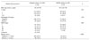
Descriptive statistics for the KGOG data set are summarized and compared with those for the original-development data set in Table 1. One of the main differences between the two cohorts was a higher rate of positive resection margin, defined as 3 mm or less, in the KGOG cohort (p<.001). In addition, a slightly higher percentage of patients in the KGOG cohort were diagnosed with earlier stages (p=.043).
With mean follow-up period of 47.9 months (range, 1 to 269.7 months), the 2-year and 5-year relapse-free survival rates in the KGOG cohort were 81% and 68%, respectively, corresponding to 79% and 72% in the original cohort.
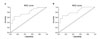
The nomogram concordance index (CI) in the KGOG data set was 0.79. When the application was limited to the 82 patients with squamous cell carcinoma, the concordance index improved to 0.82 (Fig. 1). This was similar to the CI of the original cohort (CI=0.83), and also superior to the CI of the FIGO staging system (CI=0.78).
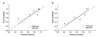
To assess the calibration of the nomogram, actual probabilites were plotted against the calculated predicted probabilities for each group of patients (Fig. 2). Patients were grouped according to their nomogram-predicted probabilities for relapses into 6 groups. The trend line in calibration plot showed comparable concordance with an ideal line, having a slope of 1.04 for 2-year RFS (R2=.35) and 0.98 for 5-year RFS (R2=.80), respectively.
DISCUSSION
Radical vulvectomy and en bloc groin dissection, with or without pelvic lymphadenectomy was the mainstay of treatment in vulvar cancer. However, due to the high incidence of postoperative morbidities, including physical and psychological morbidity, a number of attempts have been made to provide conservative therapy on an individual basis with minimal risk of recurrence. These advances were enabled by better understanding of the prognosis or course of the disease.8-11
The conventional prognostic tool is FIGO staging system.12 This system is based on tumor size, lymph node status, and presence of distant metastasis. On the whole, the prognosis correlates with the FIGO stage. However, there are some limitations. One major problem is that stage III represents a very heterogeneous group of patients. Despite being the same stage, involvement of the inguinal lymph nodes in FIGO stage III patients carries a significantly worse prognosis compared with invasion of the lower urethra, vagina or anus alone.13 In addition, FIGO stage does not fully reflect some important prognostic factors. These include the number of positive inguinal-femoral lymph nodes14, omission of lymphadenectomy, and margin status.10
Therefore, a nomogram, which incorporates more clinical and pathologic factors than the staging system, can allow the clinician to achieve a better estimation of the prognosis of an individual patient. Another potential benefit of nomogram is that it creates a simple graphical representation of a statistical predictive model that generates a numerical probability of a clinical event. But, one should be cautious about extrapolating from regression models built on different populations.15 Because a nomogram derived from one population may not be applicable to a new population, external validation is mandatory. The vulvar cancer nomogram was built and validated in a white population. Although found to be accurate by internal and external validation methods, this nomogram had not been validated in a different ethnic group. Therefore, the current study assessed the generalizability of the nomogram in a Korean population.
The clinicopathologic variables in the two cohorts were similar, except for FIGO stage and resection margin status. The differences, in addition to race effect, enabled the validation to be operated in a separate cohort.
In this study, a comparable concordance index and a similarly good calibration plot were obtained when the vulvar cancer nomogram was applied to the Korean population. When the data set was limited to squamous cell carcinoma, the concordance index improved from 0.79 to 0.82, indicating that the nomogram might be more predictive for patients with squamous cell carcinoma of vulva. In addition, the calibration of the nomogram for the 5-year RFS was better than that for 2-year RFS.
There are some limitations to our study. First, the number of patients included in this study was small, despite of the multi-institutional study. But, this limitation is inherent to rarely occurring cancer such as vulvar cancer. Second, the study design was retrospective. Since the clinicopathologic data were collected retrospectively from different institutions, the data might not be uniform. But, this limitation did not affect the study analysis because the aim of this study was to assess the generalizability of the nomogram. However, prospective randomized trials, incorporating the nomogram in stratification of patients, are still warranted to evaluate the role of nomogram.
In conclusion, the current study confirmed that the vulvar cancer nomogram is a valuable tool for individual prognostic assessment. The resulting risk estimates can help clinicians to counsel patients on an individual basis and to provide more tailored adjuvant therapy.
 XML Download