

 PDF
PDF ePub
ePub Citation
 Print
Print


INTRODUCTION
Uterine cervical cancer is the second most common type of cancer and is the third most frequent cause of cancer death in women worldwide, with 493,243 new cases and 273,505 deaths in 2002.1 Cervical cancer is more common in developing countries, where 83% of cervical cancer occurs and where it accounts for 15% of female cancers.1 In contrast, cervical cancer constitutes only 3.6% of new cancers in developed countries.1 Recently, there have been substantial decreases in both the incidence and mortality of cervical cancer in developed countries where effective screening programs are in place, while the incidence and mortality have increased in developing countries.1,2
The diagnosis of cervical cancer is based on the results of cytological and histological examinations. Although the Pap smear for cytological examination is effective for screening cervical cancer, the evaluation of the Pap smear relies on subjective diagnostic skills and experience, which may result in sampling errors or errors in the interpretation of abnormal cells. In addition, the infrastructure for Pap smear screening is not well established in most developing countries.3-5 Although colposcopy-guided biopsy and cone biopsy for histological examination can be used to diagnose cervical cancer, they are expensive procedures that require specific skills to perform.6 Therefore, the discovery of biomarkers for simple, inexpensive, and widespread testing in developing countries is needed for the screening, early detection, and post-treatment surveillance of cervical cancer.
Human blood plasma and serum are a useful source of disease biomarkers that likely contain information related to cancer development and progression.7-9 Therefore, plasma and serum are the preferred type of specimen for screening, early detection, and post-treatment surveillance of cancer, because plasma and serum samples are the most accessible human body fluid from which a sample can be acquired and are readily available using less invasive, inexpensive methods. Also, use of plasma and serum specimen for biomarker of cervical cancer may supplement problems of the Pap smear which has a lower compliance for screening and follow up in developing countries. Between plasma and serum, plasma tends to be preferable for many laboratory investigators, as the components of plasma better reflect the pathological status of a patient than serum10,11
To identify new cancer biomarkers, proteomic analysis with two dimensional gel electrophoresis (2-DE) and matrix assisted laser desorption/ionization mass spectrometry (MALDI-MS) have been used to examine the differential expression of protein in serum or plasma specimens from different tumors, such as nasopharyngeal carcinoma, gastric cancer, and hepatocellular carcinoma.12-14 The advantage of 2-DE combined with MALDI-MS is that it can detect differences in the expression levels of various proteins between healthy normal controls and cancer patients both qualitatively and quantitatively.15
This study was performed to evaluate the expression of plasma proteins using 2-DE combined with MALDI-MS in patients with squamous cell carcinoma (SCC) of the uterine cervix and patients with benign gynecologic disease as normal controls.
MATERIALS AND METHODS
1. Plasma sample collection and purification
Plasma samples were obtained from six patients with primary cervical cancer treated at Busan Paik Hospital, Inje University, Busan, Korea. The diagnosis was confirmed as SCC histologically in all cases. None of the patients received any treatment before the primary treatment described here. The stage of cervical cancer was established according to the International Federation of Gynecology and Obstetrics (FIGO) criteria: 2, 2, 1, and 1 cancers were classified as FIGO stage Ib, IIa, IIb, and IIIb, respectively. As controls, plasma samples were obtained from six patients with normal cervices who were treated for benign gynecological diseases, including uterine adenomyosis (2 cases), leiomyoma (1 cases), and benign ovarian tumor (3 cases). All patients provided informed consent before collection of the plasma samples. The collection and use of the samples was approved by the Institutional Review Board of Busan Paik Hospital (IRB number 06-14).
To improve the 2-DE of human plasma samples, the albumin and immunoglobulin G in the collected plasma samples were depleted using an Albumin and IgG Removal Kit (GE Healthcare, Buckinghamshire, UK) in accordance with the manufacturer's instructions.
2. Plasma protein preparation
The albumin and immunoglobulin G-depleted plasma samples were mixed with 4 volumes of pre-cooled acetone (-20℃, 15 min) and incubated for 2 h at -20℃ for protein precipitation. The protein pellets were obtained after high speed centrifugation (13,000 × g for 10 min, at 4℃), and then air-dried for 5-10 min at room temperature. The pellets were stored at -80℃ until analysis.
3. 2-DE of plasma proteins
The plasma protein pellets were dissolved in lysis buffer (7 M urea, 2 M thiourea, 4% CHAPS, 40 mM Tris base, 1% DTT, 0.5% IPG buffer, 0.5% Triton X-114, and protease inhibitor cocktail) and kept at room temperature for 1 h. The protein content was assayed using a 2-D Quant kit (GE Healthcare, Buckinghamshire, UK) with serum BSA (0-50 µg/µL) as a standard.
Dried 24-cm (pH 3-10NL) immobilized pH gradient (IPG) strips were rehydrated overnight in a rehydration tray with 450 µL of Destreak (tm) rehydration solution (GE Healthcare, Buckinghamshire, UK) containing 2% IPG buffer (v/v). Following rehydration, aliquots of 100 µg of the soluble plasma proteins in 100 µL of sample solution were loaded using the cup -loading method. Isoelectric focusing (IEF) was carried out at 60,000 V/h at 20℃ as follows: 500 V for 1 h, 1,000 V for 1 h, and finally 8,000 V incremented to 60,000 V/h. The IPG strips were placed in 5 ml of equilibration solution (50 mM Tris-HCl, pH 8.8, containing 6 M urea, 30% glycerol, 2% SDS, and bromophenol blue) containing 1% DTT (v/v) during the first equilibration step and 2.5% iodoacetamide (v/v) during the second equilibration step (15 min per equilibration step). The 2-D separation was performed using the Ettan DALT twelve system (GE Healthcare, Buckinghamshire, UK). The IPG strips were loaded onto a 12.5% gel for sodium dodecyl sulfate-polyacrylamide gel electrophoresis (SDS-PAGE), running buffer (25 mM Tris, 192 mM glycine, 3.5 mM SDS, pH 8.3) was added, and a constant current (5 W/gel) was applied for 6 h. The gels were then stained with silver nitrate. The stained gels were scanned on a PowerLook 1100 flatbed scanner (UMAX, Fremont, CA, USA), and the images were analyzed using commercial software (Image Master 2D Platinum; GE Healthcare, Buckinghamshire, UK).
4. Protein identification using MALDI-MS
For MS fingerprinting, the stained portions of the 2-D gels were excised and then digested with trypsin as described by Rosenfeld et al.16 Isolated protein spots were destained with 100 mM sodium thiosulfate and 30 mM potassium ferricyanide. After washing with 50% acetonitrile (AcN), the gel fragments were dried in a vacuum centrifuge. The dried gel fragments were rehydrated in 20 mL of 25 mM NH4HCO3 containing 0.5 mg of sequencing-grade trypsin (Promega, Madison, WI, USA), and incubated overnight at 37℃. The remaining peptides were extracted twice with 30 mL of a 50 mM NH4HCO3:AcN (1:1) mixture. The extracts were evaporated in a vacuum centrifuge for further drying. Aliquots of the peptide-containing samples were applied to a target disk and left to evaporate. Spectra were obtained using a Voyager DE PRO MALDI-MS (Applied Biosystems, Foster City, CA, USA). Protein databases were searched with MASCOT using monoisotope peaks. A mass tolerance within 50 ppm was allowed initially, after which recalibration was performed using the list of proteins obtained within a tolerance of 20 ppm.
5. Western blotting analysis
Aliquots of 20 µg of plasma protein were separated by 10% SDS-PAGE. The proteins were transferred onto polyvinylidene fluoride (PVDF) membranes, which were blocked overnight in TBS (20 mM Tris and 150 mM NaCl, pH 8.0) containing 5% non-fat dry milk, and then probed with antibodies for ceruloplasmin, complement C3, afamin, and alpha-1-B glycoprotein at a dilution of 1 mg/mL for 1 h at room temperature. The membranes were incubated with horseradish peroxidase (HRP)-conjugated secondary antibody at a dilution of 1:2000 for 1 h at room temperature. Immunoreactivity was visualized with an ECL Western blotting detection kit (Amersham Biosciences, Piscataway, NJ, USA) using an LAS 3000 image detector (Fuji Film, Miyagi, Japan).
6. Immunoturbidimetric assay
A new set of plasma samples for validation was collected that included 24 normal controls treated for benign gynecological diseases with normal cervices, 26 patients with carcinoma in situ (CIS) of the uterine cervix, and 27 patients with SCC of the cervix at FIGO stages I to IV; the recurrent cervical cancer was in 14, 8, 2, 2, and 3 cases, respectively. These plasma samples for validating the immunoturbidimetric assay were obtained from Busan Paik Hospital, Inje University, Busan, South Korea. All subjects provided informed consent before collection of plasma samples. The collection and use of the samples were approved by the Institutional Review Board of Busan Paik Hospital (IRB number 06-14). The immunoturbidimetric assays for ceruloplasmin and complement C3 were performed using a Cobas Integra device (Roche, Basel, Switzerland) at Seoul Medical Science Institute (SCL) with ceruloplasmin regent (Roche, Basel, Switzerland) or complement C3 reagent, respectively. No assays were performed for afamin or alpha-1-B glycoprotein because commercial assay kits are not available.
7. Statistical analysis
Statistical analysis was performed using MedCalc® version 9.5.2 (MedCalc Software, Mariakerke, Belgium). Student's t-test and analysis of variance (ANOVA) were used to analyze differences among the normal controls, CIS, and SCC cervical cancer patients. Differences were considered statistically significant at p<0.05.
RESULTS
1. 2-DE of plasma proteins in normal controls and cervical cancer
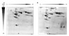
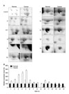
Proteome analysis was performed using 2-DE and silver staining of plasma samples from normal controls and patients with SCC of the cervix. Using an automated spot-counting algorithm provided by Image Master 2D Platinum, more than 400 protein spots were detected in each gel image. The expression patterns of the plasma proteins are shown in Fig. 1 (A, normal control; B, cervical cancer). Computer-assisted comparative analysis of the respective spot patterns matched over 30 spots and selected 15 spots that changed significantly, which showed a constant expression rate that was over 120%, or decreased to less than 80% of that in the controls (Fig. 2). In SCC of the cervix, there were 11 up-regulated spots and four down-regulated spots.
2. MALDI-MS analysis of the differentially expressed spots
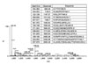
Fifteen differentially expressed spots in the 2-DE gels were isolated and subjected to MALDI-MS. The peptide mass peaks were compared to those in the NCBI database (Fig. 3). The identified protein MS data descriptions are listed in Table 1. Only eight proteins were identified from the 15 differentially expressed spots because spots from the same area were identified as one protein. Of these eight proteins, the six that were up-regulated in the plasma of SCC compared to normal controls were ceruloplasmin, complement C3, afamin precursor, alpha-1-B glycoprotein, transferrin, and alpha-fibrinogen precursor. The remaining two proteins that were down-regulated in SCC were chain A, and crystal structure of antithrombin and apolipoprotein A-IV precursor.
3. Western blotting analysis
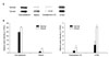
Western blotting analysis was performed to confirm the 2-DE results for the up-regulated proteins. Of the six up-regulated proteins, antibodies against four proteins were available commercially: ceruloplasmin, complement C3, afamin, and alpha-1-B glycoprotein. Consistent with the 2-DE data, Western blotting analyses of ceruloplasmin, complement C3, afamin, and alpha-1-B glycoprotein showed significant elevation of these proteins in the plasma of patients with SCC of the cervix in comparison with the normal controls (Fig. 4).
4. Immunoturbidimetric assay
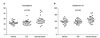
We performed an immunoturbidimetric assay with a new set of plasma samples to confirm the 2-DE and Western blotting results for the up-regulated proteins for further validation. Of the up-regulated proteins, tests for ceruloplasmin and complement C3 were available commercially. Consistent with the 2-DE and Western blotting data, the immunoturbidimetric assays of ceruloplasmin [cut-off value 22, sensitivity 74.1% (95% CI, 53.7-88.8); specificity 62.5% (95% CI, 40.6-81.2)] and complement C3 [cut-off value 112, sensitivity 51.9% (95% CI, 32.0-71.3)], specificity 83.3% (95% CI, 62.6-95.2)] showed significant elevation of these proteins in the plasma of patients with SCC of the cervix compared with normal controls and patients with CIS of the cervix (p<0.05; Fig. 5, Table 2). However, there were no associations between expression for the two proteins and clinicopathological prognostic parameters: stage, depth of stromal invasion, lymphovascular space invasion, parametrium invasion, and lymph node metastasis (p>0.05, data not shown).
DISCUSSION
In this study, we showed significant elevation of ceruloplasmin, complement C3, afamin, and alpha-1-B glycoprotein in the plasma of patients with SCC of the cervix compared to normal controls. Although we did not show associations between expression for ceruloplasmin and complement C3 and clinicopathological prognostic parameters, we demonstrated that ceruloplasmin and complement C3 were significantly elevated in the plasma of patients with SCC of the cervix compared to normal controls and CIS of the cervix using larger numbers of samples. We consider that a further, large-scale study is necessary to establish associations between expression for the two proteins and clinicopathological prognostic parameters.
Ceruloplasmin is a copper-carrying protein that acts as the major ferroxidase and stimulates iron uptake into cells, which is necessary for cell proliferation.17 Ceruloplasmin is also a hypoxia-induced gene, linking the hypoxia and copper pathways of angiogenesis.17,18 Angiogenesis is the initial step in progressive tumor development and metastasis.19 The first stage in tumor angiogenesis is the activation of endothelial cells.19 Copper stimulates the proliferation and migration of endothelial cells, and activates several proangiogenic factors including vascular endothelial growth factor, basic fibroblast growth factor, tumor necrosis factor alpha, and interleukin.19 In the absence of copper, apoceruloplasmin is secreted, but is degraded rapidly.17 Increased expression of ceruloplasmin has been reported in the serum of several cancer patients.12,20-22
Complement components play a central role as mediators of inflammation and regulate of the immune response.23 Complement C3, the third component of complement, is not only the most abundant complement protein in human serum, but also plays a central role in the complement system.24 Complement C3 is a multipotent protein that participates in different events involved in the immune response, as including complement activation, antigen presentation, cell-cell interactions, and cell proliferation.25 Complement activation with the subsequent deposition of complement components on tumor tissues has been demonstrated in cancer patients.26-30 Hanas et al. reported that complement C3 was elevated in the serum of patients with pancreas cancer.31 Lee et al. found that complement C3a was elevated in the serum of patients with hepatitis C-related hepatocellular carcinoma.32 In addition, Bjorgem et al. demonstrated that complement C3a was elevated in the ascitic fluids of ovarian cancer patients.33 Therefore, complement C3 may be a marker related to carcinogenesis.
Afamin, or alpha-albumin, is a new protein and is the fourth member of the albumin family, which comprises albumin, alpha-fetoprotein, and vitamin D binding protein.34,35 Afamin is present in the plasma/serum, cerebrospinal fluid, and follicular fluid.35,36 Afamin is considered a vitamin E binding protein, and vitamin E has been shown to play a crucial role in protection against oxidative damage and disease.35,36 Wu et al.37 reported that afamin was significantly down-regulated in hepatoma tissue. Jackson et al.38 showed that afamin was decreased in the serum of ovarian cancer patients. In contrast, we found that afamin was significantly elevated in the plasma of patients with SCC of the cervix. Therefore, the expression and role of afamin in carcinogenesis should be investigated further.
Alpha-1-B glycoprotein is a plasma protein of unknown function.39 Given its internal duplication and its sequence homology to immunoglobulin-like proteins, alpha-1-B glycoprotein appears to have evolved from an ancestral gene similar to that of the immunoglobulin supergene family.39 Alpha-1-B glycoprotein is present in normal adult plasma at an average concentration of 22 mg/dl.39 However, no biological function has yet been proposed for this protein. There has been one previous study regarding alpha-1-B glycoprotein in the blood of cancer patients. Abdul-Rahman et al. reported that alpha-1-B glycoprotein level was elevated in the serum of patients with endometrial and cervical cancer.40 As the function of alpha-1-Bglycoprotein is unknown, the biological function of alpha-1-B glycoprotein and its association with cancer should be examined in future studies.
In conclusion, we identified ceruloplasmin, complement C3, afamin, and alpha-1-B glycoprotein in plasma as potential biomarkers related to SCC of the cervix using 2-DE combined with MALDI-MS. A further validation study of afamin and alpha-1-B glycoprotein, as biomarkers of SCC of the cervix using larger numbers of samples is needed. In addition, further functional studies of these proteins will provide more information on their roles in the development and progression of cervical cancer.


 XML Download