

 PDF
PDF ePub
ePub Citation
 Print
Print


INTRODUCTION
Cervical cancer is the most common gynecologic malignancy in Korea.1 Cervical cancer is one of the malignancies whose stage can be clinically decided. The management of cervical cancer, particularly its surgical treatment, differs largely depending on the stage of the disease. Early cervical cancer can be cured with surgery. However, postoperative adjuvant therapy should be considered if pathologic findings demonstrate risk factors including lymph node metastases, deep cervical invasion, positive resection margin, parametrial invasion and bulkiness of tumor.2-4 Among them, parametrial invasion and tumor size are evaluated by preoperative pelvic examination.
If clinicians perform an exact pelvic examination, patients can avoid unnecessary surgery. Thus, pelvic examination is important for the decision of further management because pelvic examination is the major diagnostic tool for cervical cancer. Despite the importance of pelvic examination, pelvic examination is often ignored due to the development of imaging studies such as computed tomography (CT) or magnetic resonance imaging (MRI).
The objective of this study was to determine whether pelvic examination by a gynecologic oncologist could yield reliable diagnostic results as a diagnostic tool compared to imaging modality for cervical cancer.
MATERIALS AND METHODS
1. Patients
Between January 2003 and December 2005, 453 patients were diagnosed with cervical cancer. The patients' medical records were retrospectively reviewed, and the women were included if they satisfied the following criteria.
Inclusion criteria included clinically visible cervical cancer, pelvic examination prior to imaging studies, preoperative imaging studies with CT or MRI, surgical treatment as the primary therapy, and a time interval from pelvic examination to surgery of <3 weeks.
Exclusion criteria included diagnostic workups including pelvic examination or imaging at other hospitals, surgery at other hospitals, and other treatments before surgery.
2. Protocol for diagnostic work up
All patients were evaluated by a gynecologic oncologist who had worked at the gynecologic oncology clinic for more than 6 years. The examiner determined the maximum tumor diameter, the site of the tumor, the growth pattern of the tumor and the presence of vaginal or parametrial involvement. Evaluation was aided by colposcopy.
After pelvic examination, CT imaging was performed on 58 patients, and MRI was performed on 70 patients. Both CT and MRI studies were performed on 25 patients. CT imaging was performed by using 16-slice CT equipment (SOMATOM Sensation 16, Siemens, Forchheim, Germany) through the entire abdomen and pelvis after intravenous administration of contrast. CT images were obtained in the transverse plane in 1 cm slice-thickness. MRI was performed using the Signa 1.5-T system (General Electric Medical Systems, Milwaukee, WI) through the pelvis. High-resolution sagittal, axial, transverse T1- and T2-weighted images were obtained in 3 mm slice-thickness. CT and MR images were read by 2 experienced radiologists.
The fresh radical hysterectomy pathologic specimen was examined by the same experienced pathologist (P.I.A.) and the maximum tumor diameter was recorded as the "true" value.
3. Statistical analysis
Measurements of the tumor by pelvic examination and imaging modalities were compared based on the maximum diameter measured by pathologic findings. We categorized the maximum tumor diameter as ≤1 cm, 1.1-2 cm, 2.1-3 cm, 3.1-4 cm, and >4 cm. Differences in accuracy were tested for statistical significance using the McNemar χ2 tests at p=0.05. The linear correlation between the categorized diameters as determined by pathologic findings and the diameters determined by pelvic examination and imaging modality was evaluated using the Spearman correlation coefficient, and agreement was estimated using simple κ statistics. All statistical analyses were performed using SPSS 12.0 (SPSS Inc, Chicago, IL).
RESULTS
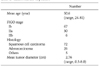
Among 453 patients, 103 met the criteria for this study. Table 1 indicates the characteristics of the patients. Most of the patients belonged to stage Ib or IIa, however, 6 women had findings consistent with stage IIb. Since the patients with advanced cervical cancer (stage IIb) were less than 50 years of age, the clinicians tried to perform surgical treatment combined with postoperative adjuvant therapy for these patients. Tumor size was 2.76±1.76 cm (mean of the maximum diameter±standard deviation), and ranged from 0.5 cm to 8.0 cm at pathologic evaluation. Histologic examination of the majority of patients revealed squamous cell carcinoma.
Experience of gynecologic oncologists was as follows: 6 to 10 years in 2 oncologists; 11 to 15 years in 1 oncologist; and ≥16 years in 2 oncologists. Tumor size measured by pelvic examination ranged from 0.5 to 6.0 cm. Tumor size measured by imaging modality ranged from 0 cm to 7.0 cm. We defined no definite mass as 0 cm.
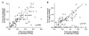
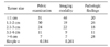
Table 2 depicts the accuracy of pelvic examination and imaging modality including CT and MRI in the measurement of tumor size compared with the "true" value. Based on pathologic findings, accuracy was estimated by the degree of agreement with a difference of 0.5 or 1.0 cm. Pelvic examination was more accurate in the evaluation of tumor size than imaging modality with a difference of both 0.5 and 1.0 cm, and the accuracy rate of pelvic examination was significantly higher than that of imaging modality (72.8% vs. 55.3%, p=0.004). Descriptive statistics for tumor size measurement categorized at 1.0-cm intervals are shown in Table 3. The correlation with pathologic findings was higher for pelvic examination than for imaging modality (rs=0.680 vs. rs=0.410, p<0.0001) (Fig. 1).
The accuracy of each method in the measurement of tumor size is shown in Table 4. The accuracy of both pelvic examination and imaging modality for the evaluation of tumor size become lower as tumor size decreased. However, the accuracy of pelvic examination for the evaluation of tumor size declined more than that of imaging modality. Moreover, imaging modality was accurate especially in the evaluation of bulky tumor.
DISCUSSION
Apart from other gynecologic malignancies such as ovarian cancer and endometrial cancer whose stages are surgically determined, the stage of cervical cancer is clinically determined. Thus, there are numerous prognostic factors which are not determinants of stage in cervical cancer. Since some pathologic parameters are related to patients' prognosis, these parameters affect the plan of further treatment.4-6 This means that pathologic findings of surgical specimens are important for the management of patients with cervical cancer. However, clinicians should consider preoperative diagnosis, because the stage of cervical cancer is clinically determined and clinicians can get much information related with prognosis from preoperative diagnosis. Tumor size has been suggested as an important prognostic factor in patients with cervical cancer.7-11 Although there are several diagnostic methods including pelvic examination, intravenous pyelogram, cystoscopy, proctoscopy and others, pelvic examination is the only single method for evaluating the size, location and parametrial invasion of the cervical mass. Thus, exact pelvic examination is important for the management of patients with cervical cancer.
Recent development of imaging techniques has made it possible to evaluate not only the characteristics of cervical mass but also the status of lymph nodes.12,13 Imaging diagnosis using CT or MRI plays an important role for cancer patients in Korea because these imaging machineries are popular and also because National Health Insurance covers a large portion of medical fee for CT or MRI in Korea. Thus, CT or MRI has progressively replaced standard diagnostic methods of cervical cancer. Moreover, some clinicians make a decision for the management of cervical cancer using imaging tools such as CT or MRI instead of the standard pelvic examination.
Could these imaging tools replace pelvic examination? The current study demonstrates the accuracy of pelvic examination and the imaging modality including CT or MRI for the evaluation of cervical mass in 103 cervical cancer patients. The real size of cervical mass ranged from 0.5 to 8.0 cm in pathologic findings. Compared with the size of the cervical mass evaluated by pathologic findings, pelvic examination estimated the size of the cervical mass more accurately than imaging modality. Many previous studies have focused on the value of imaging modality as new diagnostic method for evaluating tumor size, parametrial invasion and lymph node involvement.14-16 Although quite a few studies have reported the value of pelvic examination in cervical cancer, it has been demonstrated that the accuracy of pelvic examination is 50% with a difference of <1.0 cm from the actual values and that it is 52% with a 25% difference between clinical and pathologic measurements of tumor diameter.17,18 On the contrary, some other investigators have reported the accuracy of pelvic examination ranged from 64% to 68%.19,20 Our results showed similar accuracy rates compared with these latter reports. However, the accuracy of pelvic examination with a difference of <0.5 cm was 46.6%, and the accuracy of pelvic examination with a difference of 1.0 cm was 57.9% in the patients with the cervical mass >2.0 cm. These results were similar to findings of the former previous studies.
The accuracy of imaging modality was about 55% in agreement within 1.0 cm for tumor diameter. Many investigators have reported the value of CT or MRI in patients with cervical cancer. Nonetheless, most of them focused on the lymph node status and parametrial invasion. However, some investigators have found the accuracy of MRI ranged about from 30% to 70% according to the tumor size.21,22 Some reports have demonstrated the accuracy of MRI in the evaluation of the stage of cervical cancer ranged from 70% to 85%.23-25 These results indicate higher accuracy rate of imaging modality than those of our study. However, a comparative study has reported the accuracy of CT and MRI to be 51% and 75%, respectively in the evaluation of cervical cancer.24 Above all, a prospective study with a large sample size has demonstrated that the accuracy of CT and MRI is overestimated in the diagnosis of cervical cancer, with the accuracy of CT and MRI being 42% and 53%, respectively.26 This result was in accord with that of our study.
However, imaging modality was more accurate than pelvic examination in cases where the size of the cervical mass exceeds 4.0 cm. In addition, if a cervical tumor existed at the endocervix, the discordance between tumor diameters measured by pelvic examination and pathologic findings appears to become larger. Thus, imaging studies could be good methods for evaluating bulky masses which are >4.0 cm and are difficult to find at the exocervix. Mayr et al. also has reported MRI was more useful in tumors larger than 4.0 cm in cervical cancer patients.22
In conclusion, pelvic examination is an accurate and reliable method for evaluating the tumor size of cervical cancer. However, this study has some limitations because it was not a prospective study with a standardized examination method, and the difference between CT and MRI was not considered. Further prospective studies for evaluating pelvic examination with the standard technique are needed to confirm these findings.


 XML Download