Abstract
Background
Meningitis is a clinically important disease because of its high mortality and morbidity. The epidemiology of this disease has changed remark-ably due to the introduction of pneumococcal vaccines and Haemophilus influenzae type b (Hib) conjugate vaccine. Therefore, it is required to continuously monitor and research the organisms isolated from cerebrospinal fluid (CSF) cultures.
Methods
We analyzed trends of bacteria and fungi isolates obtained from CSF cultures between 1997 and 2016 in a tertiary care hospital according to year, month, gender, and age.
Results
Out of a total of 38,450 samples, we identified 504 (1.3%) isolates. The isolation rate in the first tested decade (1997–2006) ranged from 1.3% to 3.1%, while that in the second decade (2007–2016) ranged from 0.4% to 1.5%. The most common organisms was coagulase-negative staphylococci (CoNS) (31.9%), followed by Staphylococcus aureus (9.5%), Streptococcus pneumoniae (7.5%), Acinetobacter baumannii (5.8%), and Mycobacterium tuberculosis (5.8%).
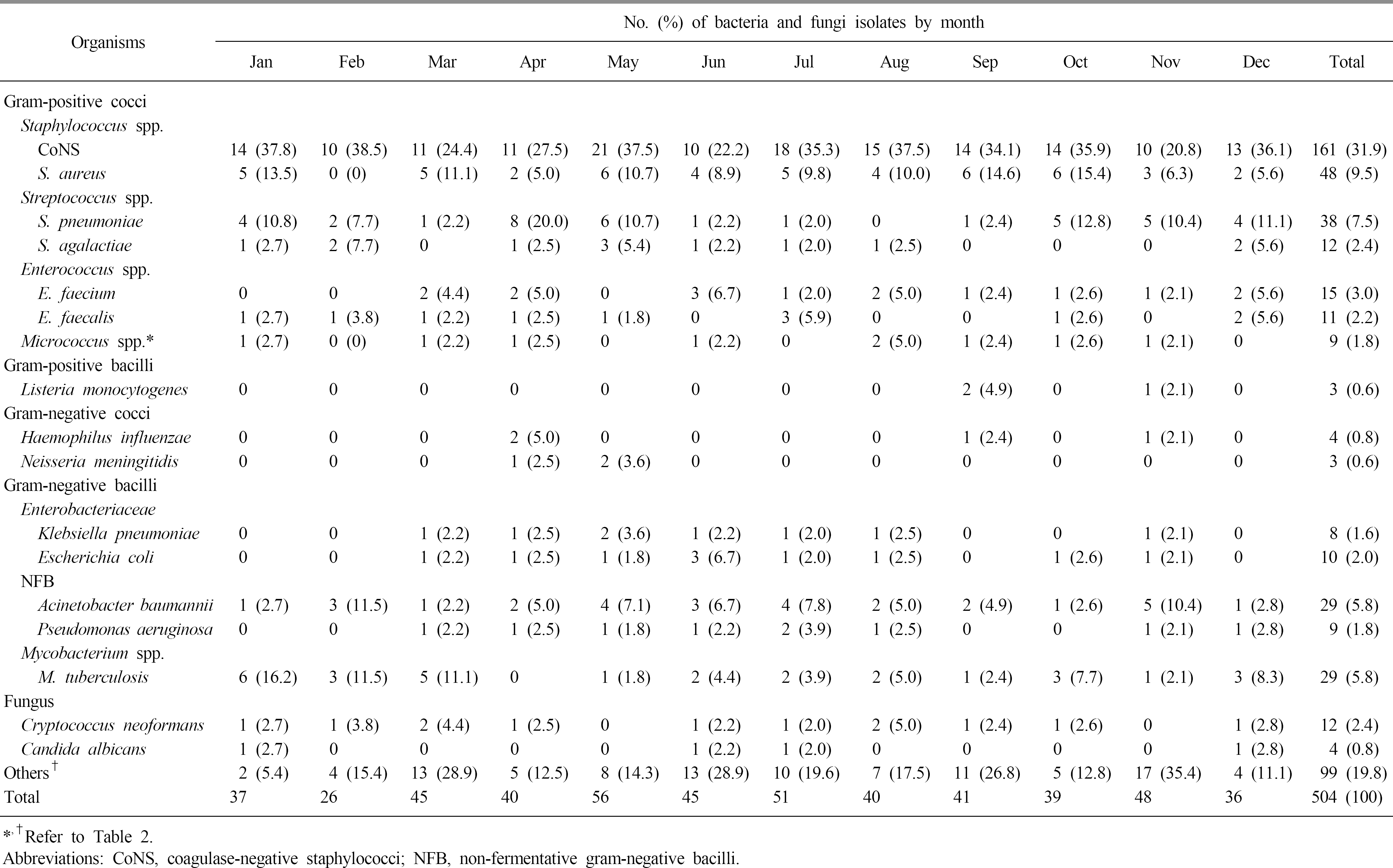
Monthly isolation rates were highest in May and July and lowest in February and December. Male to female ratio was 1.5:1. The isolation rates of S. pneumoniae, Enterococcus faecium, and Escherichia coli were similar in children and adults, but those of S. aureus, E. faecalis, A. baumannii, Pseudomonas aeruginosa, M. tuberculosis, and Cryptococcus neoformans were higher in adults than in children.
Conclusion
During the last two decades, the isolation rate of CSF culture per year has decreased, with monthly isolation rates being highest in May and July. CoNS, S. aureus, and S. pneumoniae were most common in males, whereas CoNS, S. pneumoniae, and M. tuberculosis were most common in females. While Group B Streptococcus was most common in infants younger than 1 year, S. aureus and C. neoformans were more common in adults.
Go to : 
References
1. Kim KS. Pathogenesis of bacterial meningitis: from bacteraemia to neuronal injury. Nat Rev Neurosci. 2003; 4:376–85.

2. Lepur D and Barsić B. Community-acquired bacterial meningitis in adults: antibiotic timing in disease course and outcome. Infection. 2007; 35:225–31.

3. The Korean Society of Infectious Diseases; The Korean Society for Chemotherapy; The Korean Neurological Association; The Korean Neurosurgical Society; The Korean Society of Clinical Microbiology. Clinical practice guidelines for the management of bacterial meningitis in adults in Korea. Infect Chemother. 2012; 44:140–63.
4. Scheld WM, Koedel U, Nathan B, Pfister HW. Pathophysiology of bacterial meningitis: mechanism(s) of neuronal injury. J Infect Dis. 2002; 186(Suppl 2):S225–33.

5. Cho HK, Lee H, Kang JH, Kim KN, Kim DS, Kim YK, et al. The causative organisms of bacterial meningitis in Korean children in 1996–2005. J Korean Med Sci. 2010; 25:895–9.

6. Brouwer MC, Tunkel AR, van de Beek D. Epidemiology, diagnosis, and antimicrobial treatment of acute bacterial meningitis. Clin Microbiol Rev. 2010; 23:467–92.

7. Song W, Kim YJ, Uh Y, Park JA, Lee K. Trends of isolation of organisms from cerebrospinal fluid during 1981–1990. J Wonju Med Coll. 1991; 4:139–47.
8. Kim MJ, Moon SM, Park TS, Suh JT, Lee HJ. Clinical aspects of bacterial meningitis in cerebrospinal fluid culture positive patients in a tertiary care university hospital. Korean J Clin Microbiol. 2011; 14:1–6.

9. von Eiff C, Peters G, Heilmann C. Pathogenesis of infections due to coagulase-negative staphylococci. Lancet Infect Dis. 2002; 2:677–85.

10. Huang CR, Lu CH, Wu JJ, Chang HW, Chien CC, Lei CB, et al. Coagulase-negative staphylococcal meningitis in adults: clinical characteristics and therapeutic outcomes. Infection. 2005; 33:56–60.

11. Aguilar J, Urday-Cornejo V, Donabedian S, Perri M, Tibbetts R, Zervos M. Staphylococcus aureus meningitis: case series and literature review. Medicine (Baltimore). 2010; 89:117–25.
Go to : 
Table 1.
No. (%) of isolates from cerebrospinal fluid culture by year
Table 2.
Distribution and isolation trend of bacteria and fungi isolates from cerebrospinal fluid culture by year

Table 4.
No. (%) of bacteria and fungi isolates from cerebrospinal fluid culture by gender
Table 5.
No. (%) of bacteria and fungi isolates from cerebrospinal fluid culture by age group
| Organisms | No. (%) of organisms isolates by age group (year) | ||||
|---|---|---|---|---|---|
| <1 | 1–19 | 20–69 | ≥70 | Total | |
| Gram-positive cocci | |||||
| Staphylococcus spp. | |||||
| CoNS | 22 (28.9) | 28 (28.9) | 93 (33.3) | 18 (34.6) | 161 (31.9) |
| S. aureus* | 2 (2.6) | 9 (9.3) | 31 (11.1) | 6 (11.5) | 48 (9.5) |
| Streptococcus spp. | |||||
| S. pneumoniae | 3 (3.9) | 15 (15.5) | 16 (5.7) | 4 (7.7) | 38 (7.5) |
| S. agalactiae† | 10 (13.2) | 0 | 1 (0.4) | 1 (1.9) | 12 (2.4) |
| Enterococcus spp. | |||||
| E. faecium | 4 (5.3) | 3 (3.1) | 8 (2.9) | 0 | 15 (3.0) |
| E. faecalis | 2 (2.6) | 1 (1.0) | 8 (2.9) | 0 | 11 (2.2) |
| Micrococcus spp.‡ | 1 (1.3) | 2 (2.1) | 6 (2.2) | 0 | 9 (1.8) |
| Gram-positive bacilli | |||||
| Listeria monocytogenes | 0 | 0 | 3 (1.1) | 0 | 3 (0.6) |
| Gram-negative cocci | |||||
| Haemophilus influenzae | 1 (1.3) | 3 (3.1) | 0 | 0 | 4 (0.8) |
| Neisseria meningitidis | 0 | 2 | 1 (0.4) | 0 | 3 (0.6) |
| Gram-negative bacilli | |||||
| Enterobacteriaceae | |||||
| Klebsiella pneumoniae | 2 (2.6) | 1 (1.0) | 5 (1.8) | 0 | 8 (1.6) |
| Escherichia coli | 3 (3.9) | 2 (2.1) | 4 (1.4) | 1 (1.9) | 10 (2.0) |
| NFB | |||||
| Acinetobacter baumannii | 4 (5.3) | 2 (2.1) | 21 (7.5) | 2 (3.8) | 29 (5.8) |
| Pseudomonas aeruginosa | 2 (2.6) | 0 | 5 (1.8) | 2 (3.8) | 9 (1.8) |
| Mycobacterium spp. | |||||
| M. tuberculosis | 2 (2.6) | 4 (4.1) | 18 (6.5) | 5 (9.6) | 29 (5.8) |
| Fungus | |||||
| Cryptococcus neoformans* | 0 | 0 | 9 (3.2) | 3 (5.8) | 12 (2.4) |
| Candida albicans | 2 (2.6) | 0 | 2 (0.7) | 0 | 4 (0.8) |
| Others§ | 16 (19.7) | 25 (19.6) | 48 (17.2) | 10 (19.2) | 99 (19.8) |
| Total | 76 | 97 | 279 | 52 | 504 (100) |


 PDF
PDF ePub
ePub Citation
 Print
Print


 XML Download