초록
Background
The intestinal microflora varies according to the factors such as age, diet and environment. It is debated whether the changes of microbiota after birth are associated with atopic disease. The purpose of this study was to investigate colonization rates of some intestinal microflora during the initial 9 months after birth, and their association with the development of atopy.
Methods
Stool specimens were collected at 1, 3, 7 days and at 1, 2, 4, 6, 9 months after birth, and Escherichia coli, Lactobacillus, Bifidobacterium, Staphylococcus aureus were cultured with selective media. Diagnosis for atopy was accomplished via clinical history of atopy, serum total IgE, and skin prick test.
Results
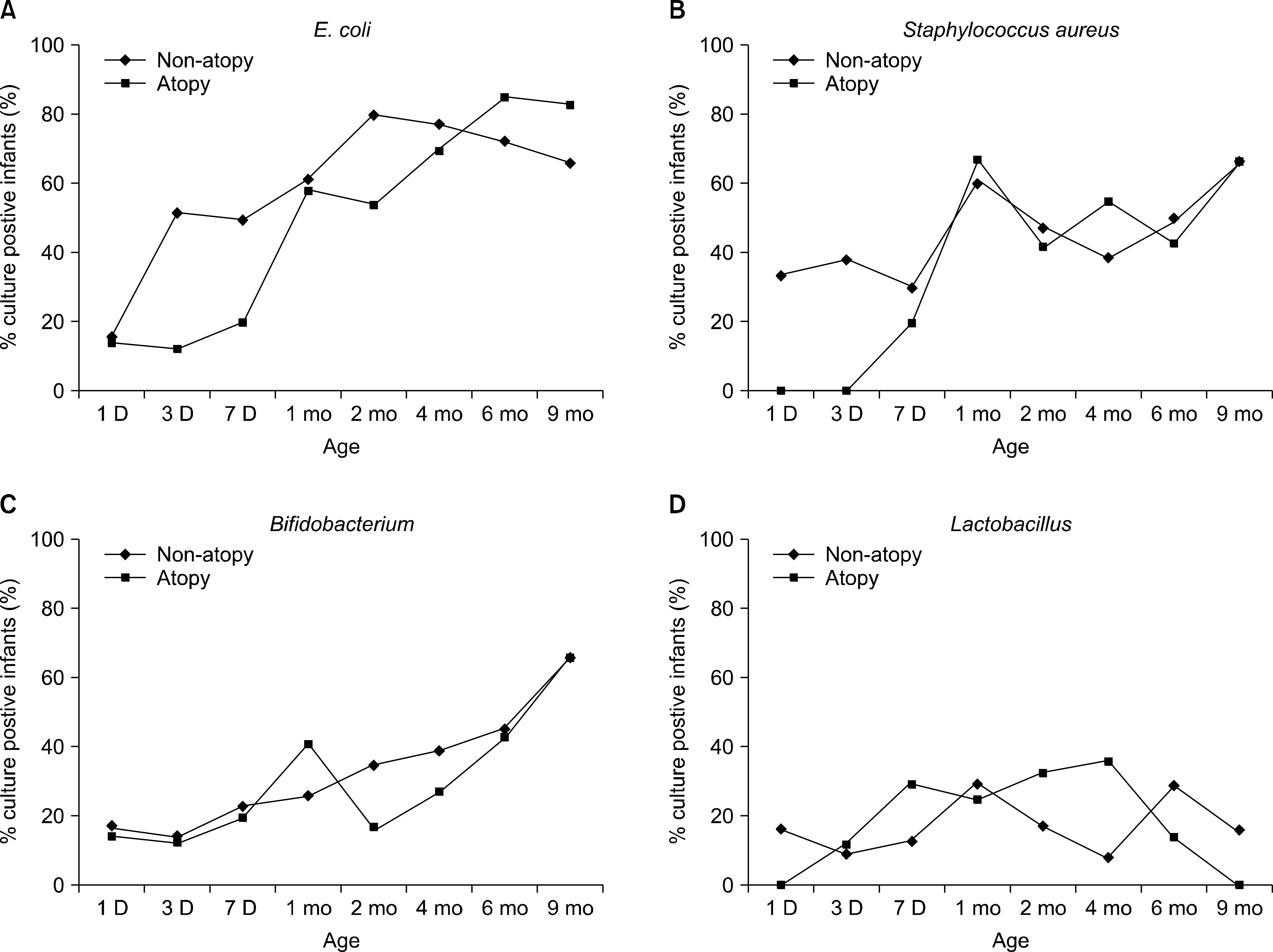
By 12 months of age, among 48 infants, 36 (75.0%) were non-atopic while 12 (25.0%) had devel-oped atopy. Although not statistically significant, the intestinal microflora of infants with atopy vs. non-ato-py was characterized by being less often colonized with E. coli (12.5% vs. 52.4%; P=0.093) and S. aureus (0% vs. 38.1%; P=0.066) at three days after birth. Colonization rates of E. coli reached 50% after 3 days of birth in non-atopy group whereas this rate was not achieved until after 1 month in the atopy group.
Conclusion
The intestinal colonization rates of bacteria in this study were not statistically different between atopy and non-atopy groups. Rapid colonization of E. coli and S. aureus was observed within 1 week after birth in the non-atopy group. The exact association between atopy and the bacterial colonization and/or diversity in the early days after birth has yet to be determined.
Go to : 
REFERENCES
1.Johnson CL., Versalovic J. The human microbiome and its potential importance to pediatrics. Pediatrics. 2012. 129:950–60.

2.Qin J., Li R., Raes J., Arumugam M., Burgdorf KS., Manichanh C, et al. A human gut microbial gene catalogue established by metagenomic sequencing. Nature. 2010. 464:59–65.

3.Penders J., Stobberingh EE., van den Brandt PA., Thijs C. The role of the intestinal microbiota in the development of atopic disorders. Allergy. 2007. 62:1223–36.

5.Bisgaard H., Li N., Bonnelykke K., Chawes BL., Skov T., Paludan- Müller G, et al. Reduced diversity of the intestinal microbiota during infancy is associated with increased risk of allergic disease at school age. J Allergy Clin Immunol. 2011. 128:646–52.e1-5.

6.Abrahamsson TR., Jakobsson HE., Andersson AF., Björkstén B., Engstrand L., Jenmalm MC. Low diversity of the gut microbiota in infants with atopic eczema. J Allergy Clin Immunol. 2012. 129:434–40. 440.e1-2.

7.Hanski I., von Hertzen L., Fyhrquist N., Koskinen K., Torppa K., Laatikainen T, et al. Environmental biodiversity, human microbiota, and allergy are interrelated. Proc Natl Acad Sci U S A. 2012. 109:8334–9.

8.Park YH., Kim KW., Choi BS., Jee HM., Sohn MH., Kim KE. Relationship between mode of delivery in childbirth and prevalence of allergic diseases in Korean children. Allergy Asthma Immunol Res. 2010. 2:28–33.

9.Lee YJ., Jee HM., Kim BJ., Kim HB., Yu J., Lee SY, et al. Prevalence of allergic diseases in children according to mode of delivery. Pediatr Allergy Respir Dis. 2011. 21:197–206.

10.Pyun BK., Kim WK, et al. eds. Pediatric Allergy Immunology Pulmonology. 2nd ed.The Korean Academy of Pediatric Allergy and Respiratory Disease;2013. p. 69–72.
11.Guarner F. Hygiene, microbial diversity and immune regulation. Curr Opin Gastroenterol. 2007. 23:667–72.

12.Björkstén B., Sepp E., Julge K., Voor T., Mikelsaar M. Allergy development and the intestinal microflora during the first year of life. J Allergy Clin Immunol. 2001. 108:516–20.

13.Kalliomäki M., Isolauri E. Role of intestinal flora in the development of allergy. Curr Opin Allergy Clin Immunol. 2003. 3:15–20.

14.Lee HB. Effects of breastfeeding on the development of allergies. Hanyang Med Rev. 2010. 30:49–59.

15.Nowrouzian F., Hesselmar B., Saalman R., Strannegard IL., Aberg N., Wold AE, et al. Escherichia coli in infants' intestinal microflora: colonization rate, strain turnover, and virulence gene carriage. Pediatr Res. 2003. 54:8–14.
16.Yi J., Kim EC. Microbiological characteristics of methicillin- resistant Staphylococcus aureus. Korean J Clin Microbiol. 2010. 13:1–6.
17.Oh HR., Moon DS., Jang SJ., Li XM., Kim DM., Park SG, et al. Epidemiological investigation of an outbreak of Escherichia coli infections in neonatal intensive care unit of a university hospital. Korean J Clin Microbiol. 2008. 11:123–8.
18.Björkstén B., Naaber P., Sepp E., Mikelsaar M. The intestinal microflora in allergic Estonian and Swedish 2-year-old children. Clin Exp Allergy. 1999. 29:342–6.

19.Grönlund MM., Arvilommi H., Kero P., Lehtonen OP., Isolauri E. Importance of intestinal colonisation in the maturation of humoral immunity in early infancy: a prospective follow up study of healthy infants aged 0-6 months. Arch Dis Child Fetal Neonatal Ed. 2000. 83:F186–92.
20.Hartemink R., Rombouts FM. Comparison of media for the detection of bifidobacteria, lactobacilli and total anaerobes from faecal samples. J Microbiol Methods. 1999. 36:181–92.

21.Grönlund MM., Lehtonen OP., Eerola E., Kero P. Fecal microflora in healthy infants born by different methods of delivery: permanent changes in intestinal flora after cesarean delivery. J Pediatr Gastroenterol Nutr. 1999. 28:19–25.
Go to : 
 | Fig. 1.Intestinal colonization rate in infants with allergic or nonallergic patients. At 3 days of age, the colonization rates of (A) Escherichia coli and (B) Staphylococcus aureus in nonallergic group (52.4% and 38.1%) was higher than those of allergic group (12.5% and 0%), without statistical significance (P=0.093 and P=0.066). No significant differences were observed in (C) Bifidobacterium and (D) Lactobacillus colonization. |
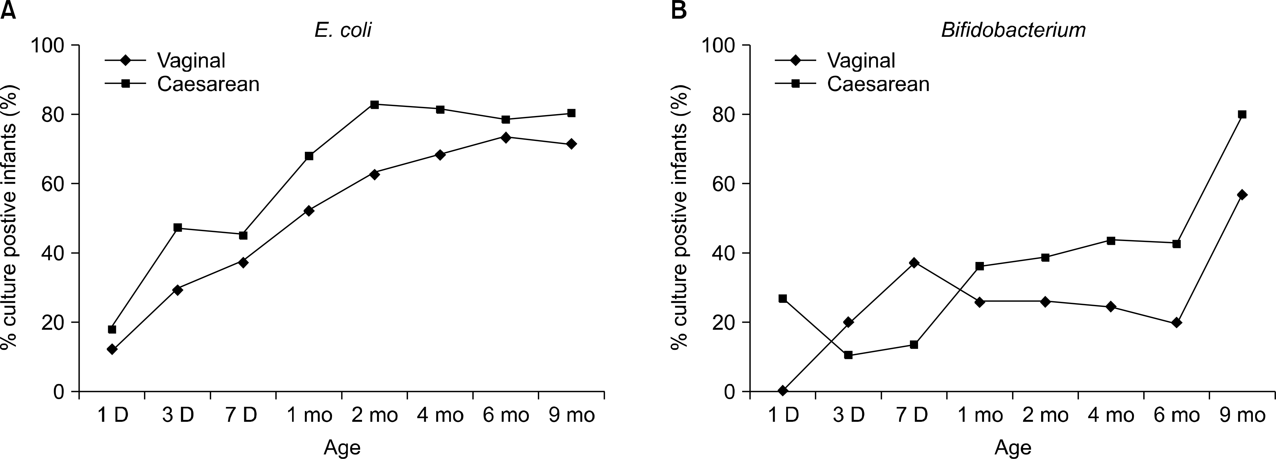
 | Fig. 2.Intestinal colonization rate of (A) E. coli and (B) Bifidobacterium in infants vaginally delivered and by caesarean section. Half of the infants had E. coli after 1 months of age, while half had Bifidobacterium at 9 months of life. There was no difference in colonization rate between these two groups during 9 months. |
Table 1.
Description of children included in the study
Table 2.
Intestinal colonization rate in all the infants after birth


 PDF
PDF ePub
ePub Citation
 Print
Print


 XML Download