초록
We report a suspicious case of spontaneous bacterial peritonitis (SBP) caused by Providencia rettgeri and Clostridium perfringens in a patient with alcoholic cirrhosis. The patient presented with altered mentality and was taken to the emergency room. He was diag-nosed with SBP after abdominal paracentesis and computed tomography and was treated with ceftriaxone and metronidazole. The pathogens were identified under sus-picion of polymicrobial infection because of Gram-stain-ing discrepancies between broth from blood culture bot-tles and colonies on solid media. He died of septic shock despite transfer to the intensive care unit. Although we could not conclude which organism had the leading role in this case of SBP and septicemia, we did verify the importance of Gram staining in a microbiology laboratory in terms of quality assurance.
Go to : 
REFERENCES
1.Conn HO. Spontaneous peritonitis and bacteremia in Laennec's cirrhosis caused by enteric organisms. A relatively common but rarely recognized syndrome. Ann Intern Med. 1964. 60:568–80.
2.Dore MP., Casu M., Realdi G., Piana A., Mura I. Helicobacter infection and spontaneous bacterial peritonitis. J Clin Microbiol. 2002. 40:1121.
3.Lata J., Stiburek O., Kopacova M. Spontaneous bacterial peritonitis: a severe complication of liver cirrhosis. World J Gastroenterol. 2009. 15:5505–10.
4.Young PE., Dobhan RR., Schafer TW. Clostridium perfringens spontaneous bacterial peritonitis: report of a case and implications for management. Dig Dis Sci. 2005. 50:1124–6.
5.Garcia-Tsao G. Current management of the complications of cirrhosis and portal hypertension: variceal hemorrhage, ascites, and spontaneous bacterial peritonitis. Gastroenterology. 2001. 120:726–48.

6.Sugihara T., Koda M., Maeda Y., Matono T., Nagahara T., Mandai M, et al. Rapid identification of bacterial species with bacterial DNA microarray in cirrhotic patients with spontaneous bacterial peritonitis. Intern Med. 2009. 48:3–10.

7.Koreishi AF., Schechter BA., Karp CL. Ocular infections caused by Providencia rettgeri. Ophthalmology. 2006. 113:1463–6.
8.Kim BN., Kim NJ., Kim MN., Kim YS., Woo JH., Ryu J. Bacteraemia due to tribe Proteeae: a review of 132 cases during a decade (1991-2000). Scand J Infect Dis. 2003. 35:98–103.

9.Lee G., Hong JH. Xanthogranulomatous pyelonephritis with nephrocutaneous fistula due to Providencia rettgeri infection. J Med Microbiol. 2011. 60:1050–2.
10.Unverdi S., Akay H., Ceri M., Inal S., Altay M., Demiroz AP, et al. Peritonitis due to Providencia stuartii. Perit Dial Int. 2011. 31:216–7.
11.Gloor HJ. 20 years of peritoneal dialysis in a mid-sized Swiss hospital. Swiss Med Wkly. 2003. 133:619–24.
12.Yang CC., Hsu PC., Chang HJ., Cheng CW., Lee MH. Clinical significance and outcomes of Clostridium perfringens bacteremia--a 10-year experience at a tertiary care hospital. Int J Infect Dis. 2013. 17:e955–60.
13.Targan SR., Chow AW., Guze LB. Role of anaerobic bacteria in spontaneous peritonitis of cirrhosis: report of two cases and review of the literature. Am J Med. 1977. 62:397–403.
14.Conn HO., Fessel JM. Spontaneous bacterial peritonitis in cirrhosis: variations on a theme. Medicine (Baltimore). 1971. 50:161–97.
Go to : 
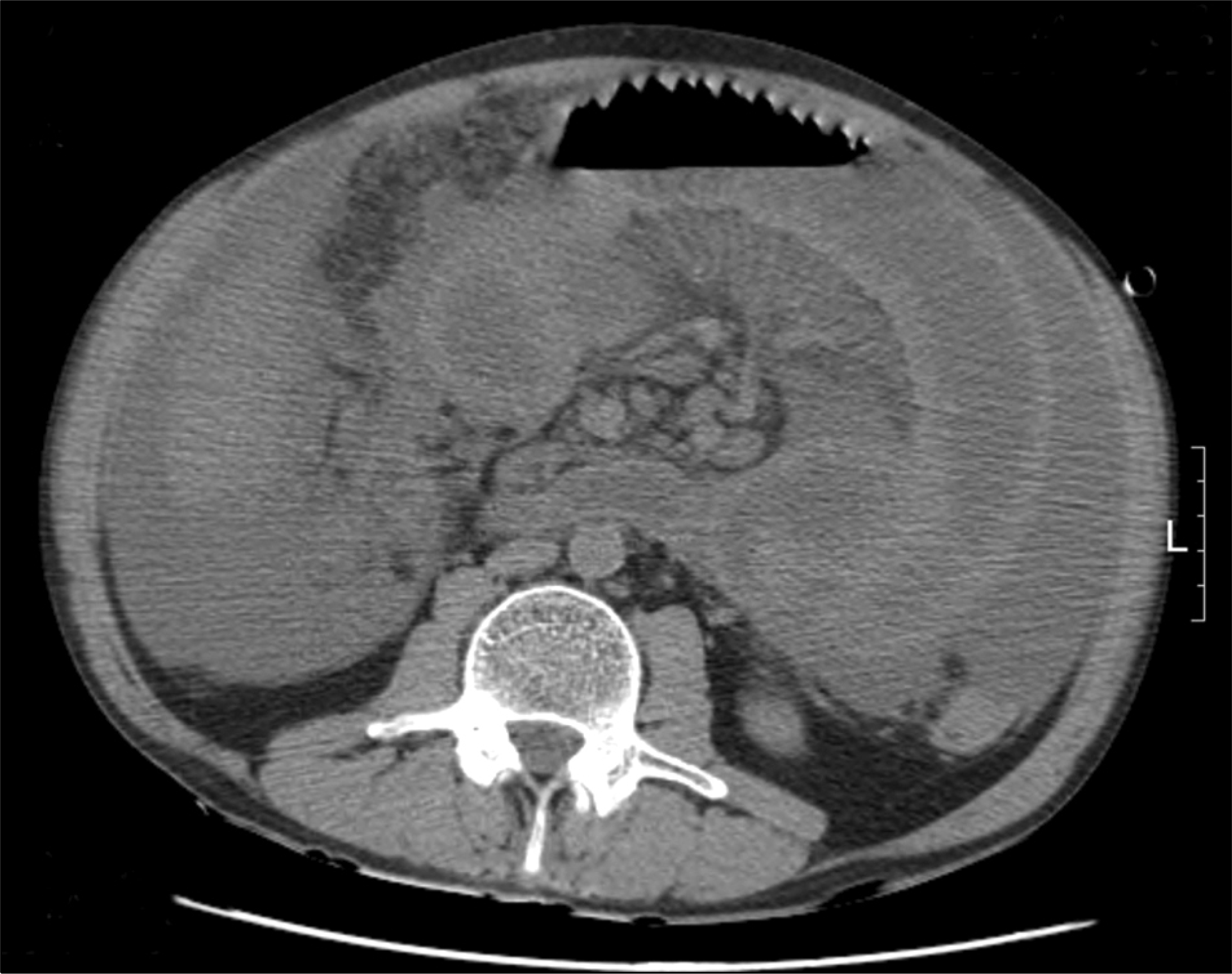
 | Fig. 1.Noncontrast abdominal computed tomography image showing ascites, hepatomegaly and diffuse wall thickening of the small bowels. |
 | Fig. 2.(A) Gram-positive and gram- negative rods from smear preparations of the positive aerobic blood culture bottle (Gram stain, ×1,000). (B) Gram-positive and gram-negative rods from smear preparations of the positive anaerobic blood culture bottle (Gram stain, ×1,000). (C) White and mucous colonies of Providencia rettgeri incu-bated for 24 hr at 35 o C in a 5% CO2 atmosphere on a blood agar plate. (D) Gram stain of P. rettgeri from smear preparations of the blood agar plate (×1,000). (E) Flat colonies with beta-hemolysis of C. perfringens incu-bated for 48 hr anaerobically on a Brucella agar plate. (F) Gram stain of Clostridium perfringens from smear preparations of the Brucella agar plate (×1,000). |


 PDF
PDF ePub
ePub Citation
 Print
Print


 XML Download