

 PDF
PDF ePub
ePub Citation
 Print
Print


초록
We report a case of the isolation of the Aspergillus versicolor complex, initially misidentified by morpho-logical characteristics as the Scopulariopsis species, from a homograft with a bicuspidalized pulmonary valve. An eighteen-month-old female, who had critical pulmonary stenosis, underwent pulmonary valve re-placement. On postoperative day 8, she developed a fever, which did not respond to empiric broad-spec-trum antibiotics. While no definitive source was identified, a filamentous fungus was isolated from the thawed homograft tissue culture prior to implantation on the operation day. The colonies were powdery green with white edges on Sabouraud dextrose agar. Microscopic examination showed septate hyphae with branched conidiophores and chains of spiny conidia, which suggested Scopulariopsis species. After direct sequencing of the internal transcribed spacer (ITS) regions, the fungus was identified as the A. versicolor complex. To our knowledge, the isolation of the A. versicolor complex from a homograft valve has not been previously described. This case shows that laboratory staff should be aware that microscopic morphology of the A. versicolor complex can re-semble that of a number of other genera, including Scopulariopsis species.
Go to : 
REFERENCES
1.Jurjevic Z., Peterson SW., Horn BW. Aspergillus section Versicolores: nine new species and multilocus DNA sequence based phylogeny. IMA Fungus. 2012. 3:59–79.
2.Van Der Linden JW., Warris A., Verweij PE. Aspergillus species intrinsically resistant to antifungal agents. Med Mycol. 2011. 49(Suppl 1):S82–9.
3.Clinical and Laboratory Standards Institute. Interpretive Criteria for Microorganism Identification by DNA Target Sequencing; Approved Guideline. Document MM18-A. Wayne PA. Clinical and Laboratory Standards Institute. 2008.
4.Wang S., Zinderman C., Wise R., Braun M. Infections and human tissue transplants: review of FDA MedWatch reports 2001-2004. Cell Tissue Bank. 2007. 8:211–9.

5.Fan YD., Van Hoeck B., Holovska V., Jashari R. Evaluation of decontamination process of heart valve and artery tissues in European Homograft Bank (EHB): a retrospective study of 1,055 cases. Cell Tissue Bank. 2012. 13:297–304.
6.Villalba R., Mirabet V., Rendal E., González AI., Solves P., Andión C, et al. Microbiological analysis of cryopreserved human heart valves after storage: a survey of 3 banks in Spain. Cell Tissue Bank. 2009. 10:345–9.
7.Soo A., Healy DG., El-Bashier H., Shaw S., Wood AE. Quality control in homograft valve processing: when to screen for microbiological contamination? Cell Tissue Bank. 2011. 12:185–90.
8.Alastruey-Izquierdo A., Mellado E., Cuenca-Estrella M. Current section and species complex concepts in Aspergillus: recommendations for routine daily practice. Ann N Y Acad Sci. 2012. 1273:18–24.
9.Larone DH. Medically important fungi. a guide to identification. 5th ed.Washington DC: ASM Press;2011.
Go to :
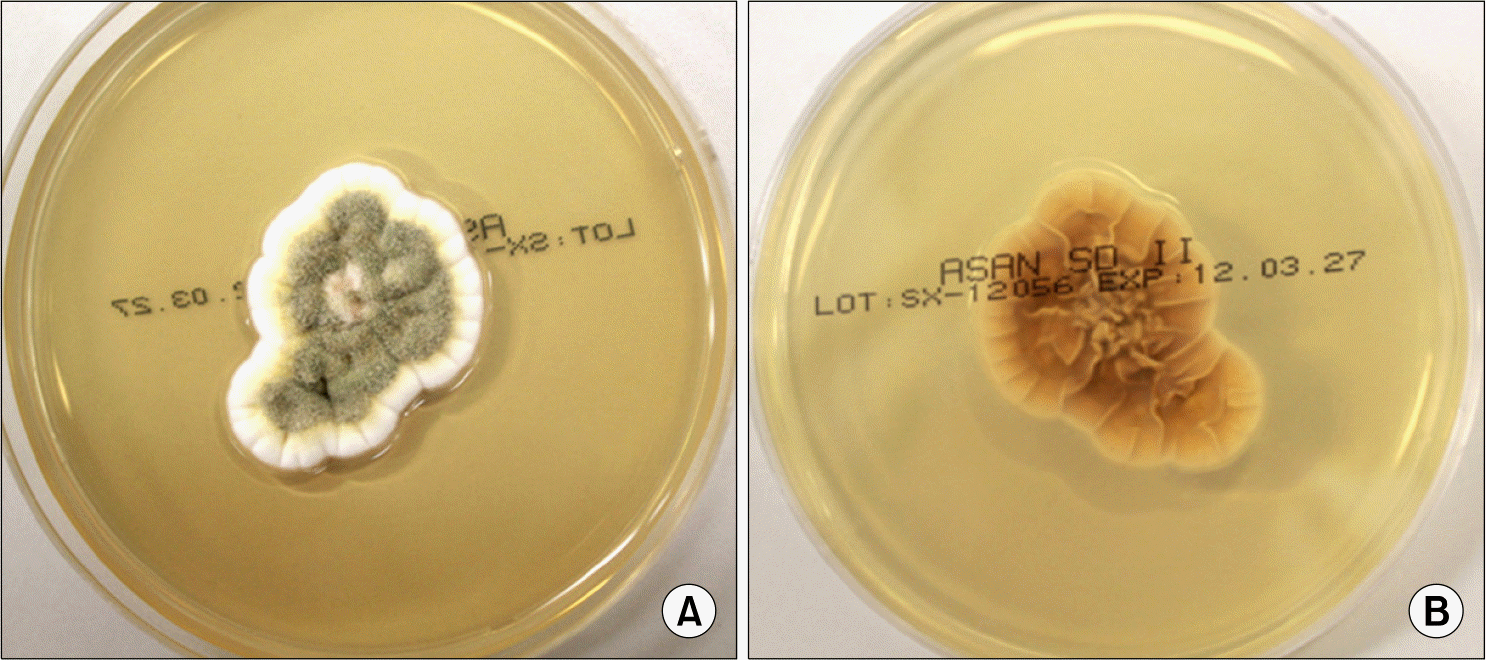 | Fig. 1.
A. versicolor colonies on a SDA plate. (A) The grayish green surface with white edges (B) Reverse of colony. |
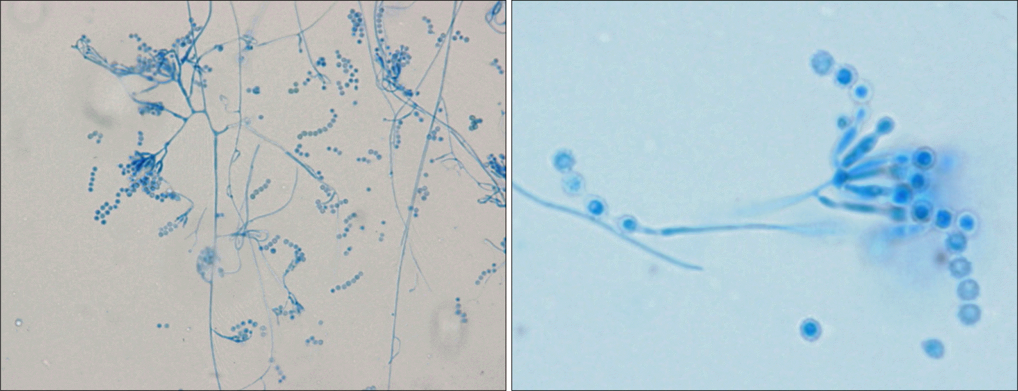 | Fig. 2.Microscopic morphology of A. versicolor isolated from this case resembling Scopulariopsis spp. (lactophenol cotton blue stain, ×400 and ×1,000, respectively). |
Table 1.
Differentiating characteristics of species of Scopulariopsis, Penicillium, Aspergillus versicolor and A. sydowii.
 XML Download