Journal List > J Gastric Cancer > v.18(1) > 1077097

Seo, Jung, Park, Song, and Jung: IgG4-related Disease in the Stomach which Was Confused with Gastrointestinal Stromal Tumor (GIST): Two Case Reports and Review of the Literature

Abstract

Immunoglobulin G4-related disease (IgG4-RD) is an immune-mediated fibro-inflammatory disorder characterized by specific pathological findings and elevated serum IgG4 level. IgG4-RD in the stomach is rare, and occasionally diagnosed as gastric subepithelial tumor (SET) by endoscopy or computed tomography scan. Two female patients in the age group of 40–50 years were diagnosed with 4 cm sized gastric SET. One underwent laparoscopic gastric wedge resection. Another one had a history of subtotal gastrectomy for early gastric cancer and idiopathic thrombocytopenic purpura with oral steroids administration. She underwent a completion total gastrectomy with splenectomy for the gastric SET and ITP. The pathology showed storiform fibrosis, and IgG4 was positive in immunohistochemistry (IHC) stain. IgG4-RD is known as a medical disease that could be treated with oral steroids. The difficulty in preoperative diagnosis of the disease occasionally causes unnecessary gastric resection. Thus, preoperative diagnostic methods for IgG4-RD such as deep biopsy with IHC stain or magnetic resonance imaging are needed.

INTRODUCTION

Immunoglobulin G4-related disease (IgG4-RD) is an immune-mediated fibro-inflammatory disorder characterized by specific pathological finding such as dense tissue infiltration of IgG4 positive lymphoplasmacytic cells, dense storiform fibrosis, obstructive phlebitis and elevated serum IgG4 level [1]. These pathological findings were first described in autoimmune pancreatitis (AIP) by Kamisawa et al. [2] in 2003 and the term IgG4-RD has been used since then.
A painless organ swelling without specific symptom is a main characteristic of IgG4-RD. The pathogenesis of this disease remains unclear, but type 2 T-helper cells, regulatory T-cell cytokines and B-cell activating factor have been suggested to be associated with development of IgG4-RD [1]. Diagnosis of IgG4-RD can be made on the basis of serological, imaging and histopathological findings. It is well known that IgG4-RD responds well to glucocorticoid. It can involve in any of the organs such as pancreas, bile duct, gallbladder, retroperitoneum, gastrointestinal tract, kidney, salivary gland, lung, brain, and prostate gland and the patients present with various symptoms depending on the involving organs [345678]. Pancreas is the most commonly involved organ, which of the disease is diagnosed as AIP, but various extrapancreatic organ can be involved with or without AIP [9]. Studies of the disease involving gastrointestinal tract, especially gastric mass lesion caused by IgG4-RD, were rarely reported.
Herein, we report the 2 cases of IgG4-RD in the stomach which were diagnosed with subepithelial tumor (SET) on endoscopy.

CASE REPORT

Case 1

The patient was a 40-year-old female who was diagnosed with gastric SET found on endoscopy by routine medical examination and visited our hospital in October 2015 for further evaluation. She presented with no specific abdominal symptom such as pain, vomiting, hematemesis, dysphagia, heartburn, abdominal distension, melena or change in bowel habit. She was not diabetic or hypertensive or allergic. She had no medication history or family history of malignant disease or autoimmune disorder. The body temperature was 36.3°C with blood pressure 120/80 mmHg, and pulse was 68 beats/min. The abdomen was soft and flat without tenderness.
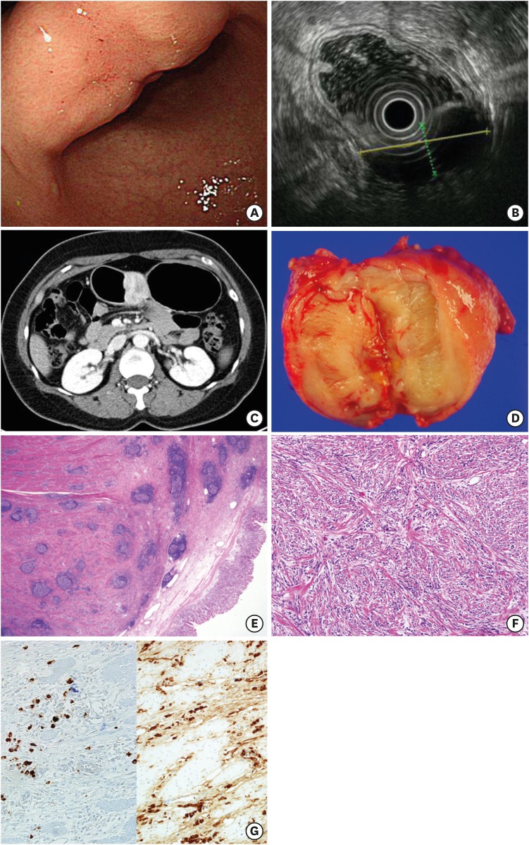
The result of the patient's endoscopy showed 4 cm sized fixed, hard submucosal lesion with central dimpling and erosion at gastric angle (Fig. 1A). The patient then underwent endoscopic ultrasonography (EUS) which revealed a large submucosal mixed echoic lesion with posterior acoustic shadowing, measuring 4.3×2.7 cm arising from muscularis propria (Fig. 1B).
Fig. 1
Case 1. (A) Endoscopy, a 4 cm sized fixed and hard submucosal lesion at gastric angle; (B) EUS, a 4.3 cm sized mixed echoic submucosal lesion; (C) CT, a well-defined heterogeneously enhancing wall mass; (D) Gross specimen, an ill-demarcated round tan-brown colored firm mass; (E) H&E stain, ×12.5, transmural diffuse fibrosis with dense lymphoplasmacytic infiltrate prominent lymphoid follicles; (F) H&E stain, ×100, storiform fibrosis with numerous plasma cells and no definite obliterative phlebitis; (G) IHC stain, ×200, IgG4 and IgG positive cells.
EUS = endoscopic ultrasonography; CT = computed tomography; H&E = hematoxylin and eosin; IHC = immunohistochemistry; Ig = immunoglobulin.
jgc-18-99-g001
The computed tomography (CT) scan showed well defined heterogeneously enhancing wall mass at antrum, lesser curvature side (Fig. 1C), most likely to be malignant gastrointestinal stromal tumor (GIST) without nodal enlargement.
The laboratory results and urinalysis were unremarkable and the proportion of serum eosinophil was 2.5% (reference range: 0%–5%). Because we did not suspect IgG4-RD from preoperative studies, we did not check the serum IgG4 level or other immunoglobulin before the surgery. The serum IgG4 level at 16 days after the surgery was 482.0 mg/L (reference range: 30–2,010 mg/L).
The microscopic result of endoscopic forceps biopsy showed chronic gastritis with regenerative foveolar epithelium. Laparoscopic wedge resection was performed using Harmonic scalpel (Ethicon endo-Surgery Inc., Cincinnati, OH, USA) and the defect was closed with continuous running suture using V Loc™ (Covidien, New Haven, CT, USA).
Histopathological examination of the lesion revealed that an ill demarcated round tan-brown colored firm mass, measuring 4.5×3.6×1.6 cm, was located from submucosa to subserosa (Fig. 1D). On cut section, it showed whitish gray rubbery cut surface. Microscopically, transmural fibrosis from submucosa to subserosa, storiform dense fibrosis, numerous lymphoid follicles and dense lymphoplasmacytic infiltrate with prominent plasma cells were shown on the hematoxylin and eosin (H&E) stain (Fig. 1E and F). The definite obliterative phlebitis was not found. IgG4 and IgG were positive on immunohistochemistry (IHC) stain. The ratio of IgG4 and IgG was heterogenous from 0.2–0.4 through the area by area. In the most hot spot, the ratio met 0.4 (Fig. 1G). The other type of SET such as GIST could be ruled out with IHC stain (Table 1). The final pathological diagnosis was IgG4-RD.
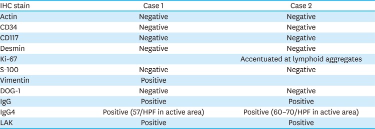
Table 1

IHC stain in 2 cases

jgc-18-99-i001
IHC stain Case 1 Case 2
Actin Negative Negative
CD34 Negative Negative
CD117 Negative Negative
Desmin Negative Negative
Ki-67 Accentuated at lymphoid aggregates
S-100 Negative Negative
Vimentin Positive
DOG-1 Negative Negative
IgG Positive Positive
IgG4 Positive (57/HPF in active area) Positive (60–70/HPF in active area)
LAK Positive Positive
IHC = immunohistochemistry; DOG-1 = discovered on gastrointestinal stromal tumor; HPF = high power field; Ig = immunoglobulin; LAK = lichenoid actinic keratosis.

Case 2

The patient was a 44-year-old female who was diagnosed with gastric SET found on endoscopy for the routine follow up examination after gastrectomy and visited our hospital in June 2016 for further evaluation. She presented no specific abdominal symptom. She underwent Billroth-II subtotal gastrectomy due to early gastric cancer 9 years ago, and 2 times of Cesarean sections 11 and 13 years ago. She had been taking oral steroid pills, 16 tablets of solondo per day for idiopathic thrombocytopenic purpura (ITP) since May 2016 though she suddenly stopped taking oral steroid by herself after a month of medication. She was diagnosed with sick sinus syndrome 2 months ago with no specific medication. She also had hypertension with medication. Her father past away due to lung cancer. On physical examination, body temperature was 36.4°C with blood pressure 130/70 mmHg, and pulse 79 beats/min. The abdomen was soft and flat without tenderness, and had midline scar due to previous gastrectomy.
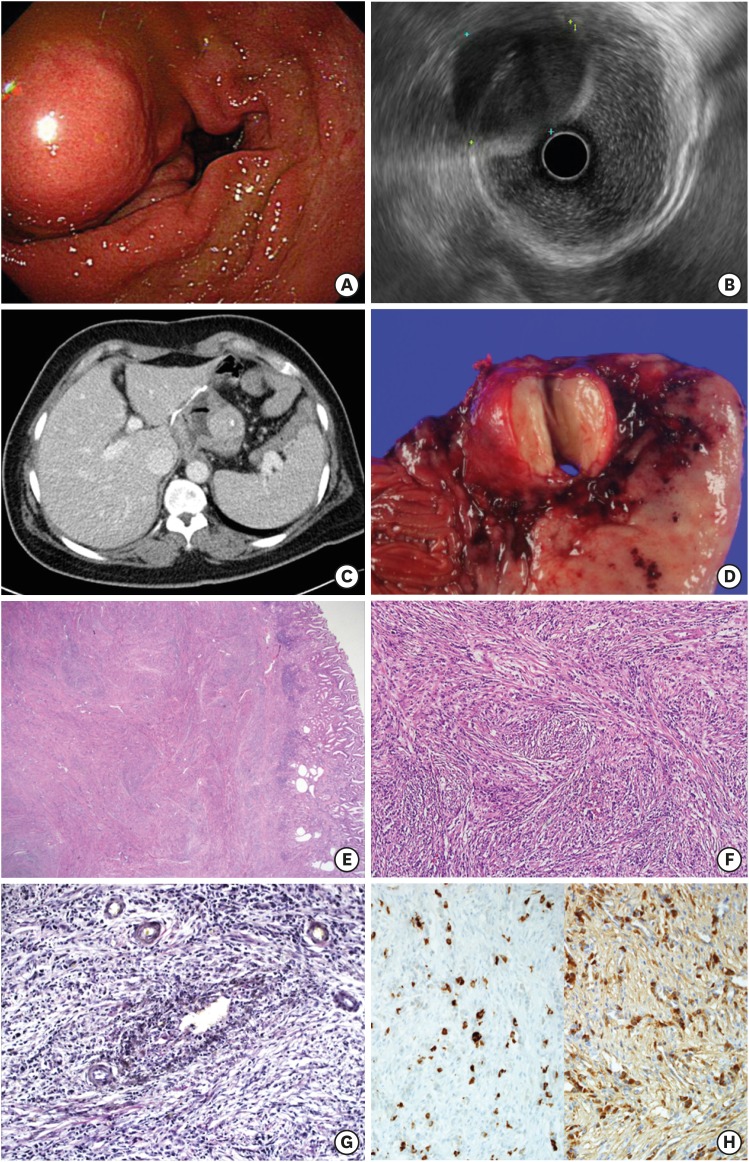
Endoscopy showed 4 cm sized fixed, hard submucosal lesion at remnant body (Fig. 2A). EUS showed a 4.1×3.0 cm sized well-defined smooth border delineated heterogeneous hypoechoic mass arising from muscularis propria at greater curvature side of remnant body (Fig. 2B). CT scan showed 2.7 cm sized mass at wall of gastric fundus, most likely to be recurrent gastric cancer without nodal enlargement, differential diagnosis was GIST. The spleen was enlarged to 11.0×5.9 cm (Fig. 2C).
Fig. 2
Case 2. (A) Endoscopy, a 4 cm sized fixed and hard submucosal lesion at remnant body; (B) EUS, a 4.1 cm sized heterogeneous hypoechoic mass; (C) CT, a 2.7 cm sized mass at wall; (D) Gross specimen, a well-demarcated round tan-brown colored firm mass; (E) H&E stain, ×12.5, transmural diffuse fibrosis with dense lymphoplasmacytic infiltrate prominent lymphoid follicles; (F) H&E stain, ×100, storiform fibrosis with numerous plasma cells; (G) Elastic stain, ×200, non-obliterative phlebitis; (H) IHC stain, ×200, IgG4 and IgG positive cells.
EUS = endoscopic ultrasonography; CT = computed tomography; H&E = hematoxylin and eosin; IHC = immunohistochemistry; Ig = immunoglobulin.
jgc-18-99-g002
Laboratory results and urinalysis were unremarkable except that the platelet count was 32,000 due to ITP. Serum IgG4 level or other immunoglobulin was not checked likewise case 1. Bone marrow examination showed about 80% hypercellularity. The microscopic result of endoscopic forceps biopsy showed mild chronic gastritis with foveolar epithelial hyperplasia.
A provisional diagnosis of SET, which could be most likely GIST, and ITP were made and the patient underwent completion total gastrectomy with R-Y reconstruction with splenectomy. Actually, we planned the operation of gastric wedge resection. However, total gastrectomy with splenectomy should be performed due to severe adhesion and very small remnant stomach because of previous subtotal gastrectomy.
Histopathological examination of the lesion revealed that a well demarcated round tan-brown colored firm mass, measuring 4.×4.0×3.0 cm, was protruded from the submucosal area. On cut section, it showed lobulated and whorled-silk appearance grossly (Fig. 2D). Microscopically, transmural fibrosis from submucosa to subserosa, storiform dense fibrosis and dense lymphoplasmacytic infiltrate with prominent plasma cells were shown on the H&E stain (Fig. 2E and F). Not definite obliterative phlebitis but non-obliterative phlebitis was found in elastic stains (Fig. 2G). On the IHC staining, IgG4 and IgG were positive and the ratio of IgG4 and IgG was exceeded 0.4 (Fig. 2H). The other type of SET such as GIST could be ruled out with IHC stain (Table 1). The spleen was measured 11.0×8.0×5.0 cm and 220.5 g. The final pathological diagnosis were IgG4-RD and ITP.

DISCUSSION

IgG4-RD is a distinct immune-mediated condition that share particular pathologic, serologic and clinical manifestations [110]. The most common features include swelling or involved organ, obliterative venulitis, perineural inflammation, elevated serum concentrations of IgG4, IgG4 positive plasma cell infiltration and fibrosis characterized by storiform pattern [1]. However, as there is a wide variation in sensitivity regarding serum IgG4 level, it is not used as a single marker for diagnosing the disease. In addition, excess level of IgG4 may simply be a response to an unknown inflammatory stimulus, like and allergy, being reported that up to 40% of patients with IgG4-RD have allergic disease such as asthma, chronic sinusitis, eczema with peripheral eosinophilia [11]. Inflammation results in tissue fibrosis at affected anatomical sites which can lead to organ dysfunction or even organ failure, if not treated [12]. Thus, early detection is very important to avoid organ dysfunctions and potential complications.
The IgG4-RD is usually diagnosed on histopathology. The 3 major pathologic features of IgG4-RD are dense lymphoplasmacytic infiltration, storiform fibrosis, and obliterative phlebitis. In addition, the other features of IgG4-RD are phlebitis without obliteration and increased numbers of eosinophils [13]. In the present 2 cases, although there were no definite obliterative phlebitis, IgG4-RD could be diagnosed by other specific findings including storiform fibrosis and non-obliterative phlebitis.
The IgG4-RD can involve in multiple organ, any sites in the body synchronously or metachronously [11014]. The patients are generally well at the time of diagnosis, although some present with non-specific symptoms caused by swelling or mass lesion. For example, patients with IgG4-RD involving pancreas or biliary tree may manifest jaundice and others involving prostate mat show urinary symptoms [15].
The disease may be detected incidentally on radiological images, but can be easily misdiagnosed as malignancies. However, there is no consensus about radiologic findings characterizing IgG4-RD as the imaging features are generally nonspecific and do not permit reliable distinctions between IgG4-RD and other malignancies [161718].
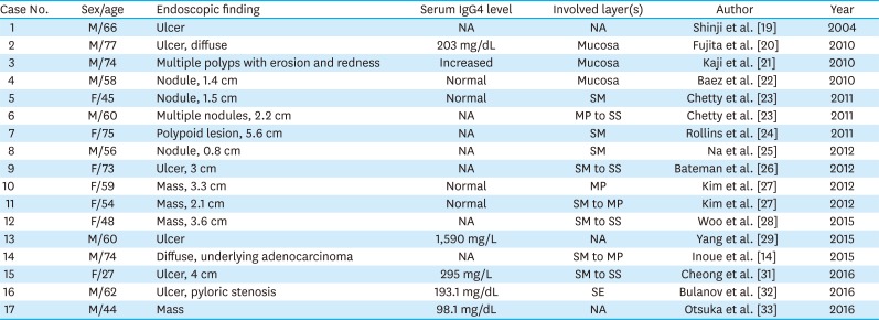
IgG4-RD involves in various organs, such as lung, pancreas, bile duct, kidney, aorta, nervous system, and retroperitoneum [345678]. IgG4-RD in stomach was first mentioned by Shinji et al. [19] in 2004. It was related with AIP and presented like gastric ulcer. Lately, several cases that IgG4-RD appeared in gastric wall were reported (Table 2) [192021222324252627282930313233]. Most cases underwent surgery as not considering IgG4-RD beforehand. In fact, IgG4-RD is a disease which could be treated by medicine such as oral steroids [1]. However, IgG4-RD is difficult to diagnose clinically without pathological examination. In addition, IgG4-RD in stomach is usually located on submucosal layer, therefore diagnosing with endoscopic forceps biopsy is difficult. Moreover, the incidence of gastric SET is 10–15 per million per year worldwide [34], while IgG4-RD in stomach is rare. For these reasons, most of the patients with IgG4-RD in gastric wall underwent surgery usually misdiagnosing as gastric SET. Despite gastric wedge resection for gastric SET is not a huge radical surgery, minor or major deformity could be generated. The deformity could cause gastric dysfunction such as dyspepsia or gastroesophageal reflux disease [35].
Table 2

Reported IgG4-related disease cases

jgc-18-99-i002
Case No. Sex/age Endoscopic finding Serum IgG4 level Involved layer(s) Author Year
1 M/66 Ulcer NA NA Shinji et al. [19] 2004
2 M/77 Ulcer, diffuse 203 mg/dL Mucosa Fujita et al. [20] 2010
3 M/74 Multiple polyps with erosion and redness Increased Mucosa Kaji et al. [21] 2010
4 M/58 Nodule, 1.4 cm Normal Mucosa Baez et al. [22] 2010
5 F/45 Nodule, 1.5 cm Normal SM Chetty et al. [23] 2011
6 M/60 Multiple nodules, 2.2 cm NA MP to SS Chetty et al. [23] 2011
7 F/75 Polypoid lesion, 5.6 cm NA SM Rollins et al. [24] 2011
8 M/56 Nodule, 0.8 cm NA SM Na et al. [25] 2012
9 F/73 Ulcer, 3 cm NA SM to SS Bateman et al. [26] 2012
10 F/59 Mass, 3.3 cm Normal MP Kim et al. [27] 2012
11 F/54 Mass, 2.1 cm Normal SM to MP Kim et al. [27] 2012
12 F/48 Mass, 3.6 cm NA SM to SS Woo et al. [28] 2015
13 M/60 Ulcer 1,590 mg/L NA Yang et al. [29] 2015
14 M/74 Diffuse, underlying adenocarcinoma NA SM to MP Inoue et al. [14] 2015
15 F/27 Ulcer, 4 cm 295 mg/L SM to SS Cheong et al. [31] 2016
16 M/62 Ulcer, pyloric stenosis 193.1 mg/dL SE Bulanov et al. [32] 2016
17 M/44 Mass 98.1 mg/dL NA Otsuka et al. [33] 2016
IgG4 = immunoglobulin G4; NA = not applicable; SM = submucosa; MP = muscularis propria; SS = subserosa; SE = serosal exposure.
The present report showed 2 cases of IgG4-RD in gastric wall that underwent gastric resection like the other reported IgG4-RD cases. Although the diagnosis of the disease was made from the surgery, a medical treatment such as oral steroid could have been better option because of several complications caused by gastrectomy. However, it is difficult to diagnose IgG4-RD in gastric wall before surgery yet. Development of the tool which could diagnose IgG4-RD without histopathology is positively necessary.

Notes

Author Contributions:

  • Conceptualization: S.H.S., S.K.Y.

  • Data curation: S.H.S., J.Y.J.

  • Formal analysis: S.H.S.

  • Funding acquisition:

  • Investigation: S.H.S., J.E.S.

  • Methodology: S.H.S., J.E.S.

  • Supervision: P.C.H., S.K.Y., J.E.S.

  • Validation: J.E.S.

  • Writing - original draft: S.H.S.

  • Writing - review & editing: S.H.S., J.E.S., S.K.Y., P.C.H., J.Y.J.

Conflict of Interest: No potential conflict of interest relevant to this article was reported.

References

1. Stone JH, Zen Y, Deshpande V. IgG4-related disease. N Engl J Med. 2012; 366:539–551. PMID: 22316447.
crossref
2. Kamisawa T, Funata N, Hayashi Y, Eishi Y, Koike M, Tsuruta K, et al. A new clinicopathological entity of IgG4-related autoimmune disease. J Gastroenterol. 2003; 38:982–984. PMID: 14614606.
crossref
3. Inoue D, Zen Y, Abo H, Gabata T, Demachi H, Kobayashi T, et al. Immunoglobulin G4-related lung disease: CT findings with pathologic correlations. Radiology. 2009; 251:260–270. PMID: 19221056.
crossref
4. Hamano H, Kawa S, Horiuchi A, Unno H, Furuya N, Akamatsu T, et al. High serum IgG4 concentrations in patients with sclerosing pancreatitis. N Engl J Med. 2001; 344:732–738. PMID: 11236777.
crossref
5. Zen Y. The pathology of IgG4-related disease in the bile duct and pancreas. Semin Liver Dis. 2016; 36:242–256. PMID: 27466794.
crossref
6. Kawano M, Yamada K. IgG4-related kidney disease and IgG4-related retroperitoneal fibrosis. Semin Liver Dis. 2016; 36:283–290. PMID: 27466797.
crossref
7. Perugino CA, Wallace ZS, Meyersohn N, Oliveira G, Stone JR, Stone JH. Large vessel involvement by IgG4-related disease. Medicine (Baltimore). 2016; 95:e3344. PMID: 27428181.
crossref
8. Williams MM, Mashaly H, Puduvalli VK, Jin M, Mendel E. Immunoglobulin G4-related disease mimicking an epidural spinal cord tumor: case report. J Neurosurg Spine. 2017; 26:76–80. PMID: 27517527.
crossref
9. Kim JH, Byun JH, Lee SS, Kim HJ, Lee MG. Atypical manifestations of IgG4-related sclerosing disease in the abdomen: imaging findings and pathologic correlations. AJR Am J Roentgenol. 2013; 200:102–112. PMID: 23255748.
crossref
10. Kamisawa T, Zen Y, Pillai S, Stone JH. IgG4-related disease. Lancet. 2015; 385:1460–1471. PMID: 25481618.
crossref
11. Aalberse RC, Stapel SO, Schuurman J, Rispens T. Immunoglobulin G4: an odd antibody. Clin Exp Allergy. 2009; 39:469–477. PMID: 19222496.
crossref
12. Khosroshahi A, Wallace ZS, Crowe JL, Akamizu T, Azumi A, Carruthers MN, et al. International consensus guidance statement on the management and treatment of IgG4-related disease. Arthritis Rheumatol. 2015; 67:1688–1699. PMID: 25809420.
crossref
13. Deshpande V, Zen Y, Chan JK, Yi EE, Sato Y, Yoshino T, et al. Consensus statement on the pathology of IgG4-related disease. Mod Pathol. 2012; 25:1181–1192. PMID: 22596100.
crossref
14. Inoue D, Yoshida K, Yoneda N, Ozaki K, Matsubara T, Nagai K, et al. IgG4-related disease: dataset of 235 consecutive patients. Medicine (Baltimore). 2015; 94:e680. PMID: 25881845.
15. Vlachou PA, Khalili K, Jang HJ, Fischer S, Hirschfield GM, Kim TK. IgG4-related sclerosing disease: autoimmune pancreatitis and extrapancreatic manifestations. Radiographics. 2011; 31:1379–1402. PMID: 21918050.
crossref
16. Takahashi N, Kawashima A, Fletcher JG, Chari ST. Renal involvement in patients with autoimmune pancreatitis: CT and MR imaging findings. Radiology. 2007; 242:791–801. PMID: 17229877.
crossref
17. Inoue D, Zen Y, Abo H, Gabata T, Demachi H, Yoshikawa J, et al. Immunoglobulin G4-related periaortitis and periarteritis: CT findings in 17 patients. Radiology. 2011; 261:625–633. PMID: 21803920.
crossref
18. Inoue D, Yoneda N, Yoshida K, Nuka H, Kinoshita J, Fushida S, et al. Imaging and pathological features of gastric lesion of immunoglobulin G4-related disease: a case report and review of the recent literature. Mod Rheumatol. 2016; 1–5.
crossref
19. Shinji A, Sano K, Hamano H, Unno H, Fukushima M, Nakamura N, et al. Autoimmune pancreatitis is closely associated with gastric ulcer presenting with abundant IgG4-bearing plasma cell infiltration. Gastrointest Endosc. 2004; 59:506–511. PMID: 15044886.
crossref
20. Fujita T, Ando T, Sakakibara M, Hosoda W, Goto H. Refractory gastric ulcer with abundant IgG4-positive plasma cell infiltration: a case report. World J Gastroenterol. 2010; 16:2183–2186. PMID: 20440861.
crossref
21. Kaji R, Okabe Y, Ishida Y, Takedatsu H, Kawahara A, Aino H, et al. Autoimmune pancreatitis presenting with IgG4-positive multiple gastric polyps. Gastrointest Endosc. 2010; 71:420–422. PMID: 19846081.
crossref
22. Baez JC, Hamilton MJ, Bellizzi A, Mortele KJ. Gastric involvement in autoimmune pancreatitis: MDCT and histopathologic features. JOP. 2010; 11:610–613. PMID: 21068496.
23. Chetty R, Serra S, Gauchotte G, Markl B, Agaimy A. Sclerosing nodular lesions of the gastrointestinal tract containing large numbers of IgG4 plasma cells. Pathology. 2011; 43:31–35. PMID: 21240062.
crossref
24. Rollins KE, Mehta SP, O'Donovan M, Safranek PM. Gastric IgG4-related autoimmune fibrosclerosing pseudotumour: a novel location. ISRN Gastroenterol. 2011; 2011:873087. PMID: 21991533.
crossref
25. Na KY, Sung JY, Jang JY, Lim SJ, Kim GY, Kim YW, et al. Gastric nodular lesion caused by IgG4-related disease. Pathol Int. 2012; 62:716–718. PMID: 23005600.
crossref
26. Bateman AC, Sommerlad M, Underwood TJ. Chronic gastric ulceration: a novel manifestation of IgG4-related disease? J Clin Pathol. 2012; 65:569–570. PMID: 22259178.
crossref
27. Kim DH, Kim J, Park DH, Lee JH, Choi KD, Lee GH, et al. Immunoglobulin G4-related inflammatory pseudotumor of the stomach. Gastrointest Endosc. 2012; 76:451–452. PMID: 21981816.
crossref
28. Woo CG, Yook JH, Kim AY, Kim J. IgG4-related disease presented as a mural mass in the stomach. J Pathol Transl Med. 2016; 50:67–70. PMID: 26420251.
crossref
29. Yang L, Jin P, Sheng JQ. Immunoglobulin G4-related disease (IgG4-RD) affecting the esophagus, stomach, and liver. Endoscopy. 2015; 47(Suppl 1 UCTN):E96–E97. PMID: 25734809.
crossref
30. Inoue K, Okubo T, Kato T, Shimamura K, Sugita T, Kubota M, et al. IgG4-related stomach muscle lesion with a renal pseudotumor and multiple renal rim-like lesions: a rare manifestation of IgG4-related disease. Mod Rheumatol. 2018; 28:188–192. PMID: 26381653.
crossref
31. Cheong HR, Lee BE, Song GA, Kim GH, An SG, Lim W. Immunoglobulin G4-related inflammatory pseudotumor presenting as a solitary mass in the stomach. Clin Endosc. 2016; 49:197–201. PMID: 26867551.
crossref
32. Bulanov D, Arabadzhieva E, Bonev S, Yonkov A, Kyoseva D, Dikov T, et al. A rare case of IgG4-related disease: a gastric mass, associated with regional lymphadenopathy. BMC Surg. 2016; 16:37. PMID: 27255154.
crossref
33. Otsuka R, Kano M, Hayashi H, Hanari N, Gunji H, Hayano K, et al. Probable IgG4-related sclerosing disease presenting as a gastric submucosal tumor with an intense tracer uptake on PET/CT: a case report. Surg Case Rep. 2016; 2:33. PMID: 27059471.
crossref
34. Søreide K, Sandvik OM, Søreide JA, Giljaca V, Jureckova A, Bulusu VR. Global epidemiology of gastrointestinal stromal tumours (GIST): a systematic review of population-based cohort studies. Cancer Epidemiol. 2016; 40:39–46. PMID: 26618334.
crossref
35. Ko SY, Lee JS, Kim JJ, Park SM. Higher incidence of gastroesophageal reflux disease after gastric wedge resections of gastric submucosal tumors located close to the gastroesophageal junction. Ann Surg Treat Res. 2014; 86:289–294. PMID: 24949319.
crossref
TOOLS
Similar articles