Journal List > J Gastric Cancer > v.18(1) > 1077088

Kim and Oh: Novel Systemic Therapies for Advanced Gastric Cancer

Abstract

Gastric cancer (GC) is the second leading cause of cancer mortality and the fourth most commonly diagnosed malignant diseases. While continued efforts have been focused on GC treatment, the introduction of trastuzumab marked the beginning of a new era of target-specific treatments. Considering the diversity of mutations in GC, satisfactory results obtained from various target-specific therapies were expected, yet most of them were unsuccessful in controlled clinical trials. There are several possible reasons underlying the failures, including the absence of patient selection depending on validated predictive biomarkers, the inappropriate combination of drugs, and tumor heterogeneity. In contrast to targeted agents, immuno-oncologic agents are designed to regulate and boost immunity, are not target-specific, and may overcome tumor heterogeneity. With the successful establishment of predictive biomarkers, including Epstein-Barr virus pattern, microsatellite instability status, and programmed death-ligand 1 (PD-L1) expression, as well as ideal combination regimens, a new frontier in the immuno-oncology of GC treatment is on the horizon. Since the field of immuno-oncology has witnessed innovative, practice-changing successes in other cancer types, several trials on GC are ongoing. Among immuno-oncologic therapies, immune checkpoint inhibitors are the mainstay of clinical trials performed on GC. In this article, we review target-specific agents currently used in clinics or are undergoing clinical trials, and highlight the future clinical application of immuno-oncologic agents in inoperable GC.

INTRODUCTION

Gastric cancer (GC) is the second leading cause of cancer-related mortality and the fourth most commonly diagnosed malignant disease, with an estimated 740,000 cancer-related deaths and 990,000 new cases annually [12]. The prevalence of GC shows large regional variations [3]. The incidence of GC has decreased in the United States over the past 80 years. However, GC in South Korea accounts for approximately 17% of all prevalent cancer cases and about 12% of all cancer-related mortalities (nearly 9,000 deaths) annually [4]. The complete surgical resection of localized GC with the removal of adjacent lymph nodes is the only curative option, and patients with pathological stage II or III GC are recommended adjuvant therapy in addition to surgery [5]. The Adjuvant Chemotherapy Trial of TS-1 for Gastric Cancer (ACTS-GC) and the Capecitabine and Oxaliplatin Adjuvant Study in Stomach Cancer (CLASSIC) trial have demonstrated survival benefits of adjuvant therapy after D2 dissection surgery [67]. Unfortunately, due to the asymptomatic early phase of the disease, many patients with GC are diagnosed in advanced stages. Several phase III trials utilizing target-specific agents for inoperable and advanced GC have been conducted with very few favorable outcomes, specifically with trastuzumab (human epidermal growth factor receptor 2 [HER2]-targeted monoclonal antibody) [8], ramucirumab (vascular endothelial growth factor receptor 2 [VEGFR2]-targeted monoclonal antibody) [9], and apatinib (VEGFR2-targeted tyrosine kinase inhibitor [TKI]) [10]. Therefore, advances made to increase the survival rate of patients suffering from GC have been much slower than those concerned with other cancers over the past decade.
GC is considered as a heterogeneous disease carrying various genetic mutations [11]. While satisfactory results were expected from various target-specific agents, most of the trials with these agents were failures. The one-size-fits-all approach to treatment is considered a key reason for the failure of these trials, and patient selection based on appropriate biomarkers is essential for successful trials [12].
In this article, we review target-specific agents currently used in clinics or are undergoing clinical trials, and highlight the future clinical application of immuno-oncologic agents in inoperable GC.

ErbB FAMILY-TARGETING AGENTS

The HER2 protein is a member of the ErbB family comprising 4 kinds of receptor tyrosine kinases (RTKs). The ErbB family of proteins includes transmembrane receptors composed of an intracellular region with a tyrosine kinase residue, an extracellular ligand-binding domain, and a single hydrophobic transmembrane segment. The HER2 protein plays a crucial role in cancer cell biology [13], and mediates apoptosis, cellular differentiation, and tumorigenesis [14]. Compared with other members of the ErbB family, HER2 lacks an identifiable ligand. The ligand-free activation of the HER2 receptor occurs via hetero- or homo-dimerization with other ErbB family members, resulting in the upregulation of downstream signaling cascades, including RAS-RAF-mitogen-activated protein kinase (MAPK), MAPK-extracellular signal-regulated kinase (MEK), and phosphatidylinositol 3-kinase (PI3K)-Akt-mammalian target of rapamycin (mTOR) pathways [1516]. HER2 gene amplification has been reported in about 15%–30% of patients with breast cancer, and is associated with poor prognosis [1718]. Its amplification also occurs in GC (approximately 7%–34% of patients), ovarian cancer, non-small cell lung cancer, endometrial carcinoma, and salivary cancer [19202122].

Trastuzumab

Trastuzumab, a monoclonal antibody specifically targeting the extracellular domain of the HER2 protein, is reported to show remarkable antitumor activity in HER2-amplified xenograft models of human GC cell lines [23]. Based on these results, a randomized phase III Trastuzumab for Gastric Cancer (ToGA) trial was conducted as the first study demonstrating the efficacy of a target-specific agent in patients with advanced or metastatic GC [8]. The ToGA study enrolled patients treated with first-line palliative chemotherapy. In this trial, 3,803 patients were evaluated for HER2 status by immunohistochemistry (IHC) as well as fluorescence in situ hybridization (FISH), and 594 patients who showed 3+ on IHC or FISH positivity (HER2: centromeric probe for chromosome 17 [CEP17] ratio ≥2) were randomly assigned to receive chemotherapy (fluoropyrimidine+cisplatin) with or without trastuzumab. The median overall survival (OS) in patients who underwent chemotherapy+trastuzumab was 13.8 months (95% confidence interval [CI], 12–16) compared with 11.1 months (95% CI, 10–13) in patients who underwent chemotherapy alone (hazard ratio [HR], 0.74; 95% CI, 0.60–0.91; P=0.005). The median progression-free survival (PFS) in patients who received chemotherapy+trastuzumab was 6.7 months (95% CI, 6–8) compared with 5.5 months (95% CI, 5–6) in the other group (HR, 0.71; 95% CI, 0.59–0.85; P<0.001). In post hoc analyses, no improvement in OS with trastuzumab was observed in patients with HER2 IHC0/FISH+ or IHC1+/FISH+, while significant improvement in OS was observed in patients with HER2 IHC2+/FISH+ or IHC3+. In cases of HER2 IHC scores of 0, 1+, or 3+, the concordance rate with FISH results exceeded 85%; however, the concordance rate with FISH results in the case of IHC2+ was only about 50%, suggesting that additional FISH tests were mandatory to determine HER2 expression levels [24]. Following the promising results of the ToGA trial in 2010, the Food and Drug Administration (FDA) approved trastuzumab for chemo-naïve HER2-overexpressing metastatic GC.

Pertuzumab

Pertuzumab is a monoclonal antibody inhibiting dimerization by targeting the HER2 ectodomain [25]. The combination of trastuzumab with pertuzumab showed superior antitumor activities compared with the individual drugs in HER2-overexpressing xenograft models of human GC [26] as well as HER2-overexpressing metastatic breast cancer [27]. A phase III study designated as the JACOB trial involved patients who were randomized to receive pertuzumab or placebo in combination with trastuzumab, cisplatin, and fluoropyrimidine. This study was conducted to evaluate the efficacy of pertuzumab in addition to survival benefits observed in the ToGA trial with the first-line treatment of HER2-positive metastatic GC. However, according to the results reported at the 2017 European Society for Medical Oncology Congress, the JACOB study failed to show a significant improvement in OS with the addition of pertuzumab, despite a 3.3-month increase in median OS (NCT01774786).

TKIs targeting HER2

Lapatinib, a small-molecule TKI targeting HER2 and epidermal growth factor receptor (EGFR), showed favorable clinical results in patients with HER2-positive metastatic breast cancer in a phase III, randomized, open-label study as well as in a meta-analysis report [2829], and the FDA approved this drug in trastuzumab-resistant breast cancer patients. However, 2 phase III clinical trials conducted in GC patients were unsuccessful. In the phase III LOGiC trial, which enrolled 545 patients with HER2-positive GC, participants were randomized to receive capecitabine+oxaliplatin (CapeOx) with or without lapatinib as the first-line regimen. There was no significant benefit in OS (HR, 0.91; 95% CI, 0.73–1.12; P=0.350) [30]. In the phase III TyTAN study, lapatinib was evaluated as a second-line treatment in HER2-positive GC. Participants in this trial were randomized to receive paclitaxel treatment with or without lapatinib. No significant improvement in OS was observed (HR, 0.84; 95% CI, 0.64–1.11; P=0.104) [31].
Afatinib, another TKI targeting EGFR, HER2, and HER4 was approved by the FDA for the treatment of non-small cell lung cancer bearing EGFR mutations. Based on a preclinical study demonstrating tumor regression by afatinib treatment in HER2-amplified GC cell xenograft models [32], 3 phase II trials of afatinib in GC are ongoing (NCT02501603, NCT01522768, and NCT01743365).

Trastuzumab-emtansine (T-DM1)

T-DM1 is a monoclonal antibody-microtubule inhibitor (maytansine) conjugate exhibiting both cytotoxic properties and antibody-dependent cytotoxicity [33]. T-DM1 exerted beneficial effects in patients with HER2-amplified metastatic breast cancer [34], and preclinical data indicated that T-DM1 is more efficacious than trastuzumab alone in GC cell xenograft models [35]. Given these results, a phase II/III trial of T-DM1, GATSBY (NCT01641939), was conducted in locally advanced or metastatic HER2-positive GC patients who exhibited disease progression following first-line treatment; however, T-DM1 did not improve OS compared with taxane (HR, 1.15; 95% CI, 0.89–1.43; P=0.860) [36].

Agents targeting EGFR

Agents targeting EGFR have been used as a standard treatment for many cancers, including colorectal cancer, non-small cell lung cancer, pancreatic cancer, breast cancer, and head & neck cancer. Anti-EGFR agents include cetuximab, panitumumab, nimotuzumab, gefitinib, and erlotinib. Since EGFR overexpression was observed in GC, which was shown to be associated with poor prognosis [3738], several trials investigating EGFR as a target for the treatment of GC were conducted.
Nevertheless, despite several favorable phase II results [3940414243], a phase III trial of cetuximab as first-line treatment (EXPAND) failed to reach the primary end point (HR, 1.09; 95% CI, 0.92–1.29; P=0.320) [44].
A phase III trial of panitumumab as first-line treatment (REAL3) reduced OS (HR, 1.37; 95% CI, 1.07–0.76; P=0.013) [45]. A phase III study of the efficacy of nimotuzumab as second-line treatment in advanced or recurrent GC was prematurely terminated because of the lack of a positive outcome in the mid-term follow-up (NCT01813253, ENRICH).

ANTI-ANGIOGENESIS

Angiogenesis is a mechanism of neovascularization from previously existing vessels known to promote carcinogenesis and cancer metastasis [4647]. VEGF gene family proteins, including VEGFA, B, C, and D, and placental growth factor are signal proteins that induce angiogenesis and lymphangiogenesis [48]. Receptors of this gene family, designated as VEGFR1, R2, and R3, are types of RTKs usually expressed in lymphatic or vascular endothelial (VE) cells [46]. Among these receptors, VEGFR2 is the main effector of RTK and mediates downstream signaling cascades, including the phospholipase C-γ-protein kinase C-MEK-MAPK pathway and PI3K-Akt-endothelial nitric oxide synthase pathway [464950]. VEGF overexpression is frequently found in GC patients and is closely associated with poor clinical outcomes [51].

Bevacizumab

Bevacizumab is a monoclonal antibody targeting VEGFA and is approved by the FDA for the treatment of colorectal cancer. In the phase III Avastin in Gastric Cancer (AVAGAST) trial, 774 patients with GC were randomized to receive fluoropyrimidine+cisplatin with bevacizumab or placebo as first-line treatment [52]. In terms of OS, there was no significant difference between the 2 groups (HR, 0.87; 95% CI, 0.73–1.03; P=0.100). However, bevacizumab was efficacious in terms of PFS (HR, 0.80; 95% CI, 0.68–0.93; P=0.004) and overall response rate (ORR) (46% vs. 37%; P=0.032). Strangely, results of this study showed a geographical variation; OS significantly improved with bevacizumab in Europe and America, but not in Asia. One of the reasons for failure in Asia is that the efficacy of bevacizumab as first-line treatment in OS was compromised as many patients in Asia receive second-line and additional treatments. Another phase III trial on bevacizumab (AVATAR), which was conducted in China, also failed to show an improvement in OS (HR, 1.11; 95% CI, 0.79–1.56; P=0.557) [53].

Ramucirumab

The efficacy of ramucirumab, a monoclonal antibody targeting VEGFR2 [54], has been investigated as second-line treatment for GC patients in 2 phase III trials (REGARD and RAINBOW) [955]. In the REGARD trial, a slight improvement in OS was obtained by ramucirumab monotherapy compared with placebo, validating VEGFR2 as a crucial target in GC (HR, 0.77; 95% CI, 0.60–0.99; P=0.047). In the subsequent RAINBOW trial, 665 GC patients who failed to respond to first-line chemotherapy, including fluoropyrimidine and platinum, were randomized to receive ramucirumab+paclitaxel or placebo+paclitaxel treatment. OS was significantly longer in the paclitaxel+ramucirumab group than in the placebo+paclitaxel group (HR, 0.80; 95% CI, 0.67–0.96; P=0.017), and the FDA approved ramucirumab in GC patients exhibiting disease progression with first-line treatment. As a result, a phase III trial evaluating the efficacy of ramucirumab as first-line treatment in metastatic GC is ongoing (NCT02314117, RAINFALL).

TKIs targeting VEGFR2

Apatinib is a small-molecule TKI inhibiting VEGFR2 tyrosine kinase. In a multicenter phase III trial conducted in China, GC patients who were refractory to at least 2 prior lines of standard chemotherapies were randomized to receive apatinib or placebo [10]. OS significantly increased in the apatinib group (HR, 0.71; 95% CI, 0.54–0.94; P<0.016), and apatinib was approved only in China. Another multicenter phase III study investigating apatinib efficacy in advanced or metastatic GC patients who were refractory to at least 2 prior treatments is ongoing (NCT03042611, ANGEL).
Regorafenib is an oral multi-kinase inhibitor, which blocks angiogenic (VEGFR2 and endothelial-specific type 2), stromal (platelet-derived growth factor-β), and oncogenic (RAF, RET, and KIT) RTKs. Regorafenib has been proven effective in GC cell xenograft models [56] and in a phase II trial (HR, 0.40; 95% CI, 0.28–0.59; P<0.001) [57]. Based on these results, a multicenter phase III trial of regorafenib efficacy in refractory advanced GC is ongoing (NCT02773524, INTEGRATEII).

OTHER TARGET-SPECIFIC AGENTS

The protein c-MET, an RTK regulating cell proliferation and invasion, and its ligand, hepatocyte growth factor (HGF), activates downstream signaling cascades, including MAPK and PI3K-Akt pathways. MET gene amplification is observed in about 10%–20% of GC patients, and increased MET protein expression is observed in approximately 50% of GC patients [5859]. Furthermore, in a phase Ib/II trial, rilotumumab, an anti-HGF antibody was shown to be effective in MET-positive GC patients [60]. However, 2 phase III trials of rilotumumab (RILOMET-1 and RILOMET-2) failed to reach the primary endpoint [6162]. A phase III study of onartuzumab, a monoclonal antibody targeting c-MET, also failed to improve OS [63].
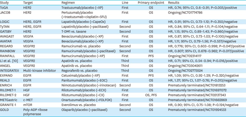
The mTOR inhibitor everolimus was shown to be effective in GC patients in a phase II study; however, a phase III study (GRANITE-1) of this drug failed to show efficacy [6465]. The combination of a TKI targeting fibroblast growth factor receptor (FGFR) 1–3, AZD4547, and paclitaxel also lacked efficacy as the second-line treatment of FGFR2-amplified GC in a phase II trial (SHINE) [66]. A poly-ADP ribose polymerase inhibitor, olaparib, was evaluated as a treatment for advanced GC in a phase III trial (NCT01924533, GOLD), but failed to show efficacy [67]. Phase III randomized clinical trials with target-specific agents are summarized in Table 1.
Table 1

Phase III randomized clinical trials of targeted therapies

jgc-18-1-i001
Study Target Regimen Line Primary endpoint Results
ToGA HER2 Trastuzumab/placebo (+XP) First OS HR, 0.74; 95% CI, 0.6–0.91; P=0.005/positive
JACOB HER2 Pertuzumab/placebo (+trastuzumab+cisplatin+5FU) First OS Ongoing/NCT01774786
LOGiC HER2, EGFR Lapatinib/placebo (+CapeOx) First OS HR, 0.91; 95% CI, 0.73–1.12; P=0.350/negative
TyTAN HER2, EGFR Lapatinib/placebo (+paclitaxel) Second OS HR, 0.84; 95% CI, 0.64–1.11; P=0.104/negative
GATSBY HER2 T-DM1 vs. taxane Second OS HR, 1.15; 95% CI, 0.89–1.43; P=0.860/negative
AVAGAST VEGFA Bevacizumab/placebo (+XP) First OS HR, 0.87; 95% CI, 0.73–1.03; P=0.100/negative
AVATAR VEGFA Bevacizumab/placebo (+XP) First OS HR, 1.11; 95% CI, 0.79–1.56; P=0.557/negative
REGARD VEGFR2 Ramucirumab vs. placebo Second OS HR, 0.776; 95% CI, 0.603–0.998; P=0.047/positive
RAINBOW VEGFR2 Ramucirumab/placebo (+paclitaxel) Second OS HR, 0.807; 95% CI, 0.678–0.962; P=0.017/positive
RAINFALL VEGFR2 Ramucirumab/placebo (+XP) First PFS Ongoing/NCT02314117
Li et al. [10] VEGFR2 Apatinib vs. placebo Third OS HR, 0.71; 95% CI, 0.54–0.94; P<0.016/positive
ANGEL VEGFR2 Apatinib vs. placebo Third OS Ongoing/NCT03042611
INTEGRATEII Multi-kinase inhibitor Regorafenib vs. placebo Third OS Ongoing/NCT02773524
EXPAND EGFR Cetuximab/placebo (+XP) First PFS HR, 1.09; 95% CI, 0.92–1.29; P=0.320/negative
REAL3 EGFR Panitumumab/placebo (+EOC) First OS HR, 1.37; 95% CI, 1.07–0.76; P=0.013/negative
ENRICH EGFR Nimotuzumab/placebo (+irinotecan) Second OS Prematurely terminated/NCT01813253
RILOMET-1 HGF Rilotumumab/placebo (+ECX) First OS Prematurely terminated/NCT01697072
RILOMET-2 HGF Rilotumumab/placebo (+CX) First OS, PFS Prematurely terminated/NCT02137343
METGastric c-MET Onartuzumab/placebo (+FOLFOX) First OS Prematurely terminated/NCT01662869
GRANITE-1 mTOR Everolimus vs. placebo Second OS HR, 0.90; 95% CI, 0.75–1.08; P=0.124/negative
GOLD Poly-ADP ribose polymerase Olaparib/placebo (+paclitaxel) Second OS Prematurely terminated/NCT01924533
HER2 = human epidermal growth factor receptor 2; XP = capecitabine+cisplatin; OS = overall survival; HR = hazard ratio; CI = confidence interval; 5FU = 5-fluorouracil; EGFR = epidermal growth factor receptor; CapeOx = capecitabine+oxaliplatin; T-DM1 = trastuzumab-emtansine; VEGFA = vascular endothelial growth factor A; VEGFR2 = vascular endothelial growth factor receptor 2; PFS = progression-free survival; EOC = epirubicin+oxaliplatin+capecitabine; HGF = hepatocyte growth factor; ECX = epirubicin+cisplatin+capecitabine; CX = cisplatin+capecitabine; FOLFOX = 5-fluorouracil+leucovorin+oxaliplatin; mTOR = mammalian target of rapamycin.

IMMUNOTHERAPY

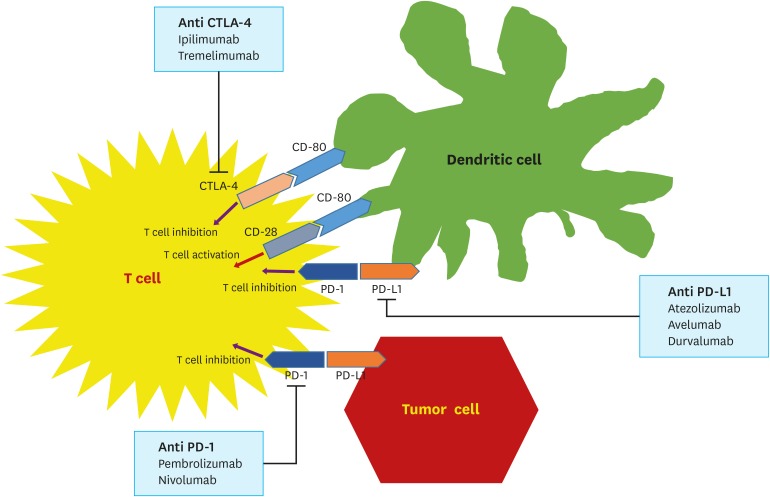
A tumor cell is recognized by and is vulnerable to the host immune system. Classical reports of animal models have indicated that the immune system recognizes and eliminates tumor cells [68]. However, via elimination, equilibrium, and finally escape, cancer cells become invisible to the host [68]. Understanding the mechanism associated with immune escape led to the advancement of the field of immuno-oncology and the development of several target-specific agents, including anti-cytotoxic T-lymphocyte antigen 4 (CTLA-4) antibodies and programmed death-1 (PD-1)/programmed death-ligand 1 (PD-L1) inhibitors (Fig. 1).
Fig. 1
Immune checkpoint inhibitors and their targets, including ligands and receptors on tumor cells, T cells, and dendritic cell surfaces.
CTLA-4 = cytotoxic T-lymphocyte antigen 4; PD-1 = programmed death 1; PD-L1 = programmed death-ligand 1.
jgc-18-1-g001
The activation of tumoricidal immune reactions requires the presentation of cancer antigens to T cells by either dendritic or cancer cells [69]. These interactions are regulated by a combination of co-stimulatory and inhibitory molecules [70]. CTLA-4 is an inhibitory receptor molecule expressed on cytotoxic T cells, and coordinates with CD80 or CD86 on antigen-presenting cells to suppress the cytotoxic activity of T cells. However, the binding of CD28, a co-stimulatory receptor on T cells, to CD80 or CD86 by competing with CTLA-4 increases the tumoricidal activity of T cells [71]. PD-1 is another inhibitory cell surface receptor expressed on antigen-specific CD8+ T cells; it binds to PD-L1 or PD-L2 on antigen-presenting cells to suppress host immune response, including T cell effector functions, migration, and proliferation [72]. Because PD-L1 is not only expressed on dendritic cells, macrophages, mast cells, and natural killer cells, it is also boosted by the cancer cell per se. Therefore, a PD-1/PD-L1 blockade is expected to enhance early immune reactions in lymphoid organs to late reactions at other locations [73].
For over a decade, the field of immuno-oncology has been in the spotlight owing to innovative practice-changing successes [74]. The therapeutic milestones involved melanomas, genitourinary cancers, lung cancers, and head & neck cancers. As a result, several trials on gastrointestinal cancers are ongoing.

Clinical trials of immune checkpoint inhibitors

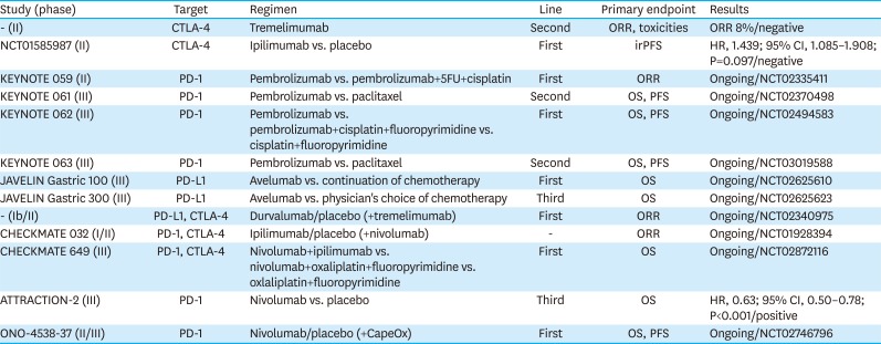
Clinical trials investigating immune checkpoint inhibitors are summarized in Table 2.
Table 2

Clinical trials investigating immune checkpoint inhibitors

jgc-18-1-i002
Study (phase) Target Regimen Line Primary endpoint Results
- (II) CTLA-4 Tremelimumab Second ORR, toxicities ORR 8%/negative
NCT01585987 (II) CTLA-4 Ipilimumab vs. placebo First irPFS HR, 1.439; 95% CI, 1.085–1.908; P=0.097/negative
KEYNOTE 059 (II) PD-1 Pembrolizumab vs. pembrolizumab+5FU+cisplatin First ORR Ongoing/NCT02335411
KEYNOTE 061 (III) PD-1 Pembrolizumab vs. paclitaxel Second OS, PFS Ongoing/NCT02370498
KEYNOTE 062 (III) PD-1 Pembrolizumab vs. pembrolizumab+cisplatin+fluoropyrimidine vs. cisplatin+fluoropyrimidine First OS, PFS Ongoing/NCT02494583
KEYNOTE 063 (III) PD-1 Pembrolizumab vs. paclitaxel Second OS, PFS Ongoing/NCT03019588
JAVELIN Gastric 100 (III) PD-L1 Avelumab vs. continuation of chemotherapy First OS Ongoing/NCT02625610
JAVELIN Gastric 300 (III) PD-L1 Avelumab vs. physician's choice of chemotherapy Third OS Ongoing/NCT02625623
- (Ib/II) PD-L1, CTLA-4 Durvalumab/placebo (+tremelimumab) First ORR Ongoing/NCT02340975
CHECKMATE 032 (I/II) PD-1, CTLA-4 Ipilimumab/placebo (+nivolumab) - ORR Ongoing/NCT01928394
CHECKMATE 649 (III) PD-1, CTLA-4 Nivolumab+ipilimumab vs. nivolumab+oxaliplatin+fluoropyrimidine vs. oxlaliplatin+fluoropyrimidine First OS Ongoing/NCT02872116
ATTRACTION-2 (III) PD-1 Nivolumab vs. placebo Third OS HR, 0.63; 95% CI, 0.50–0.78; P<0.001/positive
ONO-4538-37 (II/III) PD-1 Nivolumab/placebo (+CapeOx) First OS, PFS Ongoing/NCT02746796
CTLA-4 = cytotoxic T-lymphocyte antigen 4; ORR = overall response rate; irPFS = immune-related progression-free survival; 5FU = 5-fluorouracil; HR = hazard ratio; CI = confidence interval; PD-1 = programmed death 1; OS = overall survival; PFS = progression-free survival; PD-L1 = programmed death-ligand 1; CapeOx = capecitabine+oxaliplatin.
The multicenter open-label phase Ib KEYNOTE 012 trial of pembrolizumab was conducted in several cancers, including GC, triple-negative breast cancer, head & neck cancer, and urothelial cancer. Out of 39 patients with GC included in this study, overall response was observed in 8 patients (22%; 95% CI, 10–39). Considering that most patients in this group had failed to respond to 2 or more prior treatments, the median OS of 11 months documented in this study was a remarkable result [75]. An international open-label phase II trial of pembrolizumab in 2 cohorts (patients who had failed to respond to 2 or more prior treatments and treatment-naïve patients) was performed, and the ORR was 13.3% (95% CI, 8.2–20.2) (NCT02335411, KEYNOTE 059). Based on the study findings, the FDA approved pembrolizumab for patients with PD-L1-positive recurrent or advanced GC, who previously received 2 or more chemotherapy regimens. There are 3 phase III, randomized, open-label trials of pembrolizumab, including KEYNOTE 061, KEYNOTE 062, and KEYNOTE 063 (NCT02370498, NCT02494583, and NCT03019588, respectively). KEYNOTE 061 trial investigated the efficacy of pembrolizumab versus paclitaxel as second-line treatment in patients with PD-L1-positive advanced GC who were refractory to fluoropyrimidine and platinum doublet therapy (NCT02370498). The KEYNOTE 062 trial, which investigated pembrolizumab as the first-line treatment, enrolled patients with HER2-negative, PD-L1-positive advanced GC, and randomly assigned participants into 3 treatment arms: pembrolizumab monotherapy, perbrolizumab+platinum+fluoropyrimidine, and placebo+platinum+fluoropyrimidine (NCT02494583). The KEYNOTE 063 trial compared the efficacy of treatment with pembrolizumab versus paclitaxel in Asian, PD-L1-positive patients with advanced GC who failed to respond to any combination treatment containing a fluoropyrimidine and a platinum agent (NCT03019588).
A phase I/II trial investigating the combination of an anti-PD-1 antibody nivolumab with an anti-CTLA-4 antibody ipilimumab in patients with advanced or metastatic solid tumors is underway (NCT01928394, CHECKMATE 032). A randomized, multicenter, open-label phase III trial of nivolumab+ipilimumab or nivolumab+oxaliplatin+fluoropyrimidine versus oxaliplatin+fluoropyrimidine in patients with treatment-naïve metastatic or advanced GC is also ongoing (NCT02872116, CHECKMATE 649).
The ATTRACTION-2 study was the first randomized, multicenter, double blind, phase III trial demonstrating the efficacy of immunotherapy in unresectable or recurrent GC patients [76]. In this study, 493 patients with GC refractory or intolerant to standard therapy were randomly assigned to receive nivolumab or placebo, and a significantly longer OS was observed in patients treated with nivolumab (HR, 0.63; 95% CI, 0.50–0.78; P<0.001). A randomized, multicenter, phase II/III trial comparing nivolumab+fluoropyrimidine+oxaliplatin versus placebo+fluoropyrimidine+oxaliplatin as first-line treatment for unresectable advanced or recurrent GC is ongoing (NCT02746796).

Potential predictive markers of immunotherapy in GC

Because of defects in DNA mismatch repair genes, cancers with microsatellite instability (MSI) possess a high number of mutations, and express numerous surface antigens for presentation to immune cells [77]. In recent studies, the increased burden of somatic mutations has led to the expression of numerous neoantigens, resulting in immunostimulation; patients with cancer having these features are expected to be optimal candidates for PD-1 blockade [787980]. Indeed, MSI has been used as a predictive biomarker for immune checkpoint inhibitors in colorectal cancer [81]. However, the efficacy of immunotherapy in MSI GC, which is known to show comparatively better prognosis and is usually observed as distal GC, has not been evaluated [82]. Four patients with MSI GC were enrolled in the KEYNOTE 012 study [75]. A phase II study of nivolumab with or without ipilimumab to evaluate its efficacy in MSI-high gastrointestinal tumors is ongoing [83]. Immunotherapeutic approaches in patients with MSI GC remain to be proven.
Gene expression studies in melanoma patients enrolled in the KEYNOTE 001 trial have identified a few gene signatures, including interferon-gamma 10-gene and expanded-immune 28-gene, as potential predictive markers of clinical response to pembrolizumab [84]. Interestingly, GC patients enrolled in the KEYNOTE 012 trial, harboring these gene signatures, showed improved survival [7585]. These findings are expected to encourage further studies with larger populations to identify these signatures as potential predictive biomarkers of response to immune checkpoint inhibitors.
Epstein-Barr virus (EBV) infection is known to increase the risk of GC by 10-fold, and EBV cancers account for approximately 10% of all GC cases [8687]. Interestingly, several studies have indicated that the PD-L1 gene is amplified more frequently in EBV-positive cancers than in EBV-negative cancers, suggesting that EBV GC is another possible target of immunotherapeutic agents [8889]. In addition, PD-L1-overexpressing EBV-positive GC shows upregulated interferon adaptive immune response [90], and is associated with an increased infiltration of immune cells into tumors [89]. Furthermore, tumor infiltration of immune cells was significantly associated with disease-free intervals in EBV GC, suggesting cellular immune response as a useful prognostic marker in this group [91]. In the KEYNOTE 012 trial, no EBV infection data were collected [75].
Approximately 50% of GC cases are thought originate due to Helicobacter pylori infection [92]. H. pylori infection triggers inflammation, resulting in altered gastric microenvironment, accelerated cancer cell transformation, and immune cell infiltration. In addition, H. pylori infection induces DNA methylation in tumor suppressor genes, and increases the levels of inflammation-related proteins, including tumor necrosis factor-alpha [93]. Many studies have reported T cell anergy during H. pylori infection [949596]. Furthermore, H. pylori infection upregulates PD-L1 expression in gastric epithelial cell lines [96]. Based on these findings, it might be hypothesized that H. pylori-related GC may respond well to immune checkpoint inhibitors, which remains to be proven.
In GC with chromosomal instability (CIN) and genomic stability (GS), there is no overexpression of PD-L1/2 or high loads of mutations, which suggests that these subgroups are less likely to be responsive to immunotherapy compared with other GC subtypes [80]. GCs are not associated with hyper-mutations but show aberrations in the CHD1, ARID1A, and RHOA genes, which are related to epithelial-mesenchymal transition. CIN GC usually occurs at the esophagogastric junction and has a tendency to harbor chromosomal deletions, alterations, and amplifications, resulting in abnormalities in genes, including EGFR, HER2, MET, RAS, BRAF, and VEGFR.

Future directions in immuno-oncology

Although trials on monotherapy with anti-CTLA-4 antibodies have been unsuccessful, PD-1/PD-L1 axis inhibitors have shown promising results in patients with GC. A phase III study of nivolumab versus placebo (ATTRACTION-2) showed favorable results. CTLA-4 and PD-1/PD-L1 axis interrupt lethal immune responses in cancer via reciprocal pathways [97], and several preclinical studies have shown possible synergistic effects of the combined checkpoint blockades [9899100]. Based on these findings, clinical trials on combination treatments containing nivolumab and ipilimumab have been conducted in malignant melanoma with favorable outcomes [101102103]. Therefore, several ongoing phase III studies evaluating the efficacies of PD-1/PD-L1 axis inhibitors and their combinations with anti-CTLA-4 inhibitors in GC patients are expected to yield significant findings with clinical implications.
In addition to the integration of immune checkpoint inhibitors, immunotherapy in combination with other target-specific agents could be considered. In a phase Ib study on renal cell carcinoma, pembrolizumab in combination with axitinib, an inhibitor of VEGFRs was found to be substantially effective [104]. Therefore, a phase III KEYNOTE 426 trial was started (NCT02853331). Accordingly, treatment with ramucirumab, a monoclonal antibody targeting VEGFR2, in conjunction with immune checkpoint inhibitors is a potential therapeutic strategy for GC. Recently, MEK inhibitors in conjunction with immune checkpoint inhibitors have been suggested to exert synergistic effects in microsatellite-stable (MSS) RAS mutant colorectal cancer; this combination is also a promising intervention in GC, which often involves the activation of the RAS-MEK-ERK pathway [105].
GC is a heterogeneous disease. Therefore, subpopulation studies investigating potential predictive markers, including EBV pattern, MSI status, and PD-L1 expression, are needed. However, diagnostic challenges for the evaluation of molecular mechanisms and difficulties associated with decreased sample size after filtering participants need to be addressed.
Cost-effective studies are crucial, given the lack of consensus regarding the ideal treatment duration and exorbitant costs of immunotherapeutic agents. To identify reliable therapeutic approaches, the identification of useful predictive biomarkers with credible diagnostic tools is crucial.

NEW DIRECTION OF CLINICAL TRIALS ON TARGET-SPECIFIC AGENTS

Considering that GC harbors a diverse range of genetic mutations and tend to carry high mutational loads [12106107], various target-specific agents may be investigated in GC treatment. However, as previously noted, only a few trials have been successful, resulting in the lack of new standard therapies [89]. The possible reasons underlying the failures are summarized below.
First, patient selection was not based on validated predictive biomarkers. Several failed studies, including EXPAND, REAL3, and GRANITE-1, did not involve the selection of participants who are expected to benefit from the experimental drugs [444565]. Compared with the ToGA trial [8] in which only patients with HER2 IHC3+ or FISH+ status were included, the TyTAN study [31] enrolled all patients with HER2 FISH+ status. As a result, 34% of the enrolled patients were expected to benefit less from HER2-targeting agents, according to the criteria used in the ToGA trial. Such inappropriately broad selections of patients were also performed in trials targeting c-MET, including the RILOMET-1 trial [61]. Although MET gene amplification by FISH was observed in about 10%–20% of GC cases [5859], MET protein overexpression was found in more than 50% of patients enrolled in the RILOMET-1 study [61], in which MET protein expression was analyzed by IHC staining. MET protein level analysis by IHC is reported to be unreliable for meaningful biomarker examination [108], and any trial in which more than 50% of all screened patients are registered, is challenging to conduct successfully.
The inappropriate combination of drugs may be another reason. The REAL3 trial used triplet chemotherapy combinations, which consisted of epirubicin, oxaliplatin, and capecitabine (EOC) as a backbone regimen [45]. Clinical outcome with the combination of EOC and panitumumab was poorer than that with EOC only (HR, 1.37; 95% CI, 1.07–0.76; P=0.013). Since epirubicin-containing triplet combinations are generally associated with highly toxic side effects, target-specific agents in combination with these regimens are ordinarily thought to be inappropriate. Combinations of 2 small-molecule inhibitors or a small-molecule inhibitor along with a cytotoxic agent are also generally avoided because maximum tolerated doses of these regimens could be too low (due to high toxicities) to achieve target inhibition.
Tumor heterogeneity is the most important factor underlying the failure of single-target agents. Even in the case of trastuzumab, the only proven HER2-targeting agent, the therapeutic efficacy is markedly diversified depending on HER2 heterogeneity, and this diverse pattern of HER2 expression is more prevalent in GC than in breast cancer [24]. HER2 IHC3+ samples also showed a heterogeneous pattern of staining in more than 30% of cases [109]. Considering focal distribution, target-specific inhibitors are expected to work only against selected tumors. The disappearance of HER2 expression is also regarded as a major cause of trial failure. HER2 expression disappeared in about 30% of patients who underwent first-line treatment [110]. The GATSBY trial, which explored potential second-line treatments in GC, involved the determination of the HER2 status of participants with tumor samples obtained prior to the first-line treatment [36]. The sequential loss of HER2 expression affected the study results. A similar case of failure was reported in the SHINE study, investigating the TKI targeting FGFR1–3 [66].
A recent report demonstrated that target genes in GC are diversely distributed by using a specially-generated tissue microarray with a multitude of tissue cores from the primary tumor and various metastases [111]. This study suggested that a single clinical cancer biopsy could limit treatment decisions, and multiple biopsies might be needed for deciding the most appropriate treatment plan. Furthermore, with rapid advances in next generation sequencing technologies, systematic analyses of therapeutic targets in GC are promising [112]. In terms of concerns about sequential alterations of gene targets, the real-time monitoring of primary tumor entities by analyzing circulating tumor cells from patient blood samples could be helpful [113].

CONCLUSIONS

Clinical outcomes in GC patients may be improved via appropriately designed clinical studies. Appropriate target selection and patient inclusion criteria are essential; however, tumor heterogeneity of GC remains an obstacle even with fine targets serving as driver mutations. Therefore, clinical trials investigating a single target are of little importance in predicting GC treatment outcome; accordingly, several studies have been unsuccessful over the past decade. A combination of several target-specific agents may be considered. However, the maximum tolerated doses of combinations determined by phase I study may not be adequate for target inhibition. Nevertheless, whether successful combinations of 2 or more target-specific agents can overcome this challenge is yet to be established.
In contrast to target-specific agents, immuno-oncologic agents are designed to regulate and boost anti-cancer immunity, without the need for targets and may overcome tumor heterogeneity. Among immuno-oncologic therapies, including cancer vaccines, oncolytic viruses, and chimeric antigen receptors, immune checkpoint inhibitors are the mainstay of clinical interventions in GC trials. MSI GC, bearing high loads of mutations, and EBV-related GC, overexpressing PD-L1, may be ideal candidates for immune checkpoint inhibitors. Based on other data, a checkpoint blockade is not excluded in patients even with MSS or PD-L1-negative GC. Recent data support that checkpoint inhibition in combination with MEK inhibitors may be associated with synergistic effects in RAS mutant MSS colorectal cancer [105]. In CIN GC carrying mutations of EGFR, HER2, or VEGFR, antibodies targeting HER2 or VEGFR2 in conjunction with immunotherapy are attractive therapeutic options.
Major challenges in GC management can be overcome via appropriate patient selection, the effective combination of drugs, and proper sequential treatment. Further efforts are imperative to explore and validate useful predictive biomarkers, which are crucial for patient selection. Preclinical studies exploring ideal combinations of drugs are also needed. Despite gaps in the currently available data, further studies in this direction may establish the new frontier in GC treatment.

Notes

Author Contributions:

  • Conceptualization: K.H.J.

  • Data curation: K.H.J.

  • Methodology: K.H.J.

  • Resources: K.H.J.

  • Supervision: O.S.C.

  • Validation: K.H.J.

  • Visualization: K.H.J.

  • Writing - original draft: K.H.J.

  • Writing - review & editing: K.H.J., O.S.C.

Conflict of Interest: No potential conflict of interest relevant to this article was reported.

References

1. Jemal A, Center MM, DeSantis C, Ward EM. Global patterns of cancer incidence and mortality rates and trends. Cancer Epidemiol Biomarkers Prev. 2010; 19:1893–1907. PMID: 20647400.
crossref
2. Ferlay J, Soerjomataram I, Dikshit R, Eser S, Mathers C, Rebelo M, et al. Cancer incidence and mortality worldwide: sources, methods and major patterns in GLOBOCAN 2012. Int J Cancer. 2015; 136:E359–E386. PMID: 25220842.
crossref
3. Parkin DM. The global health burden of infection-associated cancers in the year 2002. Int J Cancer. 2006; 118:3030–3044. PMID: 16404738.
crossref
4. Oh CM, Won YJ, Jung KW, Kong HJ, Cho H, Lee JK, et al. Cancer statistics in Korea: incidence, mortality, survival, and prevalence in 2013. Cancer Res Treat. 2016; 48:436–450. PMID: 26987395.
crossref
5. Mizrak Kaya D, Harada K, Shimodaira Y, Amlashi FG, Lin Q, Ajani JA. Advanced gastric adenocarcinoma: optimizing therapy options. Expert Rev Clin Pharmacol. 2017; 10:263–271. PMID: 28094573.
crossref
6. Sakuramoto S, Sasako M, Yamaguchi T, Kinoshita T, Fujii M, Nashimoto A, et al. Adjuvant chemotherapy for gastric cancer with S-1, an oral fluoropyrimidine. N Engl J Med. 2007; 357:1810–1820. PMID: 17978289.
crossref
7. Bang YJ, Kim YW, Yang HK, Chung HC, Park YK, Lee KH, et al. Adjuvant capecitabine and oxaliplatin for gastric cancer after D2 gastrectomy (CLASSIC): a phase 3 open-label, randomised controlled trial. Lancet. 2012; 379:315–321. PMID: 22226517.
crossref
8. Bang YJ, Van Cutsem E, Feyereislova A, Chung HC, Shen L, Sawaki A, et al. Trastuzumab in combination with chemotherapy versus chemotherapy alone for treatment of HER2-positive advanced gastric or gastro-oesophageal junction cancer (ToGA): a phase 3, open-label, randomised controlled trial. Lancet. 2010; 376:687–697. PMID: 20728210.
crossref
9. Wilke H, Muro K, Van Cutsem E, Oh SC, Bodoky G, Shimada Y, et al. Ramucirumab plus paclitaxel versus placebo plus paclitaxel in patients with previously treated advanced gastric or gastro-oesophageal junction adenocarcinoma (RAINBOW): a double-blind, randomised phase 3 trial. Lancet Oncol. 2014; 15:1224–1235. PMID: 25240821.
crossref
10. Li J, Qin S, Xu J, Xiong J, Wu C, Bai Y, et al. Randomized, double-blind, placebo-controlled phase III trial of apatinib in patients with chemotherapy-refractory advanced or metastatic adenocarcinoma of the stomach or gastroesophageal junction. J Clin Oncol. 2016; 34:1448–1454. PMID: 26884585.
crossref
11. Alexandrov LB, Nik-Zainal S, Wedge DC, Aparicio SA, Behjati S, Biankin AV, et al. Signatures of mutational processes in human cancer. Nature. 2013; 500:415–421. PMID: 23945592.
12. Cristescu R, Lee J, Nebozhyn M, Kim KM, Ting JC, Wong SS, et al. Molecular analysis of gastric cancer identifies subtypes associated with distinct clinical outcomes. Nat Med. 2015; 21:449–456. PMID: 25894828.
crossref
13. Gravalos C, Jimeno A. HER2 in gastric cancer: a new prognostic factor and a novel therapeutic target. Ann Oncol. 2008; 19:1523–1529. PMID: 18441328.
crossref
14. Yarden Y, Sliwkowski MX. Untangling the ErbB signalling network. Nat Rev Mol Cell Biol. 2001; 2:127–137. PMID: 11252954.
crossref
15. Olayioye MA, Neve RM, Lane HA, Hynes NE. The ErbB signaling network: receptor heterodimerization in development and cancer. EMBO J. 2000; 19:3159–3167. PMID: 10880430.
16. Okines A, Cunningham D, Chau I. Targeting the human EGFR family in esophagogastric cancer. Nat Rev Clin Oncol. 2011; 8:492–503. PMID: 21468131.
crossref
17. Burstein HJ. The distinctive nature of HER2-positive breast cancers. N Engl J Med. 2005; 353:1652–1654. PMID: 16236735.
crossref
18. Tan M, Yu D. Molecular mechanisms of erbB2-mediated breast cancer chemoresistance. Adv Exp Med Biol. 2007; 608:119–129. PMID: 17993237.
crossref
19. Rüschoff J, Hanna W, Bilous M, Hofmann M, Osamura RY, Penault-Llorca F, et al. HER2 testing in gastric cancer: a practical approach. Mod Pathol. 2012; 25:637–650. PMID: 22222640.
crossref
20. Holbro T, Hynes NE. ErbB receptors: directing key signaling networks throughout life. Annu Rev Pharmacol Toxicol. 2004; 44:195–217. PMID: 14744244.
crossref
21. Slamon DJ, Clark GM, Wong SG, Levin WJ, Ullrich A, McGuire WL. Human breast cancer: correlation of relapse and survival with amplification of the HER-2/neu oncogene. Science. 1987; 235:177–182. PMID: 3798106.
22. Hynes NE, Stern DF. The biology of erbB-2/neu/HER-2 and its role in cancer. Biochim Biophys Acta. 1994; 1198:165–184. PMID: 7819273.
23. Fujimoto-Ouchi K, Sekiguchi F, Yasuno H, Moriya Y, Mori K, Tanaka Y. Antitumor activity of trastuzumab in combination with chemotherapy in human gastric cancer xenograft models. Cancer Chemother Pharmacol. 2007; 59:795–805. PMID: 17031648.
crossref
24. Hofmann M, Stoss O, Shi D, Büttner R, Van De Vijver M, Kim W, et al. Assessment of a HER2 scoring system for gastric cancer: results from a validation study. Histopathology. 2008; 52:797–805. PMID: 18422971.
crossref
25. Agus DB, Akita RW, Fox WD, Lewis GD, Higgins B, Pisacane PI, et al. Targeting ligand-activated ErbB2 signaling inhibits breast and prostate tumor growth. Cancer Cell. 2002; 2:127–137. PMID: 12204533.
crossref
26. Yamashita-Kashima Y, Iijima S, Yorozu K, Furugaki K, Kurasawa M, Ohta M, et al. Pertuzumab in combination with trastuzumab shows significantly enhanced antitumor activity in HER2-positive human gastric cancer xenograft models. Clin Cancer Res. 2011; 17:5060–5070. PMID: 21700765.
crossref
27. Swain SM, Kim SB, Cortés J, Ro J, Semiglazov V, Campone M, et al. Pertuzumab, trastuzumab, and docetaxel for HER2-positive metastatic breast cancer (CLEOPATRA study): overall survival results from a randomised, double-blind, placebo-controlled, phase 3 study. Lancet Oncol. 2013; 14:461–471. PMID: 23602601.
crossref
28. Amir E, Ocaña A, Seruga B, Freedman O, Clemons M. Lapatinib and HER2 status: results of a meta-analysis of randomized phase III trials in metastatic breast cancer. Cancer Treat Rev. 2010; 36:410–415. PMID: 20100635.
crossref
29. Geyer CE, Forster J, Lindquist D, Chan S, Romieu CG, Pienkowski T, et al. Lapatinib plus capecitabine for HER2-positive advanced breast cancer. N Engl J Med. 2006; 355:2733–2743. PMID: 17192538.
crossref
30. Hecht JR, Bang YJ, Qin SK, Chung HC, Xu JM, Park JO, et al. Lapatinib in combination with capecitabine plus oxaliplatin in human epidermal growth factor receptor 2-positive advanced or metastatic gastric, esophageal, or gastroesophageal adenocarcinoma: TRIO-013/LOGiC--a randomized phase III trial. J Clin Oncol. 2016; 34:443–451. PMID: 26628478.
crossref
31. Satoh T, Xu RH, Chung HC, Sun GP, Doi T, Xu JM, et al. Lapatinib plus paclitaxel versus paclitaxel alone in the second-line treatment of HER2-amplified advanced gastric cancer in Asian populations: TyTAN—a randomized, phase III study. J Clin Oncol. 2014; 32:2039–2049. PMID: 24868024.
32. Janjigian YY, Viola-Villegas N, Holland JP, Divilov V, Carlin SD, Gomes-DaGama EM, et al. Monitoring afatinib treatment in HER2-positive gastric cancer with 18F-FDG and 89Zr-trastuzumab PET. J Nucl Med. 2013; 54:936–943. PMID: 23578997.
crossref
33. Junttila TT, Li G, Parsons K, Phillips GL, Sliwkowski MX. Trastuzumab-DM1 (T-DM1) retains all the mechanisms of action of trastuzumab and efficiently inhibits growth of lapatinib insensitive breast cancer. Breast Cancer Res Treat. 2011; 128:347–356. PMID: 20730488.
crossref
34. Verma S, Miles D, Gianni L, Krop IE, Welslau M, Baselga J, et al. Trastuzumab emtansine for HER2-positive advanced breast cancer. N Engl J Med. 2012; 367:1783–1791. PMID: 23020162.
crossref
35. Barok M, Tanner M, Köninki K, Isola J. Trastuzumab-DM1 is highly effective in preclinical models of HER2-positive gastric cancer. Cancer Lett. 2011; 306:171–179. PMID: 21458915.
crossref
36. Kang YK, Shah MA, Ohtsu A, Van Cutsem E, Ajani JA, van der, et al. A randomized, open-label, multicenter, adaptive phase 2/3 study of trastuzumab emtansine (T-DM1) versus a taxane (TAX) in patients (pts) with previously treated HER2-positive locally advanced or metastatic gastric/gastroesophageal junction adenocarcinoma (LA/MGC/GEJC). J Clin Oncol. 2016; 34(suppl):abstr 5.
crossref
37. Nicholson RI, Gee JM, Harper ME. EGFR and cancer prognosis. Eur J Cancer. 2001; 37(Suppl 4):S9–S15. PMID: 11597399.
crossref
38. Kim MA, Lee HS, Lee HE, Jeon YK, Yang HK, Kim WH. EGFR in gastric carcinomas: prognostic significance of protein overexpression and high gene copy number. Histopathology. 2008; 52:738–746. PMID: 18397279.
crossref
39. Pinto C, Di Fabio F, Siena S, Cascinu S, Rojas Llimpe F, Ceccarelli C, et al. Phase II study of cetuximab in combination with FOLFIRI in patients with untreated advanced gastric or gastroesophageal junction adenocarcinoma (FOLCETUX study). Ann Oncol. 2007; 18:510–517. PMID: 17164226.
crossref
40. Han SW, Oh DY, Im SA, Park SR, Lee KW, Song HS, et al. Phase II study and biomarker analysis of cetuximab combined with modified FOLFOX6 in advanced gastric cancer. Br J Cancer. 2009; 100:298. PMID: 19127259.
crossref
41. Lordick F, Luber B, Lorenzen S, Hegewisch-Becker S, Folprecht G, Wöll E, et al. Cetuximab plus oxaliplatin/leucovorin/5-fluorouracil in first-line metastatic gastric cancer: a phase II study of the Arbeitsgemeinschaft Internistische Onkologie (AIO). Br J Cancer. 2010; 102:500. PMID: 20068568.
crossref
42. Moehler M, Mueller A, Trarbach T, Lordick F, Seufferlein T, Kubicka S, et al. Cetuximab with irinotecan, folinic acid and 5-fluorouracil as first-line treatment in advanced gastroesophageal cancer: a prospective multi-center biomarker-oriented phase II study. Ann Oncol. 2011; 22:1358–1366. PMID: 21119032.
crossref
43. Kim C, Lee JL, Ryu MH, Chang HM, Kim TW, Lim HY, et al. A prospective phase II study of cetuximab in combination with XELOX (capecitabine and oxaliplatin) in patients with metastatic and/or recurrent advanced gastric cancer. Invest New Drugs. 2011; 29:366–373. PMID: 19997960.
crossref
44. Lordick F, Kang YK, Chung HC, Salman P, Oh SC, Bodoky G, et al. Capecitabine and cisplatin with or without cetuximab for patients with previously untreated advanced gastric cancer (EXPAND): a randomised, open-label phase 3 trial. Lancet Oncol. 2013; 14:490–499. PMID: 23594786.
crossref
45. Waddell T, Chau I, Cunningham D, Gonzalez D, Okines AF, Okines C, et al. Epirubicin, oxaliplatin, and capecitabine with or without panitumumab for patients with previously untreated advanced oesophagogastric cancer (REAL3): a randomised, open-label phase 3 trial. Lancet Oncol. 2013; 14:481–489. PMID: 23594787.
crossref
46. Goel HL, Mercurio AM. VEGF targets the tumour cell. Nat Rev Cancer. 2013; 13:871. PMID: 24263190.
crossref
47. Kaplan RN, Riba RD, Zacharoulis S, Bramley AH, Vincent L, Costa C, et al. VEGFR1-positive haematopoietic bone marrow progenitors initiate the pre-metastatic niche. Nature. 2005; 438:820. PMID: 16341007.
crossref
48. Ferrara N, Gerber HP, LeCouter J. The biology of VEGF and its receptors. Nat Med. 2003; 9:669–676. PMID: 12778165.
crossref
49. Okines AF, Reynolds AR, Cunningham D. Targeting angiogenesis in esophagogastric adenocarcinoma. Oncologist. 2011; 16:844–858. PMID: 21632459.
crossref
50. Olsson AK, Dimberg A, Kreuger J, Claesson-Welsh L. VEGF receptor signalling - in control of vascular function. Nat Rev Mol Cell Biol. 2006; 7:359–371. PMID: 16633338.
crossref
51. Chen J, Zhou SJ, Zhang Y, Zhang GQ, Zha TZ, Feng YZ, et al. Clinicopathological and prognostic significance of galectin-1 and vascular endothelial growth factor expression in gastric cancer. World J Gastroenterol. 2013; 19:2073–2079. PMID: 23599627.
crossref
52. Ohtsu A, Shah MA, Van Cutsem E, Rha SY, Sawaki A, Park SR, et al. Bevacizumab in combination with chemotherapy as first-line therapy in advanced gastric cancer: a randomized, double-blind, placebo-controlled phase III study. J Clin Oncol. 2011; 29:3968–3976. PMID: 21844504.
crossref
53. Shen L, Li J, Xu J, Pan H, Dai G, Qin S, et al. Bevacizumab plus capecitabine and cisplatin in Chinese patients with inoperable locally advanced or metastatic gastric or gastroesophageal junction cancer: randomized, double-blind, phase III study (AVATAR study). Gastric Cancer. 2015; 18:168–176. PMID: 24557418.
crossref
54. Youssoufian H, Hicklin DJ, Rowinsky EK. monoclonal antibodies to the vascular endothelial growth factor receptor-2 in cancer therapy. Clin Cancer Res. 2007; 13:5544s–5548s. PMID: 17875787.
55. Fuchs CS, Tomasek J, Yong CJ, Dumitru F, Passalacqua R, Goswami C, et al. Ramucirumab monotherapy for previously treated advanced gastric or gastro-oesophageal junction adenocarcinoma (REGARD): an international, randomised, multicentre, placebo-controlled, phase 3 trial. Lancet. 2014; 383:31–39. PMID: 24094768.
crossref
56. Huynh H, Ong R, Zopf D. Antitumor activity of the multikinase inhibitor regorafenib in patient-derived xenograft models of gastric cancer. J Exp Clin Cancer Res. 2015; 34:132. PMID: 26514182.
crossref
57. Pavlakis N, Sjoquist KM, Martin AJ, Tsobanis E, Yip S, Kang YK, et al. Regorafenib for the treatment of advanced gastric cancer (INTEGRATE): a multinational placebo-controlled phase II trial. J Clin Oncol. 2016; 34:2728–2735. PMID: 27325864.
crossref
58. Carneiro F, Sobrinho-Simões M. The prognostic significance of amplification and overexpression of c-met and c-erb B-2 in human gastric carcinomas. Cancer. 2000; 88:238–239. PMID: 10618628.
crossref
59. Janjigian YY, Tang LH, Coit DG, Kelsen DP, Francone TD, Weiser MR, et al. MET expression and amplification in patients with localized gastric cancer. Cancer Epidemiol Biomarkers Prev. 2011; 20:1021–1027. PMID: 21393565.
crossref
60. Iveson T, Donehower RC, Davidenko I, Tjulandin S, Deptala A, Harrison M, et al. Rilotumumab in combination with epirubicin, cisplatin, and capecitabine as first-line treatment for gastric or oesophagogastric junction adenocarcinoma: an open-label, dose de-escalation phase 1b study and a double-blind, randomised phase 2 study. Lancet Oncol. 2014; 15:1007–1018. PMID: 24965569.
crossref
61. Cunningham D, Tebbutt NC, Davidenko I, Murad AM, Al-Batran SE, Ilson DH, et al. Phase III, randomized, double-blind, multicenter, placebo (P)-controlled trial of rilotumumab (R) plus epirubicin, cisplatin and capecitabine (ECX) as first-line therapy in patients (pts) with advanced MET-positive (pos) gastric or gastroesophageal junction (G/GEJ) cancer: RILOMET-1 study. J Clin Oncol. 2015; 33(suppl):abstr 4000.
62. Doi T, Kang YK, Muro K, Jiang Y, Jain RK, Lizambri R. A phase 3, multicenter, randomized, double-blind, placebo-controlled study of rilotumumab in combination with cisplatin and capecitabine (CX) as first-line therapy for Asian patients (pts) with advanced MET-positive gastric or gastroesophageal junction (G/GEJ) adenocarcinoma: the RILOMET-2 trial. J Clin Oncol. 2015; 33(suppl):abstr TPS226.
crossref
63. Shah MA, Bang YJ, Lordick F, Tabernero J, Chen M, Hack SP, et al. METGastric: a phase III study of onartuzumab plus mFOLFOX6 in patients with metastatic HER2-negative (HER2-) and MET-positive (MET+) adenocarcinoma of the stomach or gastroesophageal junction (GEC). J Clin Oncol. 2015; 33(suppl):abstr 4012.
crossref
64. Doi T, Muro K, Boku N, Yamada Y, Nishina T, Takiuchi H, et al. Multicenter phase II study of everolimus in patients with previously treated metastatic gastric cancer. J Clin Oncol. 2010; 28:1904–1910. PMID: 20231677.
crossref
65. Ohtsu A, Ajani JA, Bai YX, Bang YJ, Chung HC, Pan HM, et al. Everolimus for previously treated advanced gastric cancer: results of the randomized, double-blind, phase III GRANITE-1 study. J Clin Oncol. 2013; 31:3935–3943. PMID: 24043745.
crossref
66. Bang YJ, Van Cutsem E, Mansoor W, Petty R, Chao Y, Cunningham D, et al. A randomized, open-label phase II study of AZD4547 (AZD) versus Paclitaxel (P) in previously treated patients with advanced gastric cancer (AGC) with Fibroblast Growth Factor Receptor 2 (FGFR2) polysomy or gene amplification (amp): SHINE study. J Clin Oncol. 2015; 33(suppl):abstr 4014.
crossref
67. Helwick C, Goodman A. At ESMO 2016, Many Phase III Trials Fail to Meet Primary Endpoints. Huntington (NY): The ASCO Post;2016.
68. Dunn GP, Old LJ, Schreiber RD. The three Es of cancer immunoediting. Annu Rev Immunol. 2004; 22:329–360. PMID: 15032581.
crossref
69. Palucka AK, Coussens LM. The basis of oncoimmunology. Cell. 2016; 164:1233–1247. PMID: 26967289.
crossref
70. Harris TJ, Drake CG. Primer on tumor immunology and cancer immunotherapy. J Immunother Cancer. 2013; 1:12. PMID: 24829749.
crossref
71. Peggs KS, Quezada SA, Chambers CA, Korman AJ, Allison JP. Blockade of CTLA-4 on both effector and regulatory T cell compartments contributes to the antitumor activity of anti–CTLA-4 antibodies. J Exp Med. 2009; 206:1717–1725. PMID: 19581407.
crossref
72. Tumeh PC, Harview CL, Yearley JH, Shintaku IP, Taylor EJ, Robert L, et al. PD-1 blockade induces responses by inhibiting adaptive immune resistance. Nature. 2014; 515:568–571. PMID: 25428505.
crossref
73. Goode EF, Smyth EC. Immunotherapy for gastroesophageal cancer. J Clin Med. 2016; 5:84.
crossref
74. Burstein HJ, Krilov L, Aragon-Ching JB, Baxter NN, Chiorean EG, Chow WA, et al. Clinical cancer advances 2017: annual report on progress against cancer from the American Society of Clinical Oncology. J Clin Oncol. 2017; 35:1341–1367. PMID: 28148207.
crossref
75. Muro K, Chung HC, Shankaran V, Geva R, Catenacci D, Gupta S, et al. Pembrolizumab for patients with PD-L1-positive advanced gastric cancer (KEYNOTE-012): a multicentre, open-label, phase 1b trial. Lancet Oncol. 2016; 17:717–726. PMID: 27157491.
crossref
76. Kang YK, Satoh T, Ryu MH, Chao Y, Kato K, Chung HC, et al. Nivolumab (ONO-4538/BMS-936558) as salvage treatment after second or later-line chemotherapy for advanced gastric or gastro-esophageal junction cancer (AGC): a double-blinded, randomized, phase III trial. J Clin Oncol. 2017; 35(suppl):abstr 2.
crossref
77. Arzimanoglou II, Gilbert F, Barber HR. Microsatellite instability in human solid tumors. Cancer. 1998; 82:1808–1820. PMID: 9587112.
crossref
78. Colli LM, Machiela MJ, Myers TA, Jessop L, Yu K, Chanock SJ. Burden of nonsynonymous mutations among TCGA cancers and candidate immune checkpoint inhibitor responses. Cancer Res. 2016; 76:3767–3772. PMID: 27197178.
crossref
79. Topalian SL, Hodi FS, Brahmer JR, Gettinger SN, Smith DC, McDermott DF, et al. Safety, activity, and immune correlates of anti-PD-1 antibody in cancer. N Engl J Med. 2012; 366:2443–2454. PMID: 22658127.
crossref
80. Le DT, Uram JN, Wang H, Bartlett BR, Kemberling H, Eyring AD, et al. PD-1 blockade in tumors with mismatch-repair deficiency. N Engl J Med. 2015; 372:2509–2520. PMID: 26028255.
81. Van Cutsem E, Cervantes A, Adam R, Sobrero A, Van Krieken JH, Aderka D, et al. ESMO consensus guidelines for the management of patients with metastatic colorectal cancer. Ann Oncol. 2016; 27:1386–1422. PMID: 27380959.
crossref
82. Cancer Genome Atlas Research Network. Comprehensive molecular characterization of gastric adenocarcinoma. Nature. 2014; 513:202–209. PMID: 25079317.
83. ClinicalTrials.gov (US). An investigational immuno-therapy study of nivolumab, and nivolumab in combination with other anti-cancer drugs, in colon cancer that has come back or has spread (CheckMate142) [Internet]. Bethesda (MD): National Library of Medicine;2014. cited 2016 Jul 31. Available from: https://clinicaltrials.gov/ct2/show/NCT02060188.
84. Ribas A, Robert C, Hodi FS, Wolchok JD, Joshua AM, Hwu WJ, et al. Association of response to programmed death receptor 1 (PD-1) blockade with pembrolizumab (MK-3475) with an interferon-inflammatory immune gene signature. J Clin Oncol. 2015; 33(suppl):abstr 3001.
crossref
85. Shankaran V, Muro K, Bang YJ, Geva R, Catenacci DV, Gupta S, et al. Correlation of gene expression signatures and clinical outcomes in patients with advanced gastric cancer treated with pembrolizumab (MK-3475). J Clin Oncol. 2015; 33(suppl):abstr 3026.
crossref
86. Murphy G, Pfeiffer R, Camargo MC, Rabkin CS. Meta-analysis shows that prevalence of Epstein–Barr virus-positive gastric cancer differs based on sex and anatomic location. Gastroenterology. 2009; 137:824–833. PMID: 19445939.
crossref
87. Bae JM, Kim EH. Epstein-Barr Virus and gastric cancer risk: a meta-analysis with meta-regression of case-control studies. J Prev Med Public Health. 2016; 49:97. PMID: 27055546.
crossref
88. Derks S, Liao X, Chiaravalli AM, Xu X, Camargo MC, Solcia E, et al. Abundant PD-L1 expression in Epstein-Barr Virus-infected gastric cancers. Oncotarget. 2016; 7:32925. PMID: 27147580.
crossref
89. Kawazoe A, Kuwata T, Kuboki Y, Shitara K, Nagatsuma AK, Aizawa M, et al. Clinicopathological features of programmed death ligand 1 expression with tumor-infiltrating lymphocyte, mismatch repair, and Epstein–Barr virus status in a large cohort of gastric cancer patients. Gastric Cancer. 2017; 20:407–415. PMID: 27629881.
90. Derks S, Liao X, Xu X, Camargo MC, Chiaravalli AM, Solcia E, et al. PD-L1 expression in Epstein-Barr virus-infected gastric cancers. J Clin Oncol. 2016; 34(suppl):abstr 4052.
crossref
91. Kang BW, Seo AN, Yoon S, Bae HI, Jeon SW, Kwon OK, et al. Prognostic value of tumor-infiltrating lymphocytes in Epstein-Barr virus-associated gastric cancer. Ann Oncol. 2016; 27:494–501. PMID: 26673353.
crossref
92. Forman D, Burley V. Gastric cancer: global pattern of the disease and an overview of environmental risk factors. Best Pract Res Clin Gastroenterol. 2006; 20:633–649. PMID: 16997150.
crossref
93. Niwa T, Tsukamoto T, Toyoda T, Mori A, Tanaka H, Maekita T, et al. Inflammatory processes triggered by Helicobacter pylori infection cause aberrant DNA methylation in gastric epithelial cells. Cancer Res. 2010; 70:1430–1440. PMID: 20124475.
94. Anderson KM, Czinn SJ, Redline RW, Blanchard TG. Induction of CTLA-4-mediated anergy contributes to persistent colonization in the murine model of gastric Helicobacter pylori infection. J Immunol. 2006; 176:5306–5313. PMID: 16621997.
95. Strömberg E, Lundgren A, Edebo A, Lundin S, Svennerholm AM, Lindholm C. Increased frequency of activated T-cells in the Helicobacter pylori-infected antrum and duodenum. FEMS Immunol Med Microbiol. 2003; 36:159–168. PMID: 12738386.
96. Das S, Suarez G, Beswick EJ, Sierra JC, Graham DY, Reyes VE. Expression of B7-H1 on gastric epithelial cells: its potential role in regulating T cells during Helicobacter pylori infection. J Immunol. 2006; 176:3000–3009. PMID: 16493058.
97. Okazaki T, Chikuma S, Iwai Y, Fagarasan S, Honjo T. A rheostat for immune responses: the unique properties of PD-1 and their advantages for clinical application. Nat Immunol. 2013; 14:1212–1218. PMID: 24240160.
crossref
98. Curran MA, Montalvo W, Yagita H, Allison JP. PD-1 and CTLA-4 combination blockade expands infiltrating T cells and reduces regulatory T and myeloid cells within B16 melanoma tumors. Proc Natl Acad Sci USA. 2010; 107:4275–4280. PMID: 20160101.
crossref
99. Selby M, Engelhardt J, Lu LS, Quigley M, Wang C, Chen B, et al. Antitumor activity of concurrent blockade of immune checkpoint molecules CTLA-4 and PD-1 in preclinical models. J Clin Oncol. 2013; 31(suppl):abstr 3061.
crossref
100. Das R, Verma R, Sznol M, Boddupalli CS, Gettinger SN, Kluger H, et al. Combination therapy with anti–CTLA-4 and anti–PD-1 leads to distinct immunologic changes in vivo. J Immunol. 2015; 194:950–959. PMID: 25539810.
crossref
101. Wolchok JD, Kluger H, Callahan MK, Postow MA, Rizvi NA, Lesokhin AM, et al. Nivolumab plus ipilimumab in advanced melanoma. N Engl J Med. 2013; 369:122–133. PMID: 23724867.
crossref
102. Postow MA, Chesney J, Pavlick AC, Robert C, Grossmann K, McDermott D, et al. Nivolumab and ipilimumab versus ipilimumab in untreated melanoma. N Engl J Med. 2015; 372:2006–2017. PMID: 25891304.
crossref
103. Wolchok JD, Chiarion-Sileni V, Gonzalez R, Rutkowski P, Grob JJ, Cowey CL, et al. Updated results from a phase III trial of nivolumab (NIVO) combined with ipilimumab (IPI) in treatment-naive patients (pts) with advanced melanoma (MEL)(CheckMate 067). J Clin Oncol. 2016; 34(suppl):abstr 9505.
104. Atkins MB, Gupta S, Choueiri TK, McDermott DF, Puzanov I, Tarazi J, et al. Phase Ib dose-finding study of axitinib plus pembrolizumab in treatment-naïve patients with advanced renal cell carcinoma. J Immunother Cancer. 2015; 3:353.
crossref
105. Bendell JC, Kim TW, Goh BC, Wallin J, Oh DY, Han SW, et al. Clinical activity and safety of cobimetinib (cobi) and atezolizumab in colorectal cancer (CRC). J Clin Oncol. 2016; 34(suppl):abstr 3502.
crossref
106. Lee J, Ou SH. Towards the goal of personalized medicine in gastric cancer—time to move beyond HER2 inhibition. Part I: targeting receptor tyrosine kinase gene amplification. Discov Med. 2013; 15:333–341. PMID: 23819947.
107. Lee J, Ou SH. Towards the goal of personalized medicine in gastric cancer—time to move beyond HER2 inhibition. Part II: targeting gene mutations and gene amplifications and the angiogenesis pathway. Discov Med. 2013; 16:7–14. PMID: 23911227.
108. Catenacci DV, Ang A, Liao WL, Shen J, O'Day E, Loberg RD, et al. MET tyrosine kinase receptor expression and amplification as prognostic biomarkers of survival in gastroesophageal adenocarcinoma. Cancer. 2017; 123:1061–1070. PMID: 27926778.
crossref
109. Kim KC, Koh YW, Chang HM, Kim TH, Yook JH, Kim BS, et al. Evaluation of HER2 protein expression in gastric carcinomas: comparative analysis of 1414 cases of whole-tissue sections and 595 cases of tissue microarrays. Ann Surg Oncol. 2011; 18:2833–2840. PMID: 21468783.
crossref
110. Mittendorf EA, Wu Y, Scaltriti M, Meric-Bernstam F, Hunt KK, Dawood S, et al. Loss of HER2 amplification following trastuzumab-based neoadjuvant systemic therapy and survival outcomes. Clin Cancer Res. 2009; 15:7381–7388. PMID: 19920100.
crossref
111. Stahl P, Seeschaaf C, Lebok P, Kutup A, Bockhorn M, Izbicki JR, et al. Heterogeneity of amplification of HER2, EGFR, CCND1 and MYC in gastric cancer. BMC Gastroenterol. 2015; 15:7. PMID: 25649416.
crossref
112. Yamamoto H, Watanabe Y, Maehata T, Morita R, Yoshida Y, Oikawa R, et al. An updated review of gastric cancer in the next-generation sequencing era: insights from bench to bedside and vice versa. World J Gastroenterol. 2014; 20:3927–3937. PMID: 24744582.
113. Karabacak NM, Zheng Y, Emmons E, Koulopoulos M, Haber DA, Toner M, et al. Single cell signaling analysis reveals circulating tumor cell markers of drug susceptibility and tumor heterogeneity. In : 2017 AACR Annual Meeting; 2017 Apr 1–5; Washington, D.C.. Philadelphia (PA): American Association for Cancer Research;2017. p. 4953.
TOOLS
Similar articles