

 PDF
PDF ePub
ePub Citation
 Print
Print


INTRODUCTION
Gastric cancer is the fourth most common cancer and the second leading cause of cancer deaths worldwide, although its incidence and mortality rate have been decreasing for several decades [12]. In Korea, gastric cancer is the most frequent cancer in men, and the fourth most common cancer in women [3].
Currently, surgical resection including radical gastrectomy with lymph node (LN) dissection is the only curative treatment method for stomach cancer. In recent years, despite many remarkable advances in diagnostic and therapeutic techniques, the outcomes of patients with gastric cancer have shown only minor improvements. Patients diagnosed with locally advanced or metastatic gastric cancers have a very poor prognosis, and treatment primarily entails chemotherapy. These patients develop high-grade toxicity from aggressive chemotherapeutic regimens and experience severe deterioration in quality of life. Some patients may choose to receive only best supportive care.
The poor outcomes in patients with locally advanced or metastatic gastric cancer are thought to be associated with the heterogeneous pathogenesis of gastric cancer, which involves numerous different genetic mutations and molecular signaling pathways. In gastric cancer as well as many other malignancies, molecular signaling pathways have been a recent focus of investigation, and some of these pathways are being targeted with novel diagnostic tools and therapeutic agents. Accordingly, it is necessary to investigate the specialized molecular pathways and molecules associated with tumorigenesis and tumor progression.
Large tumor suppressor (LATS) is a serine/threonine-protein kinase originally isolated from Drosophila [45]. The LATS gene family, comprising LATS1 and LATS2, is a core component of the Hippo pathway, which is an essential regulator of homeostasis [6]. In mammals, the Hippo pathway has been reported to generate a tumor suppressor signal that inhibits cell proliferation and promotes apoptosis. Inactivation of the Hippo pathway — which is regulated by MST1/2, SAV1, LATS1/2, MOB, and yes-associated protein (YAP) — results in cell growth, enlargement of organ size, and malignant tumor formation [78910]. LATS is involved in several important lives sustaining processes, including cell proliferation, apoptosis, cell migration, transcriptional regulation, and maintenance of genetic stability [1112].
Abnormal expression or mutation of LATS has been found to be involved in malignant transformation and pathological progression of cervical squamous cell carcinoma, breast cancer, and hepatic malignancies [131415]. However, the expression of LATS in gastric cancer and its implications have received little attention. LATS1 expression is decreased in gastric cancer than in normal gastric epithelium and adenomas, and its expression is significantly lower in gastric cancer with LN metastasis than that without LN metastasis [16].
The aims of the present study were to confirm the expression of LATS1 and LATS2 in gastric cancer, to assess the association between expression of LATS1/2 and clinicopathological factors as well as the overall survival (OS) of patients, and to evaluate whether LATS can be used as a potential prognostic factor in patients with gastric cancer.
MATERIALS AND METHODS
Patient selection and tissue samples
Data from 264 patients who underwent surgical resection for gastric cancer at Soonchunhyang University Cheonan Hospital between July 2006 and December 2009 were retrospectively analyzed. Patient medical records were reviewed for clinicopathological information, including age; sex; tumor location; tumor, node, metastasis (TNM) stage; tumor differentiation; presence of lymphatic, vascular, or perineural invasion; and Lauren classification. Survival data were obtained from patients' medical records. All hematoxylin and eosin (H&E) stained slides were independently re-examined by 2 pathologists to confirm the diagnosis and other pathological features. The pathologists selected the most representative sections from each gastric cancer sample. Tumor stages and grades were re-classified according to the Seventh Edition of the American Joint Committee on Cancer (AJCC) Staging Manual. We excluded patients who presented with other critical medical conditions or had received neoadjuvant chemotherapy, or cases in which tissue blocks were unavailable.
Construction of tissue microarrays (TMAs)
TMAs were constructed by reviewing H&E stained slides and selecting one representative formalin-fixed paraffin-embedded archival block for each case. The most representative tumor area was carefully marked on the H&E stained slide. Tissue cores (2-mm thick) were extracted from individual formalin-fixed paraffin-embedded blocks (donor blocks) and re-arranged into recipient paraffin blocks (TMA blocks), using a trephine apparatus (Super Bio Chips Laboratories, Seoul, Korea). In addition, normal gastric mucosa specimens were included in 26 cases, using the same procedure. One section from the TMA block was stained with H&E for tissue confirmation.
Immunohistochemistry (IHC) for expression of LATS1 and LATS2
Expression of LATS1 and LATS2 was detected by IHC. Tissue sections 4 μm thick extracted from the TMA blocks were transferred to poly-L-lysine-coated glass slides and incubated in a dry oven at 60°C for 1 hour. These sections were then de-waxed in xylene (3 changes), re-hydrated in a graded series of decreasing ethanol concentration, and rinsed in Tris-buffered saline solution (pH 7.4). Endogenous peroxidase activity was inactivated with 5% hydrogen peroxide in methanol at 37°C for 15 minutes. For antibody staining, antigen retrieval was performed using a microwave treatment in an epitope retrieval solution (pH 6.0) for 20 minutes. The tissue sections were incubated with a primary antibody in a humidified chamber at 4°C for 16 hours. The primary antibodies were a rabbit polyclonal antibody against LATS1 (1:100 dilution; Abcam, Cambridge, UK), and a rabbit polyclonal antibody against LATS2 (1:200 dilution; Abcam). A secondary antibody was then applied using a Bond Polymer Refine Detection kit (Leica, Wetzlar, Germany). Diaminobenzidine was used as the chromogen and the tissue sections were counterstained using Mayer's hematoxylin solution. Positive controls, consisting of cases with known reactivity for the antibody, and negative controls obtained by omitting the primary antibody, were also included.
Immunohistochemical assessments
IHC staining was separately evaluated by 2 pathologists, and in the rare instances in which there was a discrepancy in their judgments, the 2 investigators reviewed the slides together using a multi-head microscope and reached a consensus. Semi-quantitative IHC scores were assigned for assessment of both the intensity and extent of staining. The intensity of staining was scored on a scale of 0 to 3, corresponding to negative, weak, moderate, and strong positivity (Figs. 1 and 2). The extent of staining was also scored on a scale of 0 to 3 according to the percentage of cells (0%, ≤10%, >10% and ≤50%, or >50%, respectively) that stained positive for each protein. The product of the intensity and extent scores was used as the final score (i.e., 0, 1, 2, 3, 4, 6, or 9). The IHC results were classified as follows: scores of 0–1 indicated low expression of LATS1, and scores of 2–9 indicated high expression of LATS1. For LATS2, scores of 0–3 were defined as low expression and others were defined as high expression. Similar semi-quantitative scoring systems have been successfully used for other TMA evaluations [17].


Statistical analysis
Statistical analyses were conducted using the Statistical Package for the Social Sciences (SPSS) software for Windows version 19.0 (IBM Corp., Armonk, NY, USA). Associations between LATS1 and LATS2 expression and patient clinicopathological parameters were assessed using Pearson's χ2 and Fisher's exact tests. OS was defined as the duration from the date of surgery to the date of death or last follow-up. OS rates in relationship to LATS1 and LATS2 expression were calculated using the Kaplan-Meier method. To assess the differences between Kaplan-Meier curves, a log-rank test was performed. Cox proportional hazards modeling was used to investigate the significance of prognostic factors. Statistical significance was defined as a P-value of less than 0.05.
RESULTS
Clinicopathological characteristics of gastric cancer patients
Of the 264 patients with gastric cancer included in this study, 184 were men and 80 were women. The age at diagnosis (mean±standard deviation) was 60.20±12.59 years (range, 25–85 years). The cohort comprised 103 patients with early stage and 161 patients with advanced gastric cancer. In total, there were 121 for stage I, 44 for stage II, 91 for stage III, and 8 for stage IV tumors. At the time of diagnosis, 126 patients showed signs of LN metastasis and an additional 8 patients showed signs of distant metastasis.
LATS1/2 expression was evaluated in 26 normal gastric mucosa samples and 264 gastric cancer samples. In normal gastric mucosa, weak or moderate expression of LATS1/2 was observed. However, no LATS1/2 expression was seen in foveolar epithelium. Interestingly, gastric mucosa with intestinal metaplasia demonstrated strong expression of LATS1.
Expression of LATS1 was observed in 204 patients with gastric cancer (77.3%) and expression of LATS2 was observed in 77 patients with gastric cancer (29.2%) (Table 1). The correlations between LATS1/2 expression and the clinicopathological factors of the patients are presented in Tables 2 and 3. Low expression of LATS1 was significantly associated with more advanced AJCC stage (P=0.001) and T stage (P=0.032), LN metastasis (P=0.040), perineural invasion (P=0.042), poor histologic grade (P=0.007), and diffuse-type histology by the Lauren classification (P=0.033). Weak correlations between low LATS1 expression and lymphatic invasion (P=0.061) were also observed, although these did not reach formal statistical significance.
Table 1
LATS1 and LATS2 expression in gastric cancer according to the scoring system
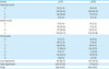
Table 2
Association with LATS1 expression and clinicopathological factors
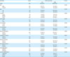
Table 3
Association with LATS2 expression and clinicopathological factors
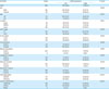
Low expression of LATS2 was significantly correlated with older age (≥65, P=0.027), more advanced AJCC stage (P=0.001) and T stage (P=0.001), LN metastasis (P=0.004), perineural invasion (P=0.004), poor histologic grade (P<0.001), and diffuse-type histology by the Lauren classification (P<0.001). Low LATS2 expression was also associated with lymphatic invasion (P=0.067) and distant metastasis (P=0.065), although these correlations did not reach statistical significance.
OS of gastric cancer patients with LATS1 and LATS2 expression
The follow-up period for patients ranged from 5 to 113 months (median interval, 60.7 months). During the follow-up period, 85 patients died. Low expression of LATS1 was significantly associated with poor OS rates, according to Kaplan-Meier analysis (P=0.037). The 5-year OS rate for patients with high expression of LATS1 was 71.2% compared with 56.9% for patients with low LATS1 expression. Patients with low LATS2 expression had significantly shorter OS rates than patients with high LATS2 expression (P=0.037). For LATS2, the 5-year OS rate was 76.5% vs. 64.4% for high expression vs. low expression, respectively (Fig. 3). In the Cox regression analysis, LATS1/2 expression was a significant factor in univariate OS analysis (LATS1, P=0.038; LATS2, P=0.038), but no significant difference was observed in multivariate OS analysis (Table 4).
Fig. 3
Kaplan-Meier survival analysis with log-rank test. Low expression of LATS1 (A) and LATS2 (B) were significantly associated with poor OS, respectively.
LATS = large tumor suppressor; OS = overall survival.
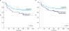
Table 4
Univariate and multivariate analysis of factors in patients with gastric cancer by Cox regression analysis

DISCUSSION
The LATS gene family is one of the core components of the Hippo pathway, an emerging signaling pathway that is an essential regulator of homeostasis. In mammals, the Hippo pathway has been reported as a tumor suppressor signal that inhibits cell proliferation and promotes cell apoptosis. Inactivation of the Hippo pathway results in cell growth, enlargement of organ size, and tumorigenesis. In 1995, lats was first identified by Xu et al. [5] as a tumor suppressor gene of Drosophila melanogaster. Subsequently, Xu's study group also isolated mouse (Lats1) and human (LATS1) genes [18]. In addition, human LATS2 was isolated after the discovery of mouse Lats2 by Yabuta et al. [19], and the researchers suggested that LATS2 may also be a tumor suppressor gene like LATS1. LATS1 and LATS2, members of the family of LATS proteins, play an important role in maintaining cellular homeostasis [6] including cell proliferation, cell apoptosis, cell migration [11], transcriptional regulation, and maintenance of genetic stability [12].
LATS1 and LATS2 have been implicated in a number of human malignant tumors. Abnormal expression or gene mutation of LATS1 contributes to malignant transformation and histologic progression in cervical squamous cell carcinoma [13]. Down-regulation of LATS1 and LATS2 is correlated in breast cancer with alterations of p53 function and cell migration [20]. LATS2 expression is lower in human prostate tumors than in normal prostate tissue, and LATS2 is a negative regulator of the androgen receptor [21]. Overexpression of LATS1 promotes YAP phosphorylation and inhibits tumorigenesis in human renal cell carcinoma [22]. It has also been reported that LATS1 contributes to better prognosis through negative regulation of YAP in non-small cell lung cancer (NSCLC) [23].
However, the expression of LATS in gastric cancer and its implications have received little investigation. In a Chinese study, LATS1 expression was found to be downregulated and negatively associated with YAP in human gastric cancer [16]. The same study reported that LATS1 expression was lower in gastric cancer than in normal gastric epithelium and adenoma, and its expression was significantly decreased in gastric cancer with LN metastasis than that without LN metastasis [16].
In the present study, we hypothesized that decreased expression of LATS1 and LATS2 would be associated with a poor prognosis in patients with gastric cancer and that overexpression of LATS1 and LATS2 would be correlated with a better prognosis through inhibition of tumor progression. We found that overexpression of LATS1 and LATS2 was significantly associated with factors indicating a good prognosis. Positive expression of LATS1 was associated with lower AJCC stage, negative LN metastasis, absence of perineural invasion, well/moderately differentiated grade, and intestinal-type histology based on the Lauren classification. LATS2 expression was also associated with a number of clinicopathological factors. In addition, the expression of LATS1 and LATS2 was associated with T stage, and the proportion of LATS2-positive cases increased as the T stage decreased (P=0.007). LATS2 expression was observed in 40/103 cases with pT1 (51.9%), 15/42 with pT2 (19.5%), 14/75 with pT3 (18.2%), and 8/44 with pT4 (10.4%).
Expression of LATS1 and LATS2 was also significantly associated with stage (LATS1/AJCC stage, p=0.014; LATS2/AJCC stage, P=0.002). LATS1 overexpression was observed in 102 of 121 stage I patients (84.3%), and lack of expression of LATS2 was found in 74 of 91 stage III patients (81.3%), but of 8 patients with stage IV, none showed expression of LATS2.
In the present study, we used different cut-off values for positive expression of LATS1 and LATS2. Because the differences in the cut-off values are presumably the result of the different dilutions of the antibodies, it is possible that there is a difference between the degree of LATS1 and LATS2 expression.
Most normal gastric mucosal tissues were weakly positive for LATS1 and LATS2. However, the expression of LATS varies in gastric cancer; as shown in the previous results, overexpression was associated with good prognostic factors as well as with tumor suppressor function. However, with loss of expression, the tumor suppressor function is weakened, and it is associated with factors indicative of a poor prognosis. These findings suggest that activation of LATS acts as a tumor suppressor in gastric cancer. Further studies will be needed to confirm the tumor suppressive activity of these proteins in non-neoplastic gastric epithelial cells or to explain the manner in which expression of LATS1 and LATS2 plays an important role.
In addition, we investigated whether the expression of LATS was associated with survival in patients with both LATS1 and LATS2 expression. The number of patients with expression of both LATS1 and LATS2 was 74/264 (28.0%), and there was a statistically significant correlation between survival and simultaneous LATS1/2 expression (P=0.016).
A potential limitation of this study was the lack of significant results in the subgroup and multivariate analyses. Thus, additional studies should be performed to support the recognition of LATS expression as an important prognostic factor.
In conclusion, the expression of LATS1 and LATS2 in patients with gastric cancer was found to be related to a number of clinicopathologic factors, and was associated with a good prognosis, including a higher survival rate. If detailed mechanisms underlying the loss or overexpression of LATS can be identified, this knowledge may have a positive effect on the treatment and clinical outcomes of various malignant tumors, including gastric cancer.
 XML Download