

 PDF
PDF ePub
ePub Citation
 Print
Print


Introduction
Gastric cancer is the fourth most common cancer and one of the leading causes of cancer-related deaths in the world1 (and the second most common cancer and the third most common cause of cancer deaths2 in Korea). Patients' survival and prognosis are largely determined by the stage of gastric cancer.3 Owing to national cancer screening programs, the detection of early gastric cancer has improved in Korea. However, mass endoscopic screening, regardless of risk factors, may not be cost-effective and can cause procedure-related complications. Therefore, it is necessary to develop better diagnostic biomarkers of early-stage gastric cancer.
MicroRNAs (miRNAs) are small noncoding RNAs that regulate gene expression through post-transcriptional silencing of target genes.4 The dysregulation of miRNAs is related to cancer development through abnormal proliferation of cells, apoptosis, and differentiation.5 In gastric cancer, various miRNAs have been shown to have tumor suppressor or oncogenic functions.6 Although many gastric cancer-related miRNAs have been identified, the reported results have been inconsistent and they require further validation. The aim of this study was to determine the expression profiles of miRNAs and to validate up- and down-regulated miRNAs in gastric adenocarcinoma.
Materials and Methods
1. Materials
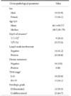
We included patients undergoing gastrectomy for potentially curable gastric cancer in Daejeon St. Mary's Hospital in 2013 and 2014. The inclusion criterion in this study was histologically confirmed adenocarcinoma of the stomach, and all patients received complete resections, with an attempt at complete tumor removal. We collected information on clinical characteristics of the patients retrospectively. Cancer staging was performed according to the 7th edition of the American Joint Committee on Cancer TNM (tumor-node-metastasis) criteria.7
All of the samples were obtained from surgical specimens of patients with gastric adenocarcinoma, and all of the patients provided written informed consent for the use of these tissues for research purposes. We obtained 34 gastric cancer tissues and 34 paired adjacent nontumorous tissues (1 pair from each patient). Among the 34 paired samples, we used 2 paired samples for GeneChip microarray analysis. The basic patient demographic characteristics are summarized in Table 1. The study protocol was approved by the Institutional Review Board of Daejeon St. Mary's Hospital.
2. RNA extraction
In total, 34 gastric cancer tissues and paired adjacent normal
gastric tissues were homogenized with Tissue Lyser2 (Qiagen, Germantown, MD, USA). TRIzol Reagent (Invitrogen, Carlsbad, CA, USA) was used for total RNA extraction. Low-molecular-weight RNAs (<200 nucleotides [nt]) were separated from total RNA using mirVana miRNA purification columns (Ambion, Austin, TX, USA) for microarray analysis and quantitative polymerase chain reaction (qPCR). The quality and quantity of each RNA sample were determined using a NanoDrop ND-1000 spectrophotometer (Agilent Technologies UK, Ltd., West Lothian, UK).
3. Microarray analysis of microRNA expression
Each total RNA sample (700 ng) was labeled and hybridized using the FlashTag Biotin HSR RNA Labeling Kit (Genisphere LLC, Hatfield, PA, USA). Total RNA was labeled using polyA polymerase. Biotin-labeled RNA was hybridized to the Affymetrix miRNA v3.0 array for 16 to 18 hours at 45℃. GeneChips were washed and stained in an Affymetrix Fluidics Station 450 (Affymetrix, Santa Clara, CA, USA) and scanned using the Affymetrix GeneChip scanner 3000 7G (Affymetrix). We analyzed the data with RMA-DABG using the normalization method. The normalized and log-transformed intensity values were analyzed using Expression Console (Affymetrix). Fold-change filters included the requirements that the miRNAs be expressed at greater than or equal to 2 fold of control levels for upregulated miRNAs and less than 2 fold of control levels for downregulated miRNAs.
4. Reverse transcription and real-time quantitative polymerase chain reaction
To validate the fold change results of the miRNA arrays, we used real-time qPCR to assess the expression of four randomly selected miRNAs including two upregulated (miR-196b-5p and miR-375) and two downregulated (miR-483-5p and miR-486-5p) miRNAs (Table 2). Total RNA was extracted from 34 paired cancer and nontumor tissues using NucleoSpin RNA II Kit (Macherey-Nagel, Düren, Germany). Reverse transcription was performed using mature miRNA-specific primer sets and the miRNA Reverse Transcription Kit (Applied Biosystems, Foster City, CA, USA). We purchased miRNA-specific TaqMan-based probes from Applied Biosystems, and real-time qPCR was performed on a 7500 Fast Real-Time PCR System (Applied Biosystems). The fold change for each miRNA was calculated using the comparative Ct (2 −ΔΔCt) method, and RNU48 (a small nuclear RNA) served as an endogenous control. We performed all of the reactions in triplicate for each sample.
5. Gene ontology analysis
We used this analysis to predict target gene function and to conduct pathway analysis of the differentially expressed miRNAs identified in GeneChip microarray analysis (Table 3). The DAVID bioinformatics resources tool was used for this purpose.
6. Statistical analysis
This analysis was performed in the PASW Statistics ver. 18.0 (IBM Co., Armonk, NY, USA). Student's t-test was used for evaluation of differences in miRNA expression between cancer and normal tissues and for measuring significance in clinicopathological parameters. Data were considered statistically significant at P<0.05.
Results
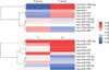
1. MicroRNA expression profiles in gastric cancer and normal gastric tissues
miRNA expression patterns significantly differed between gastric cancer and normal gastric tissues (Fig. 1). Fold-change filters included the requirements that the miRNAs be expressed at greater than or equal to 2 fold of control levels for upregulated miRNAs and less than 2 fold of control levels for downregulated miRNAs (Table 3).
Ten miRNAs satisfied the above requirements. Among these differentially expressed miRNAs, 5 (miR-196b-5p, miR-215, miR-375, miR-1, and miR-370) were significantly overexpressed, and 5 (miR-2861, miR-483-5p, miR-486-5p, miR-622, and miR-149-3p) were significantly underexpressed in gastric cancer tissues compared with normal gastric tissues.
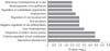
2. Gene ontology analysis
The pathways for the 10 miRNAs identified in the microarray analysis included vascular development, positive regulation of cell proliferation, regulation of protein kinase activity, tube morphogenesis, negative regulation of cell differentiation, anti-apoptosis, regulation of cell development, angiogenesis, regulation of cytoskeleton organization, morphogenesis of an epithelium, and branching morphogenesis of a tube (listed in the order of decreasing frequency; Fig. 2).
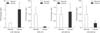
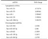
3. Validation of microarray data by real-time quantitative polymerase chain reaction
miR-196b-5p and miR-486-5p were upregulated 4.04- and 1.17-fold, respectively, in the gastric cancer tissues compared with the adjacent nontumorous gastric tissues. miR-375 and miR-483-5p were downregulated 14.2- and 3.1-fold, respectively, in the gastric cancer tissues compared with the nontumorous gastric tissues. The relative expression changes of 2 of these miRNAs (miR-196b-5p and miR-486-5p) according to qPCR analysis were consistent with the microarray analysis, but two miRNAs (miR-375 and miR-483-5p) yielded the opposite results, as noted in the histogram (P<0.05) in Fig. 3. miR-196b-5p and miR-375 showed statistically significant differences in the qPCR analysis.
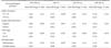
4. Relations between miRNA expression and clinical features of the patients with gastric cancer
We analyzed the relations between clinical characteristics and the qPCR results for the four miRNAs (miR-196b-5p, miR-375, miR-483-5p, and miR-486-5p). miR-375 was found to be associated with low T-stage cancers (T1+T2) and differentiated histologic grade (Table 4); the other miRNAs had no significant associations.
Discussion
Recently, aberrant miRNA expression was reported in various solid cancers, including breast, lung, pancreas, and colon cancer.89101112 Altered miRNA expression contributes to cancer initiation and progression. The relations between miRNAs and tumors have thus become the focus of many cancer studies. Numerous research articles describe the utility of miRNAs as biomarkers for early detection and for progression, recurrence, and theranostics.13 However, few researchers have assessed miRNAs in gastric cancer in Korea.
In gastric cancer, various miRNAs have been reported to be associated with tumor development, progression, metastasis, and prognosis. Overexpressed miR-296b-5p significantly accelerates gastric cancer cell growth through attenuation of Caudal-related homeobox 1-induced antigrowth effects.14 miR-223 provokes migration of nonmetastatic gastric cancer cells and invasion in vitro and in vivo by suppressing erythrocyte membrane protein band 4.1-like 3 expression.15 Li et al.16 demonstrated that a 7-miRNA signature (miR-10b, miR-21, miR-223, miR-338, let-7a, miR-30a-5p, and miR-126) can predict the clinical outcome of gastric cancer. There have been studies showing that circulating miRNA in blood could serve as noninvasive biomarkers. The plasma concentrations of miR-17-5p, miR-21, miR-106a, miR-106b, and miR-18a have been reported to be significantly higher in patients with gastric cancer than in healthy volunteers.1718 Another study showed that miR-21 has clinical value as a noninvasive biomarker and potential diagnostic value with good specificity and moderate sensitivity.19 To date, definitive noninvasive biomarkers have not been found, and larger-scale studies are required.
In this study, we identified miRNAs with significantly up-or down-regulated expression in gastric cancer tissues versus adjacent nontumorous tissues. We identified 5 upregulated and 5 downregulated miRNAs in gastric cancer tissues by GeneChip microarray analysis. Of these 10, we validated 2 upregulated and 2 downregulated miRNAs by qPCR. The 2 upregulated miRNAs are miR-196b-5p and miR-375, and the 2 downregulated miRNAs are miR-483-5p and miR-486-5p. We also determined that miR-196b-5p and miR-375 are significantly associated with gastric cancer.
The miR-196 family is encoded at three paralogous loci in the mammalian homeobox (HOX) clusters, and several HOX genes are regulated via targeting of their 3 ′untranslated region.20 The miR-196 family is composed of miR-196a and miR-196b, and mature miR-196b differs from miR-196a by 1 nt.21 miR-196 is upregulated in pancreatic adenocarcinoma, breast cancer, leukemia, esophageal adenocarcinoma, and colonic cancer,2223242526 and downregulated in melanoma.27 Several reports on miR-196 in gastric cancer have been published. Tsai et al.28 noticed that the lack of promoter methylation in miR-196b may explain its overexpression in the majority of gastric cancers and that miR-196b overexpression may be a tumor marker. This group later reported that miR-196b expression is significantly repressed by the transcription factor E26 transformation-specific sequence-2 (ETS2), whose expression is implicated in a reduced incidence of solid tumors.29 A knockdown of ETS2 significantly promotes migration and invasiveness of gastric cancer cells.29 Alterations in ETS2 and miR-196b expression in gastric cancer cell lines influence the expression of epithelial-mesenchymal-transition-related genes.29 Sun et al.30 reported that aberrant miR-196a overexpression and consequent downregulation of p27kip1 may contribute to gastric carcinogenesis. In another study, miR-196a/-196b levels were found to be frequently upregulated in human gastric cancer and significantly associated with clinically advanced stages and lymph node metastasis. In that experiment, miR-196a and miR-196b promoted gastric cancer cell migration and invasion in vitro and metastasis in vivo, and radixin was identified as a direct functional target of miR-196a and miR-196b.31
Our study revealed that miR-196b-5p expression is increased in gastric cancer tissues compared with nontumorous tissues. To date, there have been a few studies on miR-196b-5p in gastric cancer. Xie et al.32 and Liu et al.33 reported that miR-196b-5p is downregulated in a gastric cancer cell line. This result contradicts ours, but those studies were conducted on a gastric cancer cell line rather than primary tumor tissues. Another microarray study revealed that miR-196b-5p is upregulated in human gastric cancer tissues, in agreement with our results, but no validation was performed there.34
miR-375 is predominantly expressed in islets of the pancreas.35 miR-375 has an important function in the formation of insulin-secreting pancreatic islets; this result was obtained by inhibition of miR-375 maturation with morpholinos in zebrafish.36 miR-375 is downregulated in multiple types of cancer and it inhibits the hallmarks of cancer by targeting several key oncogenes, including Yes associated protein 1 (YAP1), astrocyte elevated gene-1 protein (AEG-1), 3-phosphoinositide dependent protein kinase-1 (PDK1), and insulin-like growth factor (IGF) 1R.37 Ding et al.38 reported that miR-375 is repressed in more than 90% of stomach cancer samples compared with their nontumorous counterparts and suppresses proliferation of malignant stomach cells partially by targeting Janus kinase 2 (JAK2). Tsukamoto et al.39 also reported that reduced miR-375 expression may enhance survival of gastric carcinoma tumors by activation of the PDK1/Akt survival pathway; therefore, miR-375 may be a tumor suppressor miRNA in stomach cancer. Helicobacter pylori infection is known to be a risk factor of gastric cancer. In one study, miR-375 was found to be downregulated in response to H. pylori infection and to target JAK2. JAK2 activates signal transducer and activator of transcription 3 and promotes neoplastic transformation by changing the expression of B cell lymphoma 2 (BCL-2) and twist related protein 1 (TWIST1).40 Conversely, Zhang et al.41 reported that expression of miR-375 is increased and may predict recurrence risk in patients with recurrent stomach cancer after surgical resection. Because of these discrepancies, further studies involving clinical data are needed to confirm the function of miR-375 in gastric cancer. In the present study, miR-375 was downregulated according to the qPCR analysis but upregulated in the microarray analysis.
The miR-483-5p gene is located in the chromosomal 11p15.5 region in the second intron of the IGF2 gene and expresses two mature forms (miR-483-5p and miR-483-3p).42 To date, few studies have been conducted to reveal the function of miR-483-5p in gastric cancer, and its relation to carcinogenesis remains uncertain. Wang et al.42 reported that miR-483-5p is downregulated in human gliomas compared with normal brain tissues and that miR-483-5p can repress glioma cell proliferation by directly targeting extracellular signal regulated kinase 1, which is a core controller of cell proliferation and cell differentiation. Qiao et al.43 suggested that miR-483-5p plays a role of an angiogenesis inhibitor. In contrast, miR-483-5p is reportedly upregulated in plasma samples of patients with hepatocellular carcinoma.44 miR-483-5p expression was found to be downregulated in the microarray analysis in the present study. We were unable to confirm the association of miR-483-5p with gastric cancer by qPCR analysis.
miR-486 is located in chromosomal 8p11 region, the site undergoing frequent genomic loss in various carcinomas.45 Solomides et al.46 and Tan et al.47 found that miR-486-5p is downregulated in lung cancer tissue and may serve as a new diagnostic biomarker of lung cancer. Oh et al.45 reported that miR-486 expression is decreased in gastric cancer, and that olfactomedin4 is a direct target gene. Recently, Zhu et al.48 reported that miR-486-5p concentration is consistently elevated in plasma from patients with gastric cancer. The exact roles of this miRNA in human cancers have not been fully characterized to date. In the present study, miR-486-5p expression was decreased in the microarray, but the qPCR results were not significant.
We also analyzed the relation between these miRNAs and clinical features. A lower T stage (T1+T2) and a differentiated histologic type were associated with high miR-375 expression. This result supports the previously reported function of miR-375 as a tumor suppressor.3839 Other miRNAs yielded no significant correlations.
We predicted possible genetic pathways related to the 10 above-mentioned miRNAs using bioinformatics resource tool analysis. Among the pathways involved, regulation of protein kinase activity was related to both miR-375 and miR-483-5p, and angiogenesis was related to miR-483-5p.
The interaction between dysregulated miRNAs and their potential target genes is complex, and miRNAs can be influenced by various factors, such as pathology, hypoxia, infection, and cytotoxictreatment.49 This complexity may explain the inconsistent results on miR-375 and miR-483-5p in our study.
There are a few limitations to this study. First, our study includes a relatively small number of patients. Thus, further large-scale research is required to clarify the exact roles of miRNAs in gastric cancer. Second, we could not study the functional roles of the significantly expressed miRNAs in gastric cancer. However, our study does provide basic data for future research on mi RNAs in gastric cancer. The data from this study should facilitate further research on gastric cancer aimed at elucidation of the functional roles and clinical significance of miRNAs or at the development of miRNA-based noninvasive biomarkers.
In conclusion, our results revealed that several miRNAs are significantly differentially expressed between gastric cancer tissues and nontumorous tissues. We used real-time qPCR analysis to validate the expression of 4 randomly selected miRNAs, including 2 downregulated and 2 upregulated miRNAs. Two mi RNAs (miR-196b-5p and miR-375) were significantly expressed in the qPCR validation analysis. miR-196b-5p expression was consistent with the microarray analysis, but miR-375 yielded the opposite results. We propose that these 2 validated miRNAs may serve as diagnostic biomarkers of gastric cancer.

 XML Download