

 PDF
PDF ePub
ePub Citation
 Print
Print


Introduction
Although the indication for Helicobacter pylori eradication is well known, the number of gastric cancer (GC) cases that emerge after successful eradication (post-eradication GC) has increased.1 Eradication therapy has reportedly had the limited effect of inhibiting the occurrence of post-eradication GC.23456 However, the difficulty in diagnosing post-eradication GC is well known, and the eradication may gradually result in regenerative changes.7891011 Recent studies have attempted to understand gastric carcinogenesis after eradication to develop useful biomarkers for definitive diagnosis of post-eradication GC.
There are some discrepancies in reports of the reversibility of H. pylori-induced mucosal degradation after eradication, possibly because of differences in study design, population, H. pylori strain, and measurement method.2345612 In fact, most of these studies used biopsy samples randomly taken from background mucosa using conventional endoscopy. Considering the patchy distribution of regenerative tissue after eradication, sampling errors in those studies might cause such discrepancies. According to the field carcinogenesis theory, comprehensive endoscopic imaging to reveal morphological and biological alterations in the entire stomach might be useful in early detection or prediction of gastric carcinogenesis after eradication.
Congo red chromoendoscopy (CRE) provides a comprehensive view of the extension of acid-secreting mucosa through a pH-dependent color reaction.13 In normal corpus mucosa, Congo red clearly changes from red to blue-black; in inflamed or atrophic mucosa, the lack of acid secretion does not induce a color change. By investigating regional change in the acid nonsecretory area (ANA) after eradication, as well as the GC location in the ANA remaining after eradication, previous studies demonstrated that CRE might be useful to identify ANA as a high-risk area of gastric carcinogenesis.14151617 On the other hand, autofluorescence imaging (AFI) provided real-time pseudocolor images based on biological condition in gastric mucosa, with no need for drug injection or dye spraying.18 Despite a lower resolution, AFI visualizes inflamed/atrophic/metaplastic gastric mucosa and normal corpus mucosa as bright green and purple or deep green, respectively, and some studies suggested the usefulness of AFI in prediction or early detection of GC, even after eradication.192021 However, no study reported reliable AFI findings of precancerous areas in background gastric mucosa after eradication.
Although AFI is more convenient and less invasive than CRE in a clinical setting, it remains unclear whether AFI might be an alternative to CRE in identification of high-risk areas of gastric carcinogenesis after eradication. Hence, in a single-center comparative study of patients with metachronous GC detected more than 6 years after curative endoscopic submucosal dissection (ESD) and subsequent eradication, we investigated the consistency between ANA in CRE and atrophic mucosa in AFI as a primary endpoint.
Materials and Methods
1. Patients and methods
From August 2014 to October 2015, we prospectively included 27 sequential patients (19 men and 8 women; mean age, 69.9±8.5 years old) with metachronous GC detected during surveillance endoscopy in our department after curative ESD for primary GC followed by eradication.22 Immediately after clinical interviews and 13C-urea breath tests, the patients were examined using high-resolution tri-modal endoscopy (EVIS-FQ260Z; Olympus Co. Ltd., Tokyo, Japan). The study protocol was approved by the Tohoku University Hospital Ethics Committee (2014-2-022-1, UMIN000020849), and all subjects provided written informed consent. The following exclusion criteria were applied: 1) age less than 20 or greater than 85 years; 2) history of chemotherapy, radiotherapy, or upper abdominal surgery; 3) use of nonsteroidal anti-inflammatory drugs, antibiotics, or any antisecretory drugs, such as proton pump inhibitors or H2 blockers; 4) a serious general condition; 5) mental disorders; 6) pregnancy; 7) inappropriate biopsy specimens; and 8) refusal to participation in this study.
2. Endoscopic procedure
Fifteen minutes after an intramuscular injection of pentagastrin 6 µg/kg (Sigma, St. Louis, MO, USA), an endoscopist (K.U.) performed endoscopic inspection in a sequence from white-light mode to AFI followed by CRE.13 The endoscopic findings of tumor location, macroscopic type, and tumor size in white-light mode were recorded, according to the classification for GC.23 Together with endoscopic visibility of GC, the extent of corpus atrophy was assessed and categorized by the Kimura-Takemoto classification in each endoscopic mode.24 As for the classification of corpus atrophy in AFI, cardia surrounded by purple mucosa was defined as closed type, and green mucosa on the cardia was defined as open type. A solution of 0.3% Congo red and 0.2 M sodium bicarbonate was sprayed over the entire surface of the stomach using a spray catheter. Any residual dye was carefully removed by suction. The areas of corpus mucosa with color shifting from red to blue-black (i.e., the functional area with acid secretion) were also semiquantitatively evaluated using the Kimura-Takemoto classification. Two biopsy specimens were obtained from each site from the black-colored mucosa and red-colored mucosa of the corpus within 2 cm of their boundaries. Immediately after the examination, their findings and the consistency of the extension of corpus atrophy, e.g., open-type or closed-type atrophy, between AFI and CRE were recorded on the formatted report of endoscopic examination (Fig. 1). To assess interobserver or intraobserver agreement, 10 high-quality images of corpus mucosa were selected by an independent investigator (Y.T.), and were inserted in a PowerPoint presentation with a black background as an independent testing set. Each of the image sets was numbered, and the slides were randomized in a computer-generated fashion with the image number displayed during the PowerPoint slide show. More than 2 months later, 2 of the authors (K.U. and N.A.), blinded to the clinical condition, independently reviewed endoscopic images saved in the slide show, and evaluated their extension.
3. Histological evaluation
The biopsy specimens were fixed in formalin, embedded in paraffin, serially sectioned, and stained with hematoxylin and eosin. The severity of neutrophil (activity) and lymphocytic infiltration (inflammation), glandular atrophy (atrophy), and intestinal metaplasia was graded, according to the updated Sydney system (none, 0; mild, 1; moderate, 2; and severe, 3), by an independent investigator (Y.A.) who was blinded to clinical information.25
4. Outcomes
The primary outcome was the consistency of the extension of corpus atrophy between AFI and CRE. To elucidate factors causing inconsistency in the extension of corpus atrophy, the secondary outcomes were 1) the difference in interobserver and intraobserver agreements of endoscopic findings of the extension of corpus atrophy, and 2) the difference in histological characteristics of biopsy specimens taken from the red-colored mucosa and the black-colored mucosa in the CRE findings.
5. Statistical analysis
Statistical analysis was performed with JMP Statistical Software ver. 11 (SAS Institute Inc., Cary, NC, USA). Parametric data were expressed as the mean±standard deviation, and nonparametric data were classified as the median (interquartile range). The scores of the updated Sydney system, clinical characteristics, or endoscopic evaluation of corpus atrophy were compared using the chi-square test or Fisher exact test, as appropriate. P<0.05 was considered to indicate statistical significance. Interobserver and intraobserver agreement was estimated using k statistics.26 The κ value was poor (≤0.20), fair (0.21~0.40), moderate (0.41~0.60), good (0.61~0.80), or very good (0.81~1.00).
Results
1. Demographics of post-eradication gastric cancer
The characteristics of secondary GC cases that were detected later than 82.8±50.1 months after eradication were as follows. In 4, 11, and 12 cases, the tumors were located in the upper-third, middle-third, and lower-third of the stomach, respectively. The numbers of macroscopic protruded, flat, and depressed tumor types were 1, 8, and 18, respectively. There were 20 tumors measuring <20 mm and 7 measuring ≥20 mm. All tumor staging was limited to the mucosal layer or slight invasion into the submucosa. The histotypes of the ESD specimens were of the well-differentiated, poorly differentiated, and mixed type in 24, 1, and 2 cases, respectively.
2. Endoscopic findings of post-eradication gastric cancer
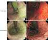
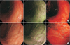
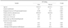
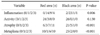
All GC lesions were detected using white-light endoscopy (Fig. 2), and all were located in the ANA in the CRE findings. Additionally, AFI clearly visualized 21 GCs in the magenta/purple colored area of the green-colored gastric mucosa. However, the remaining 6 GCs were obscurely depicted through AFI inspection, because 2 and 4 of the tumors were in the slightly pale, colored area surrounded by purple-colored or mosaic-colored corpus mucosa (Fig. 3). Thus, endoscopic evaluation of corpus mucosa may be one of the most important factors in AFI-based diagnosis of post-eradication GC. In univariate analysis, the tumor location in the upper or middle part of the stomach and the color pattern of background mucosa in AFI were identified as significant factors affecting poor visibility of GC in AFI (Table 1). Additionally, shorter duration after eradication and a heterogeneous histotype of GC seemed to be associated with poor visibility in AFI.
3. Endoscopic evaluation of atrophic change in corpus mucosa
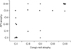
Next, to clarify the reliability of these endoscopic evaluations, we investigated the consistency of the extension of corpus atrophy between AFI and CRE and the interobserver and intraobserver agreements for each image to evaluate their extension. First, inconsistency between AFI and CRE was observed in 6 of the 27 patients (22.2%) (Fig. 4). Second, when we investigated the interobserver agreement between two individual endoscopists, the k values of CRE and AFI demonstrated good reproducibility of 0.75 (95% confidence interval [CI], 0.57~0.93), and fair reproducibility of 0.33 (95% CI, 0.14~0.53), respectively. Additionally, we investigated the intraobserver agreement, and the k value of CRE and AFI demonstrated very good reproducibility of 0.82 (95% CI, 0.12~0.59) and fair reproducibility of 0.29 (95% CI, 0.08~0.64), respectively.
4. Histological features of atrophic/non-atrophic corpus mucosa in Congo red chromoendoscopy
According to the updated Sydney score of biopsy specimens from acid secretory/non-secretory corpus mucosa, there were significant differences in inflammation, atrophy, and metaplasia (Table 2). These suggested a possible association between histological condition and the CRE findings in the post-eradicated corpus mucosa.
Discussion
We demonstrated for the first time that, compared with AFI, CRE might clearly visualize functional and histological alterations in the background mucosa of metachronous GC, which were detected more than 6 years after successful post-ESD eradication. These suggested that the evaluation of atrophic mucosa in AFI could be less reliable for identifying malignant areas after eradication than in CRE.
First, we considered the importance of CRE, rather than AFI, to directly visualize functionally degenerated mucosa with malignancy potential, even after successful eradication. We previously investigated regional change of the ANA during a 5-year follow-up after eradication, and reported that the ANA associated with marked atrophy was usually irreversible and had malignant potential.14151617 In this study, with a longer duration after eradication, all post-eradication GCs were observed in the ANA (i.e., functionally regressive mucosa), whereas 6 GC lesions were not diagnosed in the AFI findings. Further, the borderline between functional atrophy/non-atrophy of corpus mucosa in the CRE findings was revealed with higher interobserver and intraobserver agreement than with AFI. In fact, we also had data showing high consistency for the extension of corpus atrophy between AFI and white-light imaging (data not shown). In addition to previous studies demonstrating that the interobserver agreement for gastric atrophy in the white-light endoscopic findings might not be sufficient,27 the diagnostic accuracy of GC by CRE was reported to be much higher than that by routine endoscopy.28 Accordingly, these enhanced the reliability of the CRE findings in the detection of malignant mucosa after eradication.
Next, we considered a possible association between the CRE findings and the histological changes after eradication. Using CRE for subjects at a relatively late phase after eradication, we demonstrated significant differences in the scores of mononuclear inflammatory cell infiltration, atrophy, and metaplasia between the acid-secretory and non-secretory areas. Chronic H. pylori infection induces mucosal inflammation, which may lead to loss of function or existence of parietal cells, resulting in functional and histological degeneration toward malignant transformation.29 However, eradication therapy reportedly improves acid secretory function from an early phase, possibly due to removal of inhibitory factors with decreasing inflammatory changes.30 A study of the biological changes in gastric mucosa before and 12 weeks after eradication demonstrated improvement in the messenger RNA expression of H+/K+-ATPase, intrinsic factor, and M3 muscarinic receptor, while decreasing inflammatory mediators, without any increase in parietal cells.31 As for a later phase, we previously demonstrated an increase in the acid secretory area of CRE during a limited term of seven months to two years after eradication.151617 Accordingly, this study was focused on the intragastric condition at the time when the recovery of acid secretion after eradication is thought to be completed, and our results indicated that the acid secretion ability might be affected by both the degree of mononuclear cell inflammation and the histological atrophy/degeneration of parietal cells during the regeneration process after eradication. As a recent study revealed that mononuclear cell inflammation and atrophic change were gradually diminished during a 17-year follow-up after eradication,3 further studies using CRE long after eradication would clarify the mechanism of mucosal regeneration.
Recently, using in vivo confocal laser endomicroscopy (CLE) with fluorescein tracers before and after eradication in 42 patients, Ji et al.32 demonstrated that functional and morphological reversibility of a disrupted mucosal barrier by eradication was observed in atrophic gastritis but not in metaplastic mucosa which might be consistent with the point-of-no-return theory. Although the CLE findings may yield functional and morphological alterations in a point-sampling manner, such functional imaging can provide a new approach to clarify biological events during post-eradication carcinogenesis. From this point of view, we demonstrated that CRE comprehensively visualized a high-risk area of gastric carcinogenesis after eradication, which might be advantageous for a proof-of-concept of the field defect of gastric cancerization.
However, there were some potential limitations. First, we used CRE, which may have clinical issues, such as complexity of procedure and possible toxicity in a murine study.2133 However, it was difficult to assess the possibility of adverse events with only one session of CRE in this study. We routinely removed residual dye during the procedure. Moreover, when we proceeded with this study, the protocol was well considered for a balance between the risk of CRE and the benefit for the determination of gastric carcinogenesis after eradication, and subsequently for the development of future strategy for post-eradication GC. These issues could result in a limited number of subjects, which may be associated with selection bias. We did not widely enroll patients who were surveyed after gastric ESD, and we did not suggest the clinical usefulness of CRE in terms of the detection/diagnosis of metachronous GC after eradication. Second, we adopted the updated Sydney system, which was not originally designed for research. Additionally, we took biopsies from predetermined locations, instead of using a four-point scale. In fact, the distribution of acid-secretory areas after eradication was reported to be patchy (i.e., non-sequential progression of corpus atrophy), which means that the biopsy points proposed by the system may be inappropriate after eradication. However, we demonstrated different histological manifestations of targeted biopsies in the CRE findings, despite being only a few centimeters away from one another. Third, these techniques were used in a fixed sequence by the same endoscopist. The interpretation of the CRE findings might be affected by prior endoscopic findings, resulting in the possibility of information bias. However, it is impossible to conduct a cross-over trial at the same time, because the sprayed dye is not perfectly removed during the examination. Instead, we showed interobserver and intraobserver agreement for a random sequence. Therefore, it may be more advantageous to perform both examinations at the same time, which can eliminate the possibility of chronological change in the intragastric status at each of the examinations.
In conclusion, we suggest that, rather than AFI, CRE might reproducibly identify the distribution of functionally and morphologically irreversible mucosa after eradication, i.e., a cancerization field, in a site-specific manner. The combination of CRE with biopsy/marking devices may provide useful information to address the mechanism of mucosal regeneration with acid-secretion recovery, as well as gastric carcinogenesis after eradication. Further research will be needed to establish future strategies with risk stratification of post-eradication GC.

 XML Download