

 PDF
PDF ePub
ePub Citation
 Print
Print


Introduction
Gastrointestinal stromal tumors (GISTs) occur most frequently in the stomach.1234 Wedge resection of the stomach with R0 resection is regarded as a standard treatment because of the low risk of lymph node metastasis from the tumor.2567 Relatively small sized tumors have been safely treated by various laparoscopic approaches since the first report of laparoscopic wedge resection (LWR) of a gastric submucosal tumor in 1991.891011
Although laparoscopic gastric wedge resection for extraluminal tumors can be performed easily, intraluminal or small tumors are difficult to localize laparoscopically, requiring an intragastric approach or gastrotomy for tumor resection.1213141516 Consequently, the possibility of spreading cancer cells in the abdominal cavity arises due to the additional manipulation of the tumor and luminal exposure during these procedures. Thus, it could be a risk factor for peritoneal recurrence. In addition, if the tumors have ulcerations, the risk of cancer cell dissemination might increase.1718
So far, there is no report regarding the long-term outcomes of laparoscopic gastric wedge resection with gastrotomy for the treatment of gastric GISTs. In the current study, we compared the oncologic safety of LWR with gastrotomy (LWR-G) and LWR without luminal exposure. Specifically, the long-term consequences for ulcerative GISTs requiring luminal exposure during operation were investigated. Recurrence patterns were also analyzed.
Materials and Methods
1. Patients
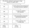
Between March 2003 and December 2013, 205 LWRs of the stomach were performed in patients with histologically confirmed gastric GISTs at the Department of Surgery at Yonsei University College of Medicine. The study included 193 patients who underwent LWR of the stomach. Of the 193 patients, 26 underwent LWR-G, while the other 167 underwent LWR without gastrotomy (LWR-C). Patients were excluded if they had a ruptured tumor at the time of diagnosis, underwent palliative resection or an endoscopic procedure before the operation, or had insufficient data regarding mitotic rate for proper staging (Fig. 1). On the basis of preoperative endoscopy, endoscopic ultrasound, and abdominopelvic computed tomography scanning, LWR was generally indicated for relatively small gastric submucosal tumors up to 5 cm in the early period, and later on, the indication was expanded to include tumors larger than 5 cm. The type of resection was selected at the surgeon's discretion according to the tumor location and size. Patients who were pathologically classified as high risk according to the National Institutes of Health-Fletcher classification were recommended for treatment with imatinib mesylate (Gleevec®; Novartis, Basel, Switzerland), whenever possible. Clinicopathologic characteristics, short-term outcomes, and long-term outcomes, including recurrence and survival status, were analyzed retrospectively.
This retrospective study to compare outcomes with another surgical techniques was approved by the Institutional Review Board (IRB) of Severance Hospital, Yonsei University College of Medicine (4-2015-0865).
2. Surgical technique
Various laparoscopic gastric wedge resection techniques have been described in the literature.1512131518 When tumors grow outward from the stomach toward the peritoneal cavity, wedge resection using endolinear staplers can be performed easily without considerable manipulation of the tumor.121314 In cases of intraluminal tumors in the posterior wall of the stomach, transgastric tumor-everting methods followed by gastrotomy of the anterior wall of the stomach facilitate tumor resection.13141920 When performing this procedure, the lesion was identified by endoscopy or laparoscopic ultrasonography, and the optimal site of the anterior stomach wall for gastrotomy was chosen. After an incision was made, the tumor was removed by transecting the inverted posterior wall using endolinear staplers. The anterior wall was closed with endolinear staplers or with a laparoscopic suture technique.1419 When relatively larger intraluminal tumors are located in the anterior wall, the eversion method can facilitate tumor resection and minimize excessive resection of the normal gastric wall.15 For this procedure, a gastrotomy was created about 1 cm from the tumor margin by using intraoperative laparoscopic ultrasonography. Then, the tumor was exteriorized via the incision and resected by endolinear staplers. The advantage of this procedure was that the gastrotomy was closed at the same time the endolinear staplers were applied.15 For intraluminal tumors near the cardia or the pylorus, the preferred procedure may be intragastric wedge resection with single incision intragastric or conventional intragastric procedures to minimize deformity of the esophagogastric junction or the pylorus.18212223 For the single incision intragastric procedure, two wound protectors were used. The anterior gastrotomy was made and pulled out of the abdominal incision, and then another wound protect was applied via gastrotomy. After laparoscopic removal of the endoluminal tumor, the anterior gastrotomy was closed with endolinear staplers.23 For small intraluminal tumors, intraoperative endoscopic guidance was sometimes required.1618
3. Statistical analysis
Clinicopathologic features, short-term outcomes, and long-term outcomes were analyzed using the IBM SPSS statistical software ver. 20 (IBM Co., Armonk, NY, USA). Categorical and continuous variables were analyzed by the χ2 (or Fisher's exact test) and Student's t test, respectively. Survival curves were depicted by the Kaplan-Meier method and compared by log-rank test. Two-sided P-values of less than 0.05 were considered to be statistically significant.
Results
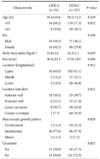
The clinicopathologic features are shown in Table 1. Of a total of 193, 26 patients (13.5%) underwent LWR-G, and 167 patients (86.5%) underwent LWR-C. The mean age did not differ between the two groups (P=0.419). The mean body mass index was similar between the two groups (P=0.659). Mean tumor size and longitudinal location between the LWR-G and LWR-C groups were comparable (P=0.696 and P=0.913, respectively). However, more anterior wall-located, intraluminal tumors were found in the LWR-G group compared to the LWR-C group (P=0.022 and P=0.029, respectively). A significantly larger number of tumors (n=13, 50.0%) in the LWR-G group than in the LWR-C group had ulcerations (P=0.021).
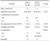
Operative outcomes are shown in Table 2. All patients included in the current study underwent complete tumor resection without gross spillage, tumor rupture, or microscopic margin involvement. No open conversion was noted in any patients. The mean operation time for LWR-G was 64.0 minutes compared with a mean of 55.9 minutes for LWR-C (P=0.313). Resumption of soft diet and postoperative hospital stay did not differ bweteen the two groups. No postoperative complications were noted in the LWR-G group, while 3 patients (1.8%) in the LWR-C group had complications.
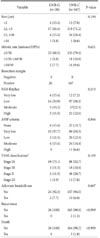
Pathologic and long-term outcomes are shown in Table 3. There was no statistically significant difference in the mean tumor size (P=0.696). When the tumor size and mitotic rate were stratified according to the current risk criteria of gastric GIST, there was no difference in proportion of the size and mitotica tre between the two groups (P=0.190 and P=0.621, respectively). Consequently, risk based on National Institutes of Health-Fletcher, American Forces Institute of Pathology criteria, and TNM classification did not differ between the groups (P=0.573, P=0.846, and P=0.193, respectively). Adjuvant therapy with imatinib mesylate was administered to 2 patients (7.7%) and 10 patients (6.0%) in the LWR-G and LWR-C groups, respectively. During the median follow-up period of 36 months, 2 patients in the LWR-C group had recurrence in the liver and in the remnant stomach at 63 and 12 months after surgery, respectively. oN patient in the LWR-G group had recurrence. The characteristics of the patients with recurrence are depicted in Table 4. During the follow-up period, 3 patients in the LWR-C group died, but there were no gastric GIST-related deaths.
Discussion
In the current study, we found that peritoneal recurrence due to potential spillage of cancer cells may not happen during LWR-G for gastric GIST. In addition, ulcerative GIST treated with the same procedures did not increase the risk of peritoneal recurrence. Gastrotomy with everting/eversion methods or intragastric procedures may not increase the rate of recurrence due to tumor spillage by luminal exposure during LWR. In addition, we observed that the pattern of recurrence, especially peritoneal recurrence, was significantly low after LWR-G or LWR-C for relatively small GISTs.
GISTs are rare tumors and a distinctive histopathological group of intestinal neoplasms of mesenchymal origin.3 They comprise fewer than 3% of all gastrointestinal cancers, and the most frequently involved site is the stomach, followed by the small intestine.324 Complete R0 resection without lymphadenectomy for primary non-metastatic GIST remains the only curative treatment.26 Since the first report of LWR of a gastric submucosal tumor, laparoscopic resection of GIST is considered to be feasible and safe from both the technical and the oncologic point of view.6817
Although LWR for gastric GIST has demonstrated acceptable oncologic outcomes, the current indication is limited to relatively small tumors due to possible rupture of the tumor into the peritoneal cavity during the procedure.101125262728 The European Society for Medical Oncology guidelines recommend laparoscopic gastric wedge resection for tumors less than 2 cm in size, while the National Comprehensive Cancer Network and Japanese guidelines recommend the procedure for tumors less than 5 cm by experienced surgeons.102728 The risk of possible tumor cell dissemination into the peritoneal cavity can be greatly increased when it is performed in conjunction with more complicated procedures such as those requiring transgastric or intragastric approaches. Transgastric everting for intraluminal masses in the posterior wall of the stomach and eversion methods for intraluminal tumors in the anterior wall have shown satisfactory short-term outcomes.1415 However, during these procedures, tumors can be manipulated more vigorously, and gastric luminal contents can be spilled out into the peritoneal cavity.1718 Furthermore, for tumors with ulcerative lesions, the potential hazard is expected to worsen.18 If the GIST ruptures into the peritoneal cavity, the recurrence rate increases by almost 100%.29
However, we experienced only 2 cases of recurrence in the LWR-C group during the median follow-up period of 36 months. The overall incidence of recurrence was 1.0% in the studied patients. The results were comparable to other reported series in the literature.2526 Even with ulcerative lesions, which comprised 50% of the lesions in the LWR-G group and had low to intermediate risk in most cases (data not shown), we did not observe any recurrence during the follow-up period. The low incidence of recurrence in the current study might have been achieved by careful manipulation of the tumors to avoid tumor rupture and significant spillage of gastric contents into the peritoneal cavity. There was no intraoperative tumor rupture in the current study. Second, the indications were limited to relatively small tumors so that only a small portion of patients were classified as high-risk. Finally, by using wound protectors to avoid direct contact of the tumors with the surgical wound when retrieving the specimens, the possibility of cancer cell dissemination to the surgical wound was minimized. In addition, all patients who received adjuvant imatinib treatment showed favorable outcomes without recurrence in the study period.
To our knowledge, this is the first study in the literature to investigate the long-term outcomes of LWR-G for gastric GISTs compared to conventional LWR. Our study revealed that gastric lumen exposure during procedures, which facilitate tumor localization and resection while avoiding excessive resection of the remnant stomach to prevent functional and structural deformities can be safely performed even in ulcerative lesions, given meticulous handling of the tumors and properly indicated patients. However, the study has several limitations. First, the current study was conducted retrospectively in a single center. Second, long-term outcomes, especially recurrence patterns, could not be properly assessed because the number of high-risk patients was small in both groups. Therefore, the results of the current study should be compared to a larger number of cases to determine the exact impact of luminal exposure during LWR for ulcerative lesions in high-risk patients.
In conclusion, LWR-G did not increase overall or peritoneal recurrence for the selected patients. This technique might be safely performed even for ulcerative gastric GISTs.

 XML Download